FIGURE 47.1 The pericranial scalp flap derives its primary blood supply from deep branches of the supraorbital and supratrochlear vessels.
HISTORY
A detailed history regarding prior cranial surgery is important to determine reconstructive options. Prior scalp incisions may interrupt the vascular supply and limit the length of the pericranial flap. Prior reconstruction with a pericranial flap may preclude the use of a new pericranial flap although it is often possible to harvest one from the other side if only one pedicle was used previously. It is also possible that a galeal flap could be used if the pericranium is not available. Caution is advised when reading operative reports since many surgeons incorrectly use the term galeopericranial flap to describe flaps that are actually pericranial flaps. Prior orbital surgery may also compromise the vascular anatomy of the pericranial flap.
A history of prior radiation is also important to ascertain since the vascularity of the scalp may be compromised. Dissection of a pericranial or galeopericranial flap may further compromise the vascularity and result in necrosis of the scalp. A history of trauma to this area is important in decision making.
PHYSICAL EXAMINATION
The scalp is examined to note the location of prior incisions or trauma (Fig. 47.2). These may limit the length and design of the pericranial flap. The sensory and motor innervation of the frontal scalp is assessed. Intact vasculature (supraorbital and supratrochlear vessels) can be confirmed using Doppler sonography.
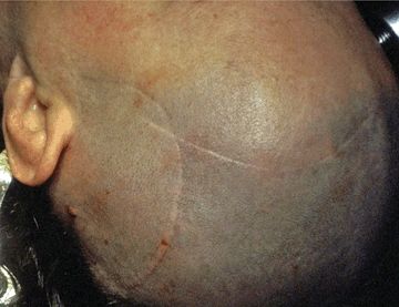
FIGURE 47.2 This patient has prior temporal and bicoronal scalp incisions that limit the placement of scalp incisions and the length of an anteriorly based pericranial flap.
INDICATIONS
The primary use of the pericranial flap is for the repair of large defects of the dura following a craniofacial resection using either a transcranial or an endonasal approach. Neoplasms commonly encountered include sinonasal malignancy (olfactory neuroblastoma, squamous cell carcinoma, adenocarcinoma, sinonasal undifferentiated carcinoma), olfactory groove meningioma, and suprasellar neoplasms (craniopharyngioma, extrasellar giant pituitary adenoma). Cerebrospinal fluid (CSF) leaks resulting from surgery or trauma or arising spontaneously can also be repaired with a pericranial flap. The pericranial flap can reach as far as the lower clivus.
Pericranial flaps can be used to provide soft tissue coverage of exposed bone following excision of a tumor or resulting from radionecrosis. The vascularity of a pericranial flap is sufficient to support a skin graft. Pericranial flaps may be used to provide protection of critical tissues such as the dura and the carotid arteries. This not only protects the structures from desiccation but also promotes vascularization and healing. Similarly, a pericranial flap can be used to cover exposed hardware or to separate the sinuses from the deep tissues.
A pericranial flap is a superior alternative to adipose tissue grafts for obliteration of the frontal sinus following excision of tumors, debridement of infected bone, or comminuted fractures. The anterior wall of the frontal sinus is excised, and all mucosa is drilled from the walls of the sinus. The pericranial flap is placed into the sinus providing vascularized tissue that facilitates rapid healing and avoids infection. In contrast to adipose tissue, there is no loss of tissue volume. The anterior wall of the sinus can be replaced with titanium mesh.
CONTRAINDICATIONS
The only absolute contraindication to the use of a pericranial flap is the absence of a blood supply. This may result from prior surgery or trauma or the needs of the current surgery (e.g., orbital exenteration). Small defects of the dura can be repaired effectively using alternative reconstructive methods. A relative contraindication is a scalp with compromised vascularity from prior radiation therapy. Lateral defects, especially those requiring bulk, are better repaired using a temporalis transposition flap.
PREOPERATIVE PLANNING
Every cranial base surgery requiring reconstruction should have a backup plan including one or two alternatives, in case the blood supply to the flap is sacrificed or the flap provides insufficient coverage. If the vascular supply to the pericranium is in doubt, Doppler sonography can be used to confirm blood flow through the vascular pedicle. The surgical approach should be planned in a manner such that the reconstructive tissues are not compromised.
SURGICAL TECHNIQUE
The patient’s head should be accessible for a bicoronal scalp incision from ear to ear. The head can be supported on a padded horseshoe headrest or immobilized with a Mayfield head holder. The pins should be placed posterior to the bicoronal plane and allow subgaleal dissection posterior to the incision. A thin strip of hair (2 cm) is shaved along the incision line, and the proposed incision is infiltrated with approximately 20 mL of 0.5% Xylocaine/1:100,000 epinephrine. Temporary tarsorrhaphy sutures are placed, and the field is prepped and draped. A large Ioban plastic drape (3 M Company) covers the entire surgical field (Fig. 47.3). This avoids the placement of bulky towels over the eyes and compression of the orbital tissues when the scalp is reflected inferiorly. Staples are placed parallel to both sides of the bicoronal incision to prevent movement of the drape and towels.
Stay updated, free articles. Join our Telegram channel

Full access? Get Clinical Tree


