FIGURE 46.1 Pericranial flap and other scalp flap options. (TPFF, temporoparietal fascia flap [superficial temporal artery]; TEMP M, temporalis muscle flap [deep temporal artery].)
The introduction of endoscopic endonasal skull base techniques has created new challenges in the reconstruction of the anterior cranial fossa. While a dural defect similar to an open craniofacial resection can be achieved endoscopically (with a dural defect from the meridian of the orbit to the meridian of the opposite orbit and from the optic chiasm to the frontal sinus), the ability to reconstruct such a defect is more challenging than in open surgery. The pedicled nasoseptal flap has produced excellent outcomes as a primary reconstructive option for endoscopic intradural skull base surgery. It has an axial blood supply based on the posterior nasoseptal artery and is readily available in the sinonasal cavity; therefore, it does not create a separate donor site defect. However, most sinonasal cancers have midline and septal involvement, and therefore the need to take widely negative margins often precludes the use of a nasal septal flap in this setting. Recently, the pericranial flap has become a useful and novel flap for endoscopic reconstructions. Operative techniques for both open and endoscopic pericranial flap use require an intimate knowledge of the vascular anatomy of the anteriorly based pericranial flap, as well as an understanding of the complex layers of the frontal and temporal scalp.
The pericranial flap is supplied by the supraorbital and supratrochlear arteries (Fig. 46.2). The main trunk and superficial branches of these arteries course from the orbit into the galea and frontalis muscle layer and give rise to deep branches that supply the pericranium. These deep branches can arise at the level of the orbit or within 10 mm of the orbital rim. It is important to understand that the deep branches that supply the pericranium may exit up to 1 cm above the exit point of the supraorbital and supratrochlear foramina; dissection of the flap beyond this level can injure the blood supply.
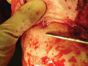
FIGURE 46.2 Supraorbital arterial supply to the pericranial flap.
HISTORY
Once the patient has been appropriately evaluated for the presence of a skull base tumor of the cribriform area or anterior cranial base, surgical planning proceeds around the goals for the primary tumor resection. Staging for distant and regional metastatic disease in the setting of sinonasal cancer is performed, and a firm histologic diagnosis is required for optimal treatment planning. Following a thorough discussion with the patient regarding operative options (whether it be an open or endoscopic craniofacial resection), the surgeon must inquire about prior procedures and prior trauma to the scalp area to know whether a pericranial flap is a viable reconstructive option. Patients with prior bicoronal approaches often have a truncated flap due to the low placement of the coronal incision. In the setting of revision surgery, the vascular pedicles of the pericranial flap may have been interrupted, or the scalp flap was elevated without regard to the potential future use of a pericranial flap; therefore, it can be scarred and/or have a significant numbers of holes. Poorly vascularized flaps that are not fully intact make for a suboptimal skull base reconstruction. It should also be noted that tumors of the sinonasal region and skull base can involve the orbit, as well as the soft tissues of the face and glabella. If an orbital exenteration has to be undertaken, sacrifice of the flap pedicle necessarily occurs on that side. If a significant amount of facial soft tissue has to be resected from the periorbital region or frontal scalp, the pedicle of the flap may be compromised. A history of prior radiation therapy is not a contraindication to the use of a pericranial flap.
PHYSICAL EXAMINATION
Physical examination should proceed to evaluating any prior trauma or scarring, as well as the presence of tumor protruding into the soft tissues of the glabella, scalp, forehead, or orbits. The ophthalmic division of the trigeminal nerve as well as the frontal branches of the facial nerve are at risk with harvesting a pericranial flap. A detailed examination of the cranial nerves, paying close attention to these two cranial nerves, should be performed. If the patient has had prior surgery or trauma, the flap pedicles can be evaluated with Doppler ultrasound to see if they are intact. This confirms vascularity at the supratrochlear and supraorbital exit points; however, it does not fully confirm vascularity of the entire axial flap.
Indications
In my opinion, a large dural defect with a sinonasal fistula is an absolute indication for reconstruction of the skull base with a vascular flap. Indications for reconstruction with a pericranial flap include an anterior skull base defect with resection of the dura resulting in an intra-arachnoidal and sinonasal CSF fistula. Relative indications include extradural resection without intraoperative CSF leak but in a patient undergoing radiation therapy.
Contraindications
Contraindications to the use of pericranial flaps include patients without an intact flap pedicle or suitable flap quality.
PREOPERATIVE PLANNING
Imaging studies, whether it be CT or MRI, should be devoted to the evaluation of the extent of the primary tumor. There are no special imaging studies that need to be devoted to planning the reconstruction with the pericranial flap. Next is preoperative planning of the primary route of access for resection of the tumor. An open craniofacial resection is going to require coronal access to the scalp in a very wide plane, and therefore the pericranial flap and the pedicles would need to be actively protected during the exposure and resection of the tumor. However, if an endoscopic transcribriform resection is planned, then the endonasal resection can proceed endoscopically until negative margins are obtained. If the operation must be converted to an open craniofacial resection to obtain clear margins, then the above principles still apply. However, if that is not the case, the operative surgeon should understand that the endonasal defect must include a Draf III frontal sinusotomy to allow for wide frontal sinus access and drainage since the pericranial flap is going to be transposed across the midline of the floor of the frontal sinus. With an endoscopic transcribriform approach, I have a separate instrument table and endoscope set that are clean for the scalp portions of the case.
SURGICAL TECHNIQUE (VIDEO 46.1)
Surgical techniques include the open extracranial pericranial flap as well as the endoscopically assisted pericranial flap for endonasal reconstruction; these will be discussed separately.
Description of the Open Anteriorly Based Pericranial Flap
Stay updated, free articles. Join our Telegram channel

Full access? Get Clinical Tree


