CHAPTER 34 The Nasal Septum
Anatomy and Embryology of the Nose
Development of the nasal airway begins during the fourth week of gestation. Collections of neural crest cells undergo proliferation and form the nasal placodes.6,7 On the fetal face, adjacent cells proliferate and give rise to the medial and lateral nasal processes. The frontal and maxillary processes fuse to give origin to the lateral two thirds of the upper lip, the superior alveolar ridges, and the palatal shelves. The medial nasal processes join the maxillary processes to form the philtrum and columella; they also fuse with the frontal prominence to form the frontonasal process, which eventually encompasses the nasal bones, the frontal bones, the cartilaginous nose, the ethmoid bones, the central incisors, and the hard palate. With the growth of the medial and lateral nasal processes, two nasal pits form that invaginate until only the nasobuccal membrane remains. This membrane eventually ruptures by the 10th week, thereby allowing communication between the nose and the nasopharynx.6,8
The nasal septum develops as a downgrowth from the merged medial nasal processes and the nasofrontal process, thus defining the right and left nasal cavities. The nasal septum and the palatine processes begin to fuse anteriorly during the ninth week, and fusion is completed posteriorly by the 12th week.6,8
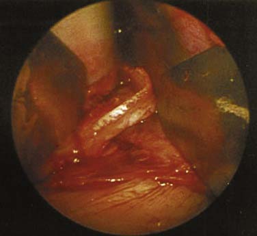
During the late embryonic period, the epithelium invaginates on each side of the nasal septum, thereby forming diverticula that are known as the vomeronasal organs. A vomeronasal cartilage develops ventral to each diverticulum. Shortly before birth, the vomeronasal organs begin to regress and usually disappear completely; the vomeronasal cartilages are usually the only adult remnants. These narrow strips of cartilage are located between the inferior edge of the cartilage of the nasal septum and the vomer.8 They are often found in conjunction with a septal spur, and they can be used as cartilage grafting material when other sources are absent or exhausted (Fig. 34-1).
The cartilaginous framework of the nose develops from three paired mesenchymal condensations in both the medial and lateral nasal swellings. Part of this cartilage begins to ossify, thereby forming the membranous bone encasing the vomer and perpendicular cartilaginous plates. The perpendicular plate of the ethmoid and the nasal bones do not completely ossify until puberty.6 Injury to the nose in the young child or teenager may not elicit a true fracture, but it may instead create growth changes in this transitioning tissue, which may ultimately result in a deviated posterior bony septum or even the formation of a spur.
The nasal septum has functional and aesthetic significance. The septum is the main support structure of the external nose.9 It divides the nose into two cavities, regulates airflow through the nose, and supports the mucosal lining of the nasal cavities.9 Where once the lining was afforded greater significance, now both the mucosa and the cartilage are recognized as codependent and necessary.
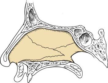
The bony components of the septum include the nasal crest of the palatine bone, the nasal crest of the maxilla and premaxilla, the vomer, the perpendicular plate of the ethmoid, the nasal crest of the frontal bone, and the spine of the paired nasal bones. The anterior septum is composed of the quadrilateral cartilage, with its caudal-most projection extending beyond the nasal spine. Because of the complicated embryologic development of the septum, any one of a number of arrangements of the bone and cartilage contributions may be encountered during surgery (Fig. 34-2).
Turbinates
The lateral nasal wall is composed of the laminae papyracea of the lacrimal bone, portions of the ethmoid bone, and the inferior and middle nasal conchae or turbinates. The turbinate bones develop from a cartilage ossification center during the fifth intrauterine month. The inferior, middle, and superior turbinates are composed of a thin bone for structural support and covered by an adherent mucoperiosteum. Stratified squamous epithelium is found on the anterior tip of the inferior turbinate, whereas pseudostratified ciliated columnar respiratory epithelium covers all other surfaces. The continually beating ciliated mucosa provides constant motion to the mucous blanket within the nose; this blanket acts as a cleaning and filtering system for the upper respiratory tract and also helps to maintain the moisture content within the nose. The turbinates maximize the effective intranasal surface area and rapidly humidify and warm the inspired air.10
Blood Supply and Innervation
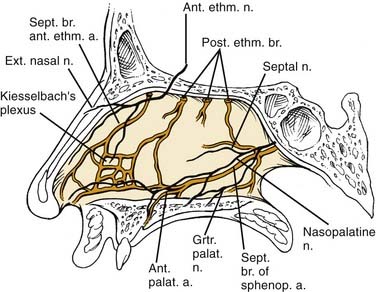
A variably located incisive artery and its associated neural fibers are found at the superior border of the vomer. This neurovascular bundle may be encountered when trimming a badly deviated maxillary crest or when elevating periosteum when a nasal floor approach is needed. Control of bleeding from this site may be obtained by infiltrating the incisive foramen from below, “plugging” the site from above, or carefully using suction Bovie cautery. After resection or trimming of the maxillary crest or work on the nasal spine, patients may complain of numbness or pain of the central incisors or of the mucosa of the hard palate just posterior to the incisors. This lack of sensation or complaint of pain is generally a short-lived phenomenon (Fig. 34-3).
Nasal Obstruction
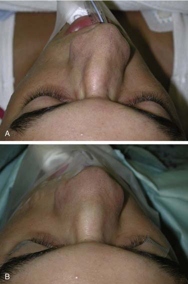
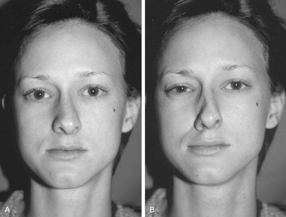
Thorough examination and visual inspection of the patient who complains of nasal airway obstruction are essential for diagnosis and treatment planning. Visual assessment of the external appearance of the nose is of utmost importance. This examination initially focuses on the size, shape, symmetry, and straightness of the nose; one should document the size of nostril openings, the thickness of the alae, and the width of the columella. Columellar widening may be seen with caudal septal cartilage deviation, splaying of the medial crura, or excess soft tissue. Septoplasty will correct airflow only if air can get into the nose. A previously fractured nose can hold the dorsal septum off the midline, and no septoplasty alone can correct this problem (Fig. 34-4). Such situations require sharply freeing up the septum from the upper lateral cartilages and performing medial osteotomies and a septoplasty with release of the septum from the maxillary crest for correction. Deformity of the nasal vault can narrow the diameter of the nasal passage. The patient with a crooked or C-shaped nose often has a severely deviated septum as well. Before examining the nose internally, one must observe the patient’s nose during normal and exaggerated nasal breathing, watching the side walls for evidence of internal or external collapse (Fig. 34-5). Care must be taken when assessing the chronic sniffer or “nasal neurotic,” however, who will show collapse as a result of an overly aggressive inspiratory force being generated, regardless of the competency of the valve. Surgery for the chronic sniffer will generally lead to poor outcomes despite adequate anatomic support and deflection correction, and it will often include frequent postoperative phone calls and second opinions obtained with regard to his or her problem, which is still perceived as uncorrected.
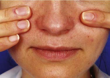
External evaluation of the patient’s nose should include performance of the Cottle maneuver, in which lateral distraction of the nasal valve is performed (Fig. 34-6). This maneuver may improve the sensation of nasal obstruction in cases of external nasal valve collapse or anterior septal deviation. A positive Cottle maneuver may suggest nasal valve compromise,11 but it is not always a reliable indicator, with many false-positive results seen. Lateralizing the upper lateral cartilage (ULC) and then assessing for improvement in the obstruction sensation may provide a more accurate assessment of the nasal valve. Lateralizing the upper lateral cartilage (ULC) with a cotton-tipped applicator or the use of a cerumen curette or similar instrument increases the internal nasal valve angle and, in those with a narrow angle, will provide improvement in airflow. The surgeon must also evaluate the nasal tip to determine whether the tip ptosis is contributing to decreased airflow (Fig. 34-7).
Deviated Nasal Septum
Deviation of the nasal septum is a common cause of unilateral nasal airway obstruction and may follow nasal and midfacial trauma. Trauma during birth, including forceps placement or passage through a narrow pelvic canal, can cause injury that may lead to early septal deviation or to deviation that is not evident until the more active growth phase of puberty. Minor trauma sustained early in life can be easily overlooked and frequently causes microfractures of the septal cartilage; healing of these microfractures leads to bending of the cartilage away from the side of injury. When this occurs early in life, it may lead to asymmetric growth of the entire nasal structure as a result of chondrocyte growth interruption. Patients with unilateral septal deviation most often complain of nasal obstruction on the contralateral side, a phenomenon called paradoxic nasal obstruction.12 Patients are confused when the physician explains that the nasal passage through which the patient feels airflow moves most freely is actually the smaller of the two sides. Decongesting the nose and then testing airflow through each nostril separately or showing the patient an endoscopic photograph of each passage can help the patient accept the explanation and understand the true problem.
Nasal Valve Angle and Nasal Valve Area
The internal nasal valve, as described before, is the narrowest portion of the nasal cavity and, therefore, any compromise of the components of the valve creates symptoms of nasal obstruction. The angle is bound medially by the septum and laterally by the inferior edge of the upper lateral cartilages and the anterior aspect of the inferior turbinate; this junction forms a trapezoidal configuration (Fig. 34-8). This angle widens and narrows with nasal muscular contraction and relaxation on inspiration and expiration. The nasal valve is normally 10 to 15 degrees in white patients and wider in nonwhite and Asian patients.13,14 Deformities of the adjacent nasal septum or loss of anatomic support structures can predispose the valve to collapse or narrowing, thereby causing nasal airway obstruction. The upper lateral cartilage at its junction with the septum may be thickened, twisted, or concave as a result of weakness or trauma or even absent if there was prior surgery. Webs of scarred mucosa may form between the septal mucosa and the lateral nasal wall or turbinates and may narrow the valve through scar contracture (Fig. 34-9). Adhesions that result in valve narrowing can create a fixed obstruction with a false-negative Cottle maneuver.
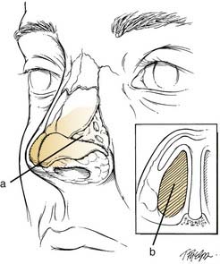
Figure 34-8. Schematic depicting (a) the external nasal valve area and angle and (b) internal nasal valve.
The external valve is a laterally based space boxed by the piriform aperture, the ULC and LLC attachments, and the caudal septum. Obstruction as a result of external valve compromise may be a postrhinoplasty phenomenon, a result of the aging process,15 or a result of caudal septal dislocation or trauma.
Nasal Physiology (Resistance and Airflow)
On inspiration, the airstream funnels through the vestibule of the nose, squeezes through the narrow valve, and disperses in the nasal cavity. Cadaver studies indicate that flow through the nasal valve of an adult at rest is accelerated to a linear velocity of 16 m/sec. As the airstream leaves the valve and enters the nasal cavity, its velocity decelerates by a factor of four; this deceleration promotes disruption of the air by allowing it to mix in the nasal cavity.16 This is essential for the effective conditioning of inspiratory air and may aid in the ability to smell. Conditioning consists of humidification, the removal of antigenic particles, and warming.
The single most important variable in nasal airflow is the diameter of the nasal passage. Dimensions and shape of the airway lumen and airflow velocity determine the magnitude of resistance to airflow. Resistance varies inversely and exponentially with lumen cross-sectional area and, because the nasal valve has the smallest lumen dimension, nasal valve resistance is very sensitive to structural or vascular displacements. In healthy noses, resistance to airflow is reduced by a third after topical decongestion, and resistance of the decongested nose is reduced by two thirds with wide alar retraction.17
The Nasal Cycle and Paradoxic Nasal Obstruction
The nasal cycle was first observed in 1927 by Heetderks,18 who described alternating turgescence of the inferior turbinates in 80% of a normal population. The turbinates in one fossa filled up while the opposite turbinates decongested. This cycle, which is controlled by the autonomic nervous system as described earlier, had a mean duration of  hours. He further observed and documented that the turbinates in the dependent nasal fossa filled when the patient was in the lateral decubitus position. Some postulate that this alternating positional obstruction has the purpose of causing a person to turn from one side to the other while sleeping. The nasal cycle is an alternating one, with the total resistance in the nose remaining constant. In patients with a fixed septal deviation and intermittent nasal obstruction, the interplay of the nasal cycle becomes evident; the sensation of obstruction frequently mirrors the congestion phase.
Some patients with a severely deviated septum learn to subconsciously eliminate the increased resistance sensation of the obstructed side. The opposite or normal side has a variable resistance as a result of the continued fluctuations of the nasal cycle. Nasal obstruction will be perceived on the open side during the turgescent phase; this phenomenon is known as paradoxic nasal obstruction.12,19 Knowledge of this phenomenon is essential to avoid misinterpretation of the significance of unilateral enlargement of one inferior turbinate, which is often present in normal noses. Surgical correction must address the deviated septum, but it must also address the etiology of the patient’s subjective complaint of increased resistance on the side on which clinical evaluation shows a more open nasal passage.
The dynamic portions of the nose (including the nostrils, vestibules, and lumen) can be narrowed due to the Venturi effect (see Fig. 34-5). Identification of collapsible nasal structures is important and should be addressed during preoperative review. Surgery can then be planned to widen the angle between the upper lateral cartilages and the septum with spreader grafts or to stiffen the nasal side wall with batten or umbrella graft placement.
Septal versus Turbinate Obstruction
When mucosal decongestion does not elicit intranasal airway changes or symptomatic improvement, bony turbinate hypertrophy, along with deviation to the septum and nasal valve compromise, should be considered. This obstruction is generally constant. One theory of bony turbinate hypertrophy is that it is based on a lack of structural resistance created by the midline nasal septum during development. The bony conchal and mucosal hypertrophy is considered compensatory and can be found in the patient with a septum that is significantly deviated away from the enlarged turbinate.10 The turbinate mucosa and underlying bone enlarge into the more open nasal passage in pursuit of normalizing nasal airway resistance.10
Stay updated, free articles. Join our Telegram channel

Full access? Get Clinical Tree


