CHAPTER 173 Surgical Anatomy of the Lateral Skull Base
Osseous Anatomy of the Lateral Skull Base
The temporal bone occupies a central position at the lateral skull base, being the most important anatomic landmark in the lateral skull base anatomy.1,2
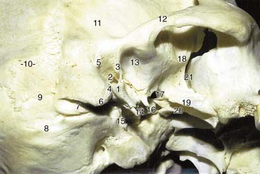
On lateral view, the squamosa presents a shape reminiscent of an open wing of a bird (Fig. 173-1). From its middle and caudal portions it contributes to the zygomatic arch and forms the superior wall of the external auditory canal. Its articulation with the tympanic bone in the anterosuperior external auditory canal forms the tympanosquamous suture. The zygomatic bone portion contributes to the roof of the glenoid fossa and the anterior part of the tympanic bone forms its posterior wall.
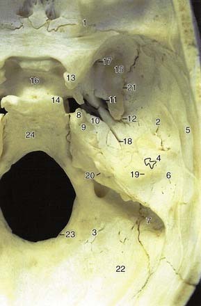
Along with the greater wing of the sphenoid and a portion of the parietal bone, the temporal bone forms the lateral boundary of the middle cranial fossa (Figs. 173-2 and 173-3). This medial aspect of the squamous bone forms the petrosquamous suture, which joins the caudal portion of the squamosa and the lateral superior portion of the petrosa. Anteriorly, at the articulation of the sphenoid with the squamous bone, is located the foramen spinosum, through which passes the middle meningeal artery into the cranium. The sulcus of this artery is easily identified on the medial surface of the squamosa (see Figs. 173-2 and 173-3).
The tympanic bone lies just inferior to the squamous portion and anterior to the mastoid and forms the tympanosquamous and tympanomastoid sutures with them (see Fig. 173-1). It is common for one edge of these suture lines (especially the tympanosquamous suture) to project as a spinous process exteding 1 to 3 mm into the lumen of the external auditory canal.1,3,4
The tympanic bone forms the anterior and inferior walls and a portion of the posterior wall of the external auditory meatus and is primarily responsible for the size and shape of the external auditory canal. As mentioned, it also composes the posterior wall of the glenoid fossa (see Fig. 173-1).
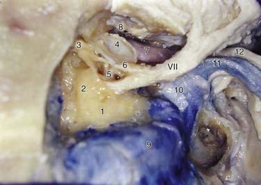
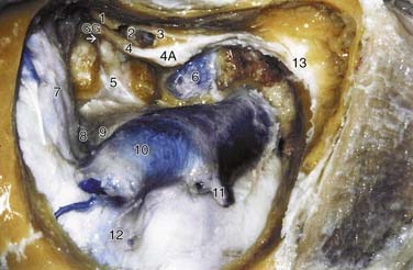
At the medial extension of the external auditory canal, the tympanic bone ends at the tympanic sulcus or annular sulcus, into which the tympanic membrane inserts (Figs. 173-4 and 173-5; see also Fig. 173-1). On its inferior surface, the tympanic bone articulates with the petrous bone, forming the petrotympanic suture. It also contributes to the formation of the internal carotid artery canal (ascending portion) (see Figs. 173-4 and 173-5). Also, on its inferior aspect and lateral to the carotid canal, the tympanic bone forms the styloid process (see Fig. 173-1). Slightly posterior and medial to this process is the stylomastoid foramen, through which the seventh cranial nerve emerges (see Fig. 173-5).
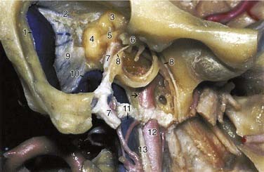
The osseous anterior wall of the external auditory canal abuts the temporomandibular joint and is of variable thickness. The bony inferior wall is thick and related to the anterior aspect of the jugular bulb (posteriorly) and carotid canal (anteriorly) (Fig. 173-6; see also Figs. 173-1, 173-4, and 173-5). The posterior wall separates the external auditory canal from the mastoid cells and the descending portion of the facial nerve (see Fig. 173-6).3,5
The mastoid is a pneumatized bony process located at the most posterior and inferior part of the temporal bone that, in reality, can be considered an extension of the squamosa and a lateral projection of the petrous portion (see Figs. 173-1 and 173-2). It has a somewhat triangular form, with the vertex directed inferiorly and the base superiorly.
Anteriorly, the mastoid process forms the tympanomastoid suture as it joins the posterior portion of the tympanic bone. Above and lateral to the tympanomastoid suture is the suprameatal spine, an osseous elevation of variable size that anchors the nonosseous portion of the external auditory canal and auricle. Immediately posterior to this spine and inferior to the temporal line is a slightly depressed region called the cribriform plate, a multiperforated region that serves as a landmark for surgical access to the mastoid antrum (see Fig. 173-1).
Medial to the mastoid tip, the posterior belly of the digastric muscle at its insertion forms an osseous depression or sulcus, running anterior to posterior. At its anterior termination, this sulcus is a landmark for the descending portion of the facial nerve and the stylomastoid foramen (see Figs. 173-1 and 173-6).
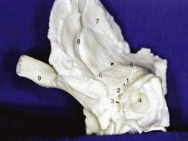
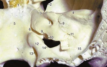
Viewed from above and from inside the cranium, the petrous bone is a key element in the osseous anatomy of the middle and posterior fossae. It has a configuration of a three-sided pyramid (see Fig. 173-2). The base of the pyramid constitutes its lateral surface and articulates with the squamous, tympanic, and mastoid portions of the temporal bone (Fig. 173-7; see also Fig. 173-2). Two sides of the pyramid are clearly recognized as medial projections: one superior, which forms much of the floor of the middle cranial fossa, and the medial surface, which faces the posterior cranial fossa, forming most of its lateral wall. The third and inferior side articulates with the occipital bone (see Figs. 173-2 and 173-3). The anterior medial limit of the pyramid (petrous apex) reaches the greater sphenoid wing laterally, the body of the sphenoid medially and superiorly, and the clivus portion of the occipital bone medially and inferiorly (see Figs. 173-2 and 173-3). The petroclival sutures lie on either side of the midline within the circumference of the foramen magnum.
Stay updated, free articles. Join our Telegram channel

Full access? Get Clinical Tree


