CHAPTER 37 Special Rhinoplasty Techniques
Aesthetic and reconstructive rhinoplasty is considered to be the most challenging and difficult of all facial plastic procedures. Every rhinoplasty is unique and presents the surgeon with a diversity of challenges for which he or she must have a variety of specialized techniques to address each patient’s anatomic and functional deformities. This chapter addresses some of the less common nasal deformities and the surgical techniques that can be used to correct them. These deformities require the inclusion of all aspects of combined septorhinoplasty, with frequent extension and modification of several techniques.1,2 These cases are particularly challenging in regard to improving the airway and gaining maximum improvement in appearance. A form of combined septorhinoplasty to correct both the external and the internal nasal deformities in one stage is virtually essential.1–4 This chapter focuses on modifications of the septum, nasofrontal angle, and nasal tip while detailing the principles and techniques needed to correct the twisted nose, the saddle nose, the noncaucasian nose, and the cleft-lip nose.
Analysis
Coordination of the preoperative analysis with the findings at the operative table can be accomplished through either the standard intranasal rhinoplasty approaches or the external rhinoplasty approach. The authors use both the standard rhinoplasty approaches and the external approach. The external approach is particularly useful in the twisted nose, the nose requiring implant materials, and the cleft-lip nose.2,5–7
Incisions and Soft Tissue Elevation
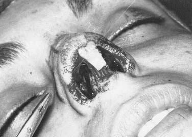
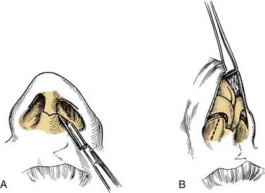
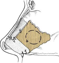
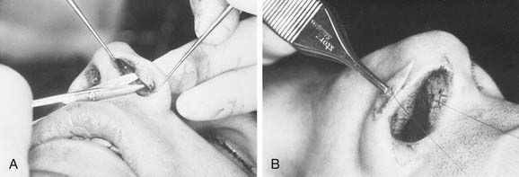
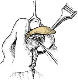
The authors generally use intranasal incisions for a deviated nose but will frequently use the open or external rhinoplasty technique when there are severe asymmetries, deviations, and tissue deficiencies.2,5–7 Some of the indications for use of the external rhinoplasty are as follows: (1) a nasal deformity that is difficult to precisely analyze; (2) one in which there is severe asymmetry of the lateral bony walls and the lower lateral cartilages; (3) when tissue deficiencies exist so that it is necessary to precisely place and suture implants in position8; (4) congenital anomalies such as the cleft-lip nose or other deformities in which profound asymmetries exist; (5) an extremely wide bony and cartilaginous dorsum, whether or not a hump is removed; and (6) for teaching purposes. The external approach involves a transverse midcolumella incision in a gull-wing curve rather than one with sharp angle. The authors use the gull-wing incision as opposed to the inverted gull-wing incision to create a single point rather than two points at the distal end of this diffuse random pattern flap. The incision is then extended upward along the caudal edge of the medial crus and laterally along the caudal margin of the lateral crus as an alar cartilage margin incision (Fig. 37-1). The elevation is initiated with sharp knife dissection, with the columella skin being elevated before the elevation is extended laterally over the lateral crura of the lower lateral cartilages. The external incision crossing the columella leaves a barely perceptible scar and offers excellent visualization by means of the “degloving” of the nose.
Septal and Columella Modifications
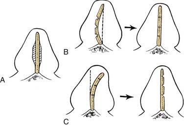
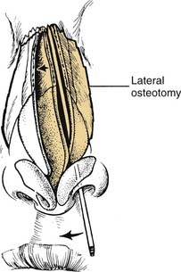
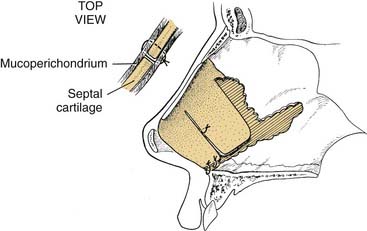
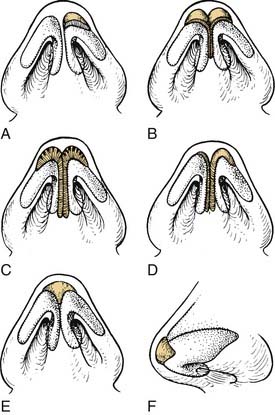
For the severely deviated or previously operated nose, it is usually necessary to elevate the mucoperichondrium on both sides of the septal cartilage, although it is preferable whenever possible to leave it attached on one side as the bone and cartilaginous angulations are corrected. Freeing fibrous contractions and septal angulations is essential in allowing the septal framework to straighten. The extent to which cartilage and bone must be removed or repositioned is dictated by the particular deformity encountered and the direction of the angulations. Generally, scoring or removal of thin strips of cartilage along the existing angulations is required to achieve straightening (Fig. 37-2). Release of all tension vectors allows a buckled septum to straighten. Determination of the final position of the septum may not be possible until osteotomies have been performed, particularly if the nasal bones and upper lateral cartilages are severely deviated from the midline. A final determination of what more, if anything, needs to be done to the septum, therefore, must sometimes await the completion of medial and lateral osteotomies, when the entire nose is positioned in the midline (Fig. 37-3) regarding deviations of the central bony complex).
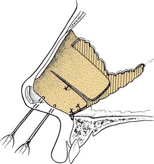
Transseptal coaptation sutures, which bring the two mucoperichondrial flaps together and pass between angulations or cartilage pedicles to prevent overlap, are great aids in this surgery (Fig. 37-4). Polyethylene splints have been used for a number of years to stabilize the septum in the early postoperative period and to prevent the development of synechiae, particularly when turbinate surgery has been performed. Two through-and-through sutures are always placed through the polyethylene splints. The sutures are placed so that one arm of the suture goes between cartilage pedicles or beneath the septal cartilage for support. The second arm of the suture goes back through the mucoperichondrium and cartilage. One may splint the septum further at a major angulation with a straight portion of thin perpendicular plate of the ethmoid.
Caudal Repositioning
When it is necessary to reposition the caudal portion of the septum, it is usually necessary to free the mucoperichondrium on both sides to release all extrinsic tension on the cartilage. The septal cartilage is then detached from the underlying anterior nasal spine and maxillary crest. Intrinsic tension of the septum is relieved by scoring, cross-hatching, and wedging. Once this caudal pedicle is freed of the tension vectors, it may be maintained in the midline by sutures that go around the anterior spine or through a burr hole in the spine (Fig. 37-5). To further prevent dislocation, an additional suture may be passed through the two mucoperichondrial flaps and between the bone of the spine and the cartilage. The cartilage pedicle is attached directly to the spine for alignment, and the two mucoperichondrial flaps when sutured together add further stability. When necessary, the caudal edge of the septum may be held in proper relation to the columella with interrupted sutures or direct through-and-through septocolumellar sutures (Fig. 37-6).
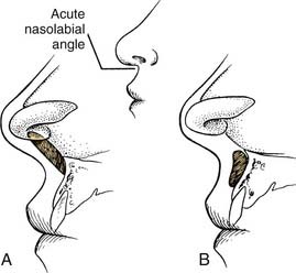
Shortening the Caudal Septum
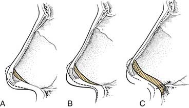
For the deformed nose with a drooping tip and columella retraction, the entire septocolumellar complex must be critically evaluated. In correcting the drooping tip, the lower lateral cartilages must be rotated cephalically, and strong tip support is needed. Both the caudal septum and the soft tissue may need modification to achieve this (Fig. 37-7). Frequently, there is an associated columella retraction, resulting in an acute nasolabial angle. Correction of the retraction requires straightening the caudal septum and placement of a columellar strut or a plumping graft (Fig. 37-8). Some limited resection of the caudal edge of the septum may be necessary to remove a portion of deformed septum that is dislocated off the anterior spine and projecting into one nostril. When the caudal edge of the septal cartilage is severely dislocated, it often creates distortion and retraction of the columella, even though adequate cartilage is present. Straightening and repositioning of the caudal septum within the columella sometimes corrects the retraction (Fig. 37-9). Rarely is it necessary to shorten the upper lateral cartilages, although reduction of the scroll along their caudal border may be necessary to reduce excessive width in that area.
Making the Final Septal Assessment
It is not until all septal reconstructive surgery and the osteotomies are completed that the final analysis of the position of the septum can be made. This position is particularly critical at the junction of the bony and cartilaginous portions of the nose, both internally and externally. At this point, the surgeon can determine whether adequate dorsal support has been maintained. If there is any tendency for the cartilaginous dorsum to sink inward, a draw suture can be passed through the skin to pick up either the septum alone or the upper lateral cartilages and the septum along the dorsum (Fig. 37-10). The suture is passed upward through the skin and held in position while all coapting and supporting sutures are placed and packing is completed. The draw suture can then be removed. In extreme cases, the draw suture can be passed through a thin external splint and tied over a bolus to remain in place for a week until the splint is removed.
The Septocolumellar Complex
These deformities can be addressed by trimming or sculpturing the medial crura and caudal septum as demonstrated in Fig. 37-11. Other structures that may contribute to the perceived or actual abnormalities of the caudal septum and columella include either ptosis of the lateral crus of the lower lateral cartilage, causing what appears to be a retracted columella, or retraction of the ala, resulting in excessive columella show (Fig. 37-12). These abnormalities must be corrected when addressing the lower lateral cartilages, either through reduction or augmentation (Fig. 37-13). Repositioning of the anterior maxillary spine, division of the depressor nasi musculi, sectioning of the frenulum, and defatting or augmentation of the nasolabial angle may also be necessary when addressing the caudal septum and columella.9
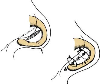
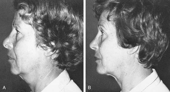
Figure 37-12. Cephalad retraction of the alar margin. A, Postoperative complications: the all-too-frequent supratip swelling or pollybeak deformity and cephalic retraction of the lateral alar margin. B, Correction is accomplished by reducing the cartilaginous nasal septum and removing supratip fibrous tissue, further sculpturing the lower lateral cartilages, freeing the lateral crus, and inserting a cartilage implant retrograde along the alar margin. (See also Fig. 37-13.)
Nasofrontal Angle Modifications
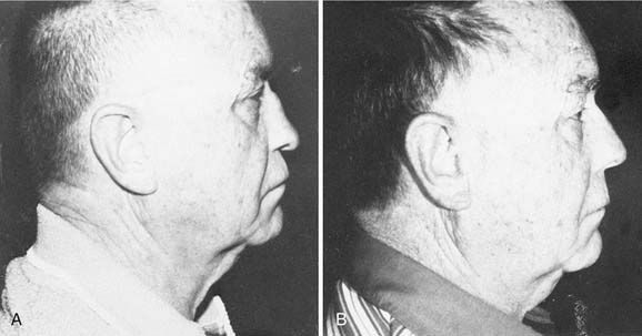
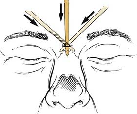
Subtle modifications in the nasofrontal angle can significantly enhance a patient’s nasal and facial profile. In patients with a shallow nasofrontal angle, the nose often appears to have excessive length. In this type of patient a 2-mm osteotome can be inserted through a stab incision in the midline over the nasal bones (Fig. 37-14). The nasal bones can be scored or weakened on either side with transverse osteotomies, thereby controlling the cephalic fracture site. This will allow the surgeon to control the site of the nasofrontal angle when the bony hump is removed. After removal, the angle may be smoothed or further deepened by the use of the rasp or rongeur.
Tip and Alar Asymmetries
Tip Grafts
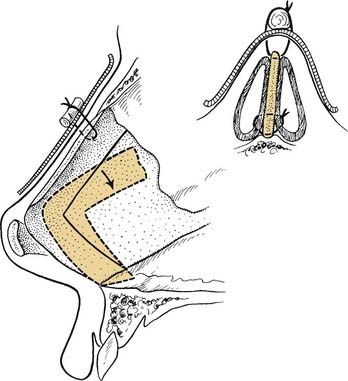
When there are severe asymmetries of the lower lateral cartilages, management usually requires modification of the more normal cartilage and the more abnormal side if one is to achieve a symmetric tip. The cephalic portion of the lower lateral cartilage is an excellent implant to be used in the nasal tip to overlie angulations, to fill small depressions, or for splinting cartilage by direct suturing. When more sturdy cartilage is necessary, sculptured or laminated septal or auricular cartilage is useful (Fig. 37-15). In the tip, these implants may be combined and sutured directly to a columella strut or to the domes of the lower lateral cartilages. If there is marked distortion with associated soft tissue contracture, it may be necessary to free the lateral crus of the lower lateral cartilage on the involved side of both dorsal and vestibular skin and then advance the lateral crus into the nasal tip between the two skin layers as in the technique used in the cleft-lip nose repair (Fig. 37-16).
The septum is an ideal source of grafts to be placed in the nasal tip. Such an implant has the effect of stabilizing the medial crura and domes in proper relationship and giving some increased tip projection. This technique is useful in both the severely traumatized tip and the previously operated nose, in which all too often too much tissue has been removed from the lower lateral cartilages. A nasal tip graft or shield graft should be sutured directly to the lower lateral cartilages. This may be accomplished readily through intranasal incisions (alar cartilage margin incisions) or can be placed with greater ease and less chance of dislodgment through an external rhinoplasty (Fig. 37-17). Migration of the shields has been the major problem with their use, and direct suturing prevents this displacement. By removal of a wedge from the inferior border of the shield, two points are created along the inferior border, assisting in stabilization to prevent movement.