Revision Rhinoplasty

CHAPTER 39 Revision Rhinoplasty


Key Points


Many consider rhinoplasty to be the most difficult facial plastic operation. The anatomy is intricate, three dimensional, and highly variable. Airway function depends on multiple factors, which are modulated with every surgical maneuver. Postoperative scar contracture and healing may alter surgical structural modifications over the lifetime of the patient. For these reasons, primary rhinoplasty frequently results in a suboptimal outcome. Moreover, the recent information explosion of the Internet has led to a patient population that has become increasingly knowledgeable and discriminating. Not surprisingly, it is estimated that 8% to 15% of primary rhinoplasty patients eventually undergo revision surgery.1


Secondary rhinoplasty poses additional challenges because the surgeon must restore nasal cosmesis and function beginning from a compromised baseline. The study of secondary rhinoplasty is the study of the complications of rhinoplasty. It is therefore critical that the surgeon undertake the process with as much information about the condition of the nose as possible.


Although many aspects of the preoperative analysis and surgical technique are similar to those used in primary rhinoplasty, these methods must be focused toward the problems and deformities most commonly found in secondary rhinoplasty. The ability to correct these problems is limited by the integrity of the existing and often compromised structures, the availability of grafting material, and the severity of the individual deformities. In many situations, the techniques themselves must be dramatically altered to restore structure to a nose that has been significantly destabilized. Frequently, secondary rhinoplasty becomes an operation of reconstruction more than of simple refinement.


Patient Interview


It is imperative that the surgeon obtain as much information as possible concerning the problems that led to the patient’s visit. A patient seeking secondary rhinoplasty surgery may be more knowledgeable, demanding, or distraught than a primary rhinoplasty patient. Because these patients have endured a period of disappointment regarding the appearance or function of their noses, they may have invested considerable energy and effort in considering revision surgery. The patients must understand that the greater the degree of baseline damage, the more limited will be the possibilities for improvement. Therefore, it is critical that the surgeon instill realistic outcome expectations in these patients. Surgery should not be performed unless common expectations are reached between patient and surgeon.


During the initial consultation, old operative notes and rhinoplasty diagrams can provide useful information. However, they may be incomplete or erroneous regarding grafting material, implants, and septal cartilage status. Preoperative and postoperative photographs may help in determining the nature and time course of the given problems. Photographs can also reveal which problems resulted from surgery and which problems predated the primary surgery. These original images are valuable because many patients may have had variant nasal anatomy that created a predisposition to a specific postoperative complication or deformity. The physician should list and prioritize patient complaints and gauge how reasonable and achievable these requests are. Functional airway problems that result from anatomic disturbances are surgically correctable. Intermittent obstruction is more likely attributable to mucosal inflammatory problems and should not represent the main impetus for surgery. Cosmetic problems should be discussed in detail with the aid of photographs and computer imaging. Each aesthetic complaint should be discussed with regard to possible etiology and prospects for repair.


Physical Examination


Cosmetic nasal analysis begins with a global assessment of the nasal deformities. Often one or two problem areas are immediately noticeable to the physician. These may include an asymmetric tip, dorsal irregularities, or a narrowed base. It is important to prioritize these deformities during surgery because surgery on each subunit of the nose affects the appearance of the others. The surgeon must modify a given structure based on the status of adjacent structures. Knowing that one aspect of the nose is particularly problematic allows the surgeon to focus on it and modify the rest of the nose around those corrections. For instance, in a patient with an overly foreshortened nose, the surgeon may first choose to correct nasal length and tip projection by resetting the medial crura onto a caudal extension graft. The dorsal height may then be modified in relation to the newly restored tip position.


Analysis should continue with a systematic assessment of each view of the nose. Whereas analysis of the patient is done in the office setting, high-quality preoperative photographs allow for more detailed study at a later time. On the frontal view, symmetry and width should be assessed in each of the vertical thirds of the nose. If the brow-tip aesthetic lines are irregular or asymmetric, the anatomic cause of the problem should be noted. Middle vault collapse may be visible as pinching in the middle third of the nose or as an inverted V deformity. Indications of previously overresected lower lateral cartilages (LLCs) include supra-alar pinching and alar retraction (revealed as excessive nostril show). Common tip deformities in the previously operated nose include the pinched tip, bossae, and asymmetric tip.


The base view provides information about the shape and size of the columella, alar base, nostrils, and lobule. Excessively narrowed or asymmetric nostrils, malposition of alar insertion, and the presence of visible scars are signs of complications from alar base reduction. Other stigmata of previous surgery on the base view include alar pinching, tip irregularities, alar notching, and persistent caudal septal deviation.


On the lateral view, the dorsum is assessed for smoothness, vertical position of the nasal starting point, convexity or concavity, and presence of a supratip break. An overreacted dorsum can lead to a scooped appearance in the presence of a projecting tip. A pollybeak deformity may be present as the result of relative supratip excess (soft tissue or cartilaginous) or a deficiency in tip projection. In the lower third, the overall projection and rotation of the nasal tip must be assessed. Using Goode’s method, nasal tip projection, as defined from the alar crease to the tip defining point, should be just over one-half (0.55) the length of the nose.2,3 The ideal length should be based on a nasal starting point near the superior palpebral fold and a tip-defining point determined by the ideal degree of tip rotation. One measure of rotation is the nasolabial angle, which in men should be between 90 and 95 degrees and in women between 95 and 105 degrees. In cases of relative tissue excess or deficiency at the premaxilla, this angle may not reflect the degree of rotation at the tip and infratip lobule. A common secondary deformity occurs after excessive caudal septal resection and cephalic trim of the LLCs. In such cases, the nose is foreshortened, the nasolabial angle overly obtuse, and the ala retracted. In other cases in which the nasal base was previously destabilized, the tip may become ptotic, resulting in a long nose with an acute nasolabial angle. Both lateral views should be compared because unequal amounts of cartilage reduction may have been performed.3


The surgeon must note the thickness and sebaceous quality of the nasal SSTE. Particularly in thick-skinned individuals, previous reduction of the underlying skeletal framework may cause significant scarring in the dead space. This can cause the SSTE to be exceptionally thick and inelastic. Further structural reduction should be avoided in these patients so that subsequent additional scar formation can be prevented. In such cases, one should augment areas of relative deficiency in the underlying structural framework to project form into the thick soft tissue envelope. An advantage of such a thick SSTE is that irregularities of the underlying nasal skeleton and grafts are camouflaged.


The SSTE may have been overly thinned, damaged, or devascularized during prior surgery. The presence of acquired cutaneous telangiectasias, purple or blue discoloration of the nasal skin with cold temperature, and visible irregularities are signs of such a condition. In these patients, the dissection of the SSTE of the underlying structural framework must be precise, because extensive soft tissue elevation will increase the risk of ischemia and wound breakdown. Although patients with thin skin may not have injury to the SSTE, it is important to remember that there is added risk of contour irregularities becoming visible or palpable. Care must therefore be taken to ensure that all existing bony and cartilaginous structures, grafts, and implants are precisely positioned and smoothly contoured. The benefit of thin skin is that leaving a small amount of dead space will have a greater tendency to contract over time and allow for greater degrees of reduction.4


It is crucial to assess each patient’s nasal airflow, which should be undertaken before and after decongestion of the nasal mucosa. The surgeon must note the external stigmata of an obstructed nose or one that is prone to develop postoperative problems. These characteristics include a thin SSTE, a narrow or collapsed middle vault, short nasal bones, supra-alar pinching, a prominent supra-alar crease, narrow nostrils, and thin lateral nasal walls. Intranasal examination may reveal a narrow internal valve angle, dynamic lateral wall collapse, septal deviation, inferior turbinate hypertrophy, mucosal synechiae, or shortage of lining from prior excision. Assessment of dynamic function should be performed by observing the lateral wall of the nose with inhalation. Obvious collapse indicates lateral wall weakness. Significant improvement of breathing by supporting the lateral wall with a small instrument may predict airway improvement with placement of a supporting graft to the lateral wall. All of these factors must be considered in formulating a surgical plan, which will restore or preserve a functional airway.


Palpation of the nose is important to determine the shape, position, and strength of the nasal structure. Dorsal irregularities may not be visible beneath a thick SSTE and may require digital palpation to be detected. An attempt should be made to trace the LLCs to assess position and stability. The resistance and recoil of the nasal tip to digital pressure will provide information of tip support. Finally, palpation of the caudal nasal septum will help determine the position and integrity of the caudal septal strut.5


General Considerations


Patients who seek revision rhinoplasty may come with any number of functional and cosmetic complaints. Small asymmetries, malposition, and irregularities can occur as the result of minor errors of technique. These problems are generally straightforward and easily corrected. Significant asymmetries, functional obstruction, and gross deformities are more likely to result from errors of judgment. In such cases, the primary surgeon may have been overly aggressive in excisional or reductive maneuvers or failed to resupport destabilized structures. These problems might not become apparent for years and might therefore escape the awareness of the original surgeon. The goal of analysis in secondary rhinoplasty is not only for the diagnosis of these problems, but also to determine the limitations and strategies for treatment.


There are several categories of complications, each of which result from different types of surgical errors. Identification, diagnosis, and correction of these problems depend on a thorough understanding of surgical pitfalls and postoperative processes. The various groups of complications are listed in Table 39-1.


Table 39-1 Examples of Surgical Errors with Resulting Deformity


Class of Surgical Error Common Examples Resulting Deformities
Minor error of technique Asymmetric skeletal modification (e.g., osteotomies, dome sutures) Asymmetric nasal skeletal
  Malpositioned graft Palpable or visible graft
  Malpositioned implant Palpable or visible implant (possible infection)
Error of omission Poor closure of columellar incision Columellar scar
  Various Persistent primary deformity (e.g., bulbous tip, cartilaginous pollybeak)
Failure to restabilize Failure to stabilize nasal base Tip ptosis and underprojection
  Failure to stabilize middle vault Pinched middle third, collapse of upper lateral cartilage, inverted V, internal valve obstruction
  Failure to stabilize lateral wall Supra-alar and alar pinching, dynamic external valve obstruction
Excessive excision Caudal septum Short nose, wide nasolabial angle, retracted columella
  Cephalic trim of lower lateral cartilage Lateral wall weakness, supra-alar and alar pinching, alar retraction
  Dorsal hump reduction Scooped dorsum, saddle deformity, bony open roof, middle vault collapse
  Alar cartilage division Palpable or visible graft
  Alar base reduction Overly narrow alar base, narrow slitlike nostrils
Gross error of judgment Various Possible severe deformity (collapse from removal of lateral crura, extruded implant from placement of alloplast in nasal tip, skin necrosis from excessive debulking to tip skin)

A common type of problem encountered is one of subtle asymmetry or malposition of existing structures, grafts, or implants caused by minor errors in technique. In thin-skinned patients, suboptimal graft placement or unequal excision of cartilage can lead to slight cosmetic imperfections. In some cases, skeletal modifications can lead to subtle abnormalities of the nasal pyramid. Imprecise closure of the columellar incision can result in a visible scar. These complications are typically minor and relatively easy to correct. An exception to this is the case of migrating or traumatized alloplastic implants that become infected. Although such problems can be remedied by removing the implant, if left untreated, the infection can become serious and lead to permanent damage to the nose. Even after an appropriate and technically sound operation, the forces of scar contracture, mechanical trauma, and edema can result in an imperfect outcome.


Errors of omission will result in variable degrees of postoperative problems depending on the severity and nature of the original problem. This type of error, often committed by an inexperienced or overly conservative surgeon, will be evident in photographs before the original operation. Common examples include a persistent caudal septal deviation, a twisted nose, an asymmetric middle vault, and various tip deformities. As the structures are left relatively undisturbed, these problems are readily corrected with application of the proper techniques.


A third type of error involves failure to reconstruct destabilized structures. Many maneuvers in primary rhinoplasty require disassembling structures of the nose. If left unsupported, these destabilized areas become more susceptible to the forces of scar contracture, gravity, and facial mimetic function. The most common problems of this type include failure to stabilize the nasal tip at the base after compromising tip support mechanisms, failure to resupport the upper lateral cartilages (ULCs) onto the dorsal nasal septum after cartilaginous hump removal, and failure to support the lateral nasal wall in patients with a lateral wall deficiency. Correction of such problems usually requires structural grafting techniques to restore strength to the nasal framework.


A more problematic type of error is one of excessive reduction and excision. Excessive caudal septal resection, lateral crural cephalic trim, dorsal hump reduction, alar cartilage division, and alar base reduction can result in an assortment of cosmetic and functional problems. The type and severity of deformity partially depends on the quality of the SSTE. Many of these problems are challenging to repair because there is a deficiency of tissue that can be difficult to replace. This is particularly true when a portion of the internal lining of the nose has been excised.


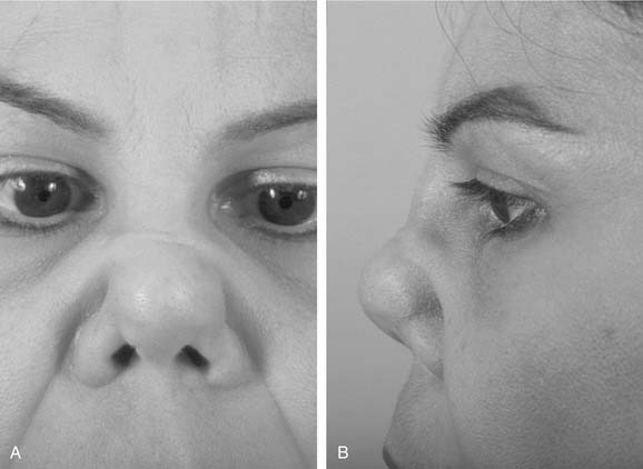
The final category of surgical error represents those deformities stemming from gross errors of judgment. Whereas the problems outlined earlier can result from the poor execution of a reasonable surgical strategy, this last class of deformity usually results from a fundamentally flawed plan. These problems can be catastrophic and may not be possible to correct. These mistakes may stem from faulty analysis, disregard for basic principles of rhinoplasty, or use of improper techniques for a given problem. The inexperienced but aggressive surgeon is most likely to commit such an error. These problems are varied and typically cause an unnatural appearance. In some cases, they can result in serious cosmetic and functional deformity. Some of the most difficult cases involve situations in which large areas of soft tissue have been excised or violated (Fig. 39-1).


Jun 5, 2016 | Posted by in OTOLARYNGOLOGY | Comments Off on Revision Rhinoplasty

Full access? Get Clinical Tree

Get Clinical Tree app for offline access