Floaters
 Floaters
In young persons, the cortical layer of the vitreous body is in close contact with the internal limiting membrane of the retina. In advanced age, the vitreous body collapses. The gel, which contains hyaluronic acid, gradually liquefies. The collagen framework of the vitreous stroma condenses and forms prominent vitreous strands. Patients often perceive fine punctate figures that appear to dance in front of a bright background. They are known as floaters. They are attributable to small vitreous densities that often go undetected by slit lamp biomicroscopy (see also p. 171).
Treatment: none.
 Unilateral Visual Impairment in Children-Amblyopia
This decrease in visual acuity occurs in childhood in unilateral concomitant strabismus. It is due to chronic suppression of images perceived with the fovea of the deviating eye (strabismic amblyopia), as a sensory adaptation of the visual system to the strabismic deviation. This means the disorder is a developmental anomaly of the central nervous system. Strabismic amblyopia can vary greatly in its severity. Congenital strabismus or strabismus in early childhood is often associated with severely impaired vision of the strabismic eye, and often patients are only able to perceive hand motions. However, one encounters the full spectrum of reduced visual acuity. The amblyopia may be so slight that it only becomes apparent when the patient slowly and haltingly reads rows of numbers during visual acuity testing.
This sort of amblyopia due to functional impairments is reversible with early treatment, provided the development of the central nervous system is not yet complete.
The fundus in sensory strabismus is completely normal. However, in a few cases of strabismus organic changes in the macula or optic nerve are found. In these cases, the reduced visual acuity is caused by the disease in the fundus, and strabismus is a secondary disorder. To rule out fundus disease in strabismic patients, careful examination is required, before initiating the classic occlusion therapy for strabismus.
Organic causes of childhood amblyopia that involve the fundus:
- macular scarring secondary to fetal toxoplasmosis (p. 30)
- retinopathy of prematurity (p. 159)
- diffuse chorioretinopathy (p. 80)
- optic disc coloboma (p. 211)
- congenital storage diseases (p. 99f.).
Treatment: amblyopia should be treated as early as possible, usually by occlusion. Patients then require the care of an ophthalmologist and an orthoptist for many years.
Strabismic Amblyopia
- strabismus
- nystagmus
- impaired vision
- disorder affects 4% of the population
- fundus without any abnormality
 Acute Visual Impairment with Normal Optic Disc
 Principal Signs of Retrobulbar Optic Neuritis
 Retrobulbar Optic Neuritis
The diagnostic situation in retrobulbar optic neuritis is best summed up by the catchphrase: “The patient doesn’t see anything, and neither does the doctor.” The optic disc initially appears perfectly normal. At the onset of retrobulbar optic neuritis, the functional impairments are the most striking features of the disorder. They also play a crucial role in the diagnostic workup. The temporal pallor indicative of incipient, partial atrophy only appears after six to eight weeks.
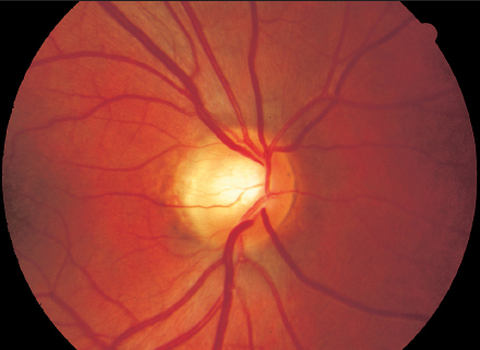
Fig. 7.1 Temporal pallor of the optic disc in a 27-year-old-man indicative of partial optic nerve atrophy secondary to retrobulbar optic neuritis.
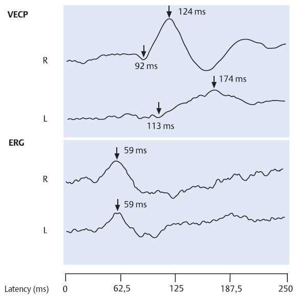
Fig. 7.2 Retrobulbar optic neuritis in the left eye. VECP (above) in the right eye (R) shows normal latencies and amplitudes, whereas in the left eye (L) the latency in the VECP is prolonged and the amplitude reduced. Below: normal pattern ERG.
Examination of the visual evoked cortical potentials (VECP) can confirm the diagnosis of optic neuritis at an early stage. VECP testing is an important diagnostic tool especially in retrobulbar optic neuritis in which the optic disc appears normal. The latencies of the VECP are prolonged and the amplitudes are reduced. The normal values for the latencies vary from one laboratory to the next. For patients with normal eyes, the latency for the Nl peak is about 99 milliseconds and for the P2 peak up to 126 milliseconds. Prolongation of the latencies in the VECP can be demonstrated from the onset of the visual impairments and remains more or less pronounced.
Similar to retrobulbar optic neuritis, there is also a posterior ischemic optic neuropathy (PION) analogous to anterior ischemic optic neuropathy (p. 190f.). The shape of the visual field defects can help to clinically distinguish this disorder from retrobulbar optic neuritis. MRI scans that show the optic nerves and help to distinguish this rare ischemic disorder of the optic nerve from multiple sclerosis.
Treatment: as described in the section on optic neuritis (p. 195).
 Visual Impairment with Normal Optic Disc
Physical compression of the optic nerve can lead to irreversible functional impairments that progress to manifest optic nerve atrophy after a latency period of at least six weeks. Findings in the optic disc are initially normal. Prompt diagnosis of such lesions decisively influences the prognosis.
Examinations to detect detrimental influences on the optic nerve in the presence of visual impairments of uncertain etiology with still normal optic discdisc findings:
- visual acuity
- visual field
- contrast sensitivity
- VECP and focal foveal cone ERG
- CT, MRI of the visual pathway.
 Graves Disease
- exophthalmos
- proliferation of orbital connective tissue
- swelling of extraocular muscles
- impaired ocular motility
- compression of optic nerve (demonstrated on CT, MRI, and ultrasound images)
- visual acuity decreases
- visual field defects
- visual evoked cortical potential: delayed latency, reduced amplitudes
- normal optic disc
- imminent optic nerve atrophy
Stay updated, free articles. Join our Telegram channel

Full access? Get Clinical Tree


