CHAPTER 188 Congenital Malformations of the Nose
Developmental Errors of the Anterior Neuropore
The anterior neuropore persists medial to the optic recesses in the third week of life, and around it the skull base develops as the frontal, ethmoidal, and nasal bones. Behind the nasal bones but in front of the nasal and septal cartilages lies a potential space, the prenasal space. The foramen caecum forms a defect in the anterior skull base at the apex of the prenasal space, where the cribriform plate ultimately condenses. This structure is closed by its fusion with the fonticulus frontalis, a fontanelle between the inferior aspect of the frontal bones and the developing nasal bones. During the third through the eighth weeks of development, a projection of dura extends through the foramen caecum, traverses the prenasal space, and apposes ectoderm at the tip of the nasal bones (the future rhinion). As the foramen closes, the dural diverticulum detaches from the overlying ectoderm and retracts into the cranium. Irrevocably apposed ectoderm may be pulled posterosuperiorly toward and even through the foramen caecum, giving rise to a dermoid fistula, cyst, or sinus. Faulty or premature closure of the foramen may enable persistence of neural tissue in the nasal cavity as isolated heterotopic glial tissue (glioma) or as a patent central nervous system (CNS) communication (meningocele or encephalocele) (Fig. 188-1). The latter can occur in a paramedian position, as a nasoethmoidal encephalocele, or, more laterally through a medial orbital wall defect, as a naso-orbital encephalocele. Basal encephaloceles herniate posterior to the cribriform plate. Similar errors may occur above the developing nasal bones at the fonticulus frontalis. Intracranial contents may herniate through the patent fonticulus frontalis until the eighth week. If the fonticulus also closes abnormally, the result may be persistence of an extranasal path, leading to nasofrontal meningoceles or encephaloceles or gliomas1 (Fig. 188-2).
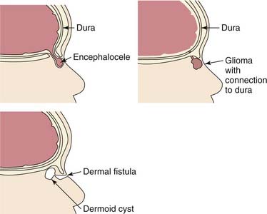
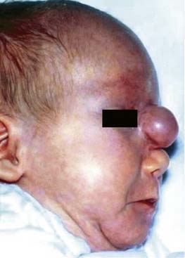
Figure 188-2. Sincipital encephalocoele. A soft, bluish compressible mass protruding from the glabellar region.
Encephaloceles
An encephalocele is an extracranial herniation of cranial contents through a defect in the skull. When the encephalocele includes meninges only, it is termed a meningocele; if it includes both brain and meninges, it is termed a meningoencephalocele. Estimates of the incidence of these lesions vary considerably, ranging from 1 in 3000 to 1 in 30,000 live births in North America and Europe. The incidence in Asian populations is higher, with a reported incidence of 1 in 6000 live births. Encephaloceles have no familial tendency or gender predilection. Approximately 40% of affected patients have other associated anomalies.2–4
Encephaloceles are divided into occipital, sincipital, and basal types (Tables 188-1 and 188-2). Occipital encephaloceles will not be discussed in this chapter as they occur outside the nose. Sincipital encephaloceles account for 25% of all encephaloceles, and they are further classified according to their location. Nasofrontal encephaloceles manifest as glabellar masses, causing telecanthus and inferior displacement of the nasal bones. Nasoethmoidal lesions manifest as dorsal nasal masses, causing superior displacement of the nasal bones and inferior displacement of the alar cartilages. Naso-orbital lesions manifest as orbital masses, causing proptosis and visual changes.1 The anatomic course of each of these sincipital encephalocele presentations is shown in Table 188-1. Basal encephaloceles are less common, arising between the cribriform plate and the superior orbital fissure or posterior clinoid fissure and manifesting as an intranasal mass (Fig. 188-3). These masses may not manifest until later in childhood, when they cause nasal obstruction and drainage. Sincipital and basal encephaloceles appear as pulsatile, bluish compressible lesions that transilluminate. Classically, these lesions expand with crying, straining, or compression of the jugular veins.
Table 188-1 Sincipital Encephaloceles
| Type | Course | Clinical Features |
|---|---|---|
| Nasofrontal | Through bone defect between orbits and forward between nasal and frontal bones to the area superficial to the nasal bones | |
| Nasoethmoidal | Through foramen caecum deep to the nasal bones, turning superficially at the cephalic end of the upper lateral cartilage to expand superficial to the upper lateral cartilage | |
| Naso-orbital | Through the foramen caecum deep to the nasal and frontal bones through a lateral defect in the medial orbital wall |
Table 188-2 Basal Encephaloceles
| Type | Course | Clinical Features |
|---|---|---|
| Transethmoidal | Through the cribriform plate into the superior meatus medial to the middle turbinate | |
| Sphenoethmoidal | Passes through a bony defect between the posterior ethmoid cells and sphenoid | |
| Transsphenoidal | Through a patent craniopharyngeal canal into the nasopharynx | |
| Spheno-orbital | Through the superior orbital fissure and out the inferior orbital fissure into the sphenopalatine fossa |
On histopathologic analysis, sincipital and basal encephaloceles have a glial component, with astrocytes surrounded by collagen, submucosal glands, and sometimes nasal septal cartilage or calcification. When it is difficult to differentiate between gliomas and encephaloceles, the presence of ependymal tissue is consistent with an encephalocele.5
High-resolution computed tomography (CT) or magnetic resonance imaging (MRI) delineates encephaloceles and additionally helps to exclude associated anomalies such as agenesis of the corpus callosum and hydrocephalus.5 CT outlines the bony cartilaginous defect, whereas MRI provides complementary information regarding the soft tissue characteristics of the mass (Fig. 188-4). MRI also differentiates meningoceles from meningoencephaloceles. Sagittal reconstructions and contrast enhancement are particularly advantageous for identifying an intracranial connection.
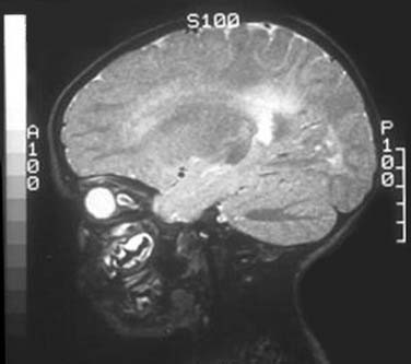
Figure 188-4. Sagittal magnetic resonance image of a basal meningoencephalocele protruding into the nasopharynx.
Once the diagnosis of encephalocele has been made, management is surgical. Most authorities advocate intervention in the first few months of life2 to minimize the risk of meningitis and cosmetic deformity, but the risk of meningitis is not decreased with prophylactic antibiotics while surgery is being planned. Additionally, early intervention makes the identification of the intracranial connection technically easier and allows more complete repair of the dural defect.2 Small lesions with minimal skull base defects may be managed endoscopically. Larger lesions require a combined approach with a craniotomy to resect condemned dura and cerebral tissue with concomitant endoscopic removal of the residual nasal tumor. The skull base defect can then be reconstructed using a pericranial flap or split-thickness calvarial bone graft.5 The most commonly encountered postoperative complications are cerebrospinal fluid (CSF) leak, meningitis, and hydrocephalus. Postoperative neurologic function reflects the patient’s preoperative performance. Encephalocele recurrence rates of 4% to10% have been reported.6
Gliomas
Gliomas consist of heterotopic glial tissue that lacks a patent CSF communication to the subarachnoid space; however, 5% to 20% of gliomas maintain a fibrous affiliation (see Fig. 188-1). Nasal gliomas are rare and show nonfamilial inheritance. They occur more commonly in males (in a ratio of 3 : 2). These benign masses manifest as extranasal (60%), intranasal (30%), or combined (10%) lesions.7 Extranasal gliomas are smooth, firm, noncompressible masses that occur most commonly at the glabella, although they may arise along the side of the nose or the nasomaxillary suture line.2 Intranasal gliomas are polypoid pale masses that may protrude from the nostril. The nasal fossa on the involved side may be obstructed. Intranasal gliomas most often arises from the lateral nasal wall near the middle turbinate and occasionally from the nasal septum.7 Nasal gliomas rarely may extend into the orbit, frontal sinus, oral cavity, or nasopharynx. Because they lack a patent CSF connection, gliomas do not change in size with crying or straining and do not transilluminate.
Proper management of gliomas requires surgical extirpation using a multidisciplinary approach involving otolaryngology and neurosurgery. Delaying intervention may lead to distortion of the septum or nasal bones, or infection. With cosmesis kept in mind, surgical access should provide exposure of the lesion and possible exploration of the skull base. For resection of extranasal gliomas, options include lateral rhinotomy, external rhinoplasty, transglabellar subcranial, bicoronal, and midline nasal approaches.8 Except in the case of large glabellar lesions, the external rhinoplasty approach provides adequate surgical exposure while minimizing the use of facial incisions.5 When a fibrous stalk is present that extends deep to the nasal bones toward the base of the skull, a nasal osteotomy is recommended to improve exposure. Following the stalk in its entirety is crucial in determining the possible presence of an intracranial extension. As a result of advancements in surgical instrumentation, image guidance, and surgical techniques, most intranasal gliomas can be removed endoscopically.9,10 Reported recurrence rates are between 4% and 10%.11
Nasal Dermoids
Nasal dermoids are frontonasal inclusion cysts or tracts related to embryologic errors localized to the anterior neuropore. Congenital midline nasal masses occur in 1 in 3000 to 1 in 40,000 live births, and dermoids are by far the most common.12 Nasal dermoids account for 1% to 3% of all dermoids and approximately 10% to12% of head and neck dermoids. Most dermoid cysts occur sporadically, with a slight male preponderance,13,14 although familial associations have been reported. Nasal dermoids may accompany aural atresia, pinna deformity, mental retardation, hydrocephalus, branchial arch anomalies, cleft lip and palate, hypertelorism, and hemifacial microsomia in 5% to 41% of presentations.13
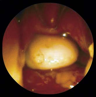
Nasal dermoids usually manifest as a midline pit or mass. In approximately 50% of clinical presentations, a dimple is present at or near the rhinion, along with a widened nasal bridge; however, the true spectrum of disease includes cysts, sinuses, or fistulas that may occur anywhere along the embryologic line from the nasal tip into the cranial space. Mass lesions are firm, lobulated, and noncompressible and may be associated with a sinus opening (Fig. 188-5) with intermittent caseous discharge or infection. Although a protruding hair is seen in only a minority of patients, it is pathognomic for a nasal dermoid. A lesion within the nasal septum will cause nasal obstruction. Intracranial extension rates range from 4% to 45%.13 Recurrent meningitis with typical skin flora may indicate an intracranial tract. Nasal dermoids do not enlarge with crying or straining and do not transilluminate.
Figure 188-5. Nasal dermoid manifesting as a firm midline nasal swelling associated with a sinus opening.
CT and MRI provide complementary information in the radiologic evaluation of nasal dermoids. Thin-slice (1 to 3 mm) CT scans with intravenous contrast are recommended to differentiate the dermoid from the surrounding nasal mucosa and to define the bony anatomy of the nose and anterior skull base (Fig. 188-6). Unique findings with intracranial dermoids include a bifid crista galli (Fig. 188-7) and an enlarged foramen caecum; a normal crista galli and foramen caecum may be used to exclude intracranial extension.15 Multiplanar, thin-section contrast-enhanced MRI is used to depict the soft tissue anatomy of the anterior skull base, and contrast-enhanced MRI distinguishes the nonenhancing dermoid from enhancing lesions such as hemangiomas and teratomas. The neonatal crista galli does not contain marrow fat, so high signal intensity on T1-weighted images is suggestive of an intracranial dermoid.
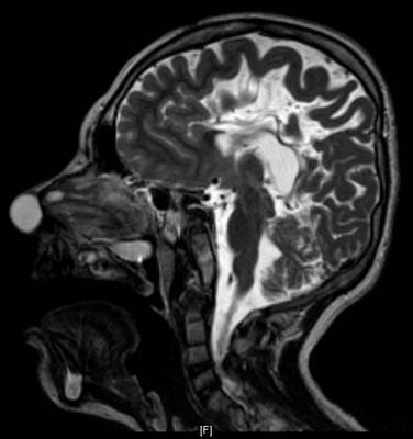
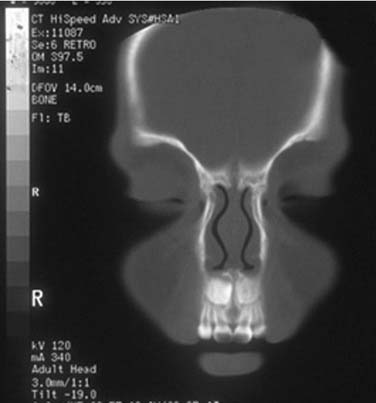
Figure 188-7. Coronal computed tomography scan showing a bifid crista galli and an enlarged foramen caecum.
As with other congenital anterior neuropore lesions, the management of nasal dermoids is surgical. Because aspiration, incision and drainage, curettage, and subtotal excision are associated with high recurrence rates, these management approaches generally are not advocated. Assessing the degree of intracranial extension is crucial, and in patients with features suggestive of intracranial extension, a combined intracranial-extracranial approach should be planned with the participating neurosurgeon. Extracranial access should fulfill four criteria: (1) excellent access to the midline; (2) access to the base of the skull; (3) adequate exposure for reconstruction of the nasal dorsum; and (4) an acceptable scar.16 Several different extracranial approaches have been described. The external rhinoplasty incision generally gives the best cosmetic result and is therefore the most widely used approach. This approach also gives access to the skull base and allows for exposure of the nasal dorsum; nevertheless, it provides limited access to lesions in the glabellar region. Alternative approaches include lateral rhinotomy and midline vertical incisions, both of which provide excellent access.13 For lesions in the glabellar area without a sinus tract, a transglabellar subcranial approach, a paracanthal incision, or a bicoronal approach is an acceptable alternative. Glabellar lesions with a sinus opening require an elliptic incision to excise the ostium, despite the possibility of a widened scar.
For lesions that extend into the cranial cavity, a multidisciplinary effort is required to perform a frontal craniotomy. This usually is carried out through a coronal incision using a combined intracranial-extracranial approach with a large frontal craniotomy; however, anterior small window craniotomies have been described.17 Sessions18
Stay updated, free articles. Join our Telegram channel

Full access? Get Clinical Tree


