CHAPTER 203 Voice Disorders
Voice production starts with the expiration phase of respiration, which provides the air pressure to vibrate the vocal folds. The lowest periodic component of vocal fold vibration is termed fundamental frequency and is perceived as pitch.1 Harmonics are integer multiples of fundamental frequency in voiced sounds. The energy or intensity in the harmonic components decreases as frequency increases.
Sound waves generated by vocal fold vibration are modified by the size, shape, and tenseness of the resonating chamber, which consists of the oropharynx, the nasopharynx, and the nasal cavities. The resonance of the vocal tract is termed formant.1 Although an infinite number of formants exist, only the first four formants (F1, F2, F3, and F4) are of clinical interest; the lowest frequency formant is F1. Each formant is characterized by its center frequency and bandwidth. Constriction of the vocal tract near a volume velocity maximum or minimum can respectively lower or raise the formant frequency. The resulting sound wave is further modified by the articulators (lips, teeth, tongue), resulting in voice and speech production. A normal voice should be pleasing in quality and have appropriate balance of oral and nasal resonance, intensity, fundamental frequency level, and prosody. Voice disorder can result in a voice that is unpleasant for the listener or can interfere with effective communication.
The underlying cause of a voice disorder can be organic or functional. Organic voice disorders result from congenital or acquired anatomic abnormalities. Functional disorders are caused by emotional or psychological problems but can lead to anatomic alterations. However, even when a voice disorder results primarily from an organic cause, there is often a psychological overlay.2
Although the reported incidence of voice disorders in children varies greatly, most voice surveys of children show a 6% to 9% incidence of voice disorders.3 Voice disorders are categorized depending on the area of problem: voice quality, resonance, loudness, and pitch. This classification is arbitrary, and a voice disorder often has several problem areas. Any anatomic abnormality involving the free edge of the vocal fold can affect voice quality and result in harshness, breathiness, or hoarseness. Disturbances in resonance can be caused by hypernasality or hyponasality. Loudness or intensity problems occur when a child speaks too loudly or too softly. Deviations in pitch occur with inappropriate speaking fundamental frequency, narrow pitch range, or excessive pitch breaks. An algorithm for the evaluation and management of voice disorders is outlined in Fig. 203-1.

Evaluation
Social and functional impact of voice impairment can be evaluated using rating scales such as Pediatric Voice Outcomes survey (PVOS), Pediatric Voice-Related Quality-of-Life Survey (PVRQOL), and Pediatric Voice Handicap Index (pVHI).4–6 These rating scales are designed to provide physicians with the parents’ perception of the severity of the child’s voice disorder and its impact on the child’s daily life; they are used to follow the child’s progress before and after therapy and surgical intervention.
Stroboscopic examination, which uses a short burst of light in synchrony with vocal fold motion, can seemingly slow down the movement of the vocal folds and allow more careful examination of the vocal folds and their motion.7 It is particularly useful in differentiating superficial from deep lesions. Laryngeal stroboscopy can delineate vocal fold symmetry, periodicity, vibratory amplitude, mucosal wave, glottal closure, and rigidity,7 but it is not possible in many children. Bouchayer and Cornut8 found that stroboscopic examination in children often had to be quick; therefore their results tended to be inconclusive. Hirschberg and colleagues9 found that stroboscopy was possible only in children older than 6 or 7 years. McAllister and others10 completed stroboscopic examination in only half of 60 patients age 10 years and older. The newer digital flexible endoscopes may allow stroboscopic examination in younger children.11 In our voice clinic, digital flexible stroboscopy has been used successfully in children as young as age 3 years. Although stroboscopic examination can be instrumental in accurate diagnosis of a voice disorder, rigid endoscopy under anesthesia may be necessary in some children to establish diagnosis and for therapeutic intervention.
There are several commonly used speaking tasks.3 The first is oral reading, which is possible only in older children. Children who can read at third grade level or better are given a specific passage to read; younger children are allowed to select the reading material. The second task is conversational speech or connected speech at least 1 minute long. The child is asked to tell a story about a picture or talk about a specific topic (e.g., pets, a vacation, a hobby). Based on these two tasks, the speech-language pathologist can perceptually and acoustically analyze the child’s speech for most voice parameters, including prosody.
The remaining speaking tasks are designed to evaluate more isolated aspects of the child’s voice. The third task consists of counting from 1 to 10, 60 to 70, and 90 to 100.3 The counting is done carefully, once slowly and then as rapidly as possible. This is repeated for three levels of loudness (soft, average, loud) and pitch (low, modal, high). This task reveals any problems with laryngeal tone, resonance, pitch, and loudness.
The fourth speaking task is the production of isolated speech sounds. This consists of sustaining certain vowel sounds (e.g., /a/, /i/) for at least 5 seconds and repeating them three times to determine laryngeal tone, resonance, and fundamental frequency. The child should then sustain /a/ at a comfortable pitch and loudness for as long as possible after a deep inspiration and repeat this three times with a break in between each attempt to determine the maximal phonation time and whether the child had adequate respiratory drive to maintain continuous voice.3 This same procedure is repeated with /s/ and /z/. In a normally functioning larynx, the s:z ratio should be close to 1. However, a lesion in the vocal fold margins (e.g., vocal fold nodules) increases the amount of airflow and decreases the time on /z/, resulting in a ratio of 1.4 or greater 95% of the time.12
The speech samples are analyzed, and a voice profile can be then developed using scales such as the Buffalo III voice profile.3 This voice profile classifies voice abnormalities as follows: 1, normal; 2, mild; 3, moderate; 4, severe; or 5, very severe. It evaluates laryngeal tone, pitch, loudness, nasal resonance, oral resonance, breath supply, muscles, voice abuse, rate, speech anxiety, speech intelligibility, and overall voice rating. Other perceptual scales such as GRBAS and Consensus Auditory-Perceptual Evaluation-Voice (CAPE-V) are also used in the evaluation of pediatric voice.13,14
Aerodynamic analysis provides objective measures of the velopharyngeal and vocal fold functions. The oral-nasal acoustic ratio and palatal efficiency rating computed instantaneously have been used to evaluate velopharyngeal function.15,16 Laryngeal airway resistance can be measured to assess the effective closure of the vocal folds to airflow. This is measured noninvasively using an anesthesia mask, a pressure-sensing catheter, and a flow-sensing pneumotachometer. Values less than 30 cm H2O/L/sec indicate inadequate closure, whereas values greater than 60 cm H2O/L/sec are associated with hyperkinetic voice disorders.17 Normative aerodynamic measures for children age 6 to 10 are available.18 These techniques of aerodynamic analysis are being replaced by computer-assisted voice analysis programs.
Computer-assisted analysis of voice disorder was introduced in 1990 and is being used with increasing frequency.19 This technology presents the opportunity to supplement perceptual evaluation of voice disorders and has replaced many traditional methods of evaluation. Evaluation of naturalness and intelligibility of speech still requires the human ear.
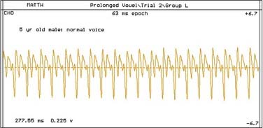
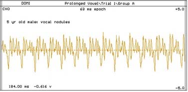
Computer-assisted voice analysis can provide the mean fundamental frequency, intensity, and amplitude of voice based on a small voice sample (0.5 second) for comparison with existing normative values for age and sex (Figs. 203-2 and 203-3).19 Until recently, no normative data for children in these analyses was available and normative data from adult studies in the literature were used20; however, in 2002, Campisi and colleagues21 published a pediatric normative database for computer-assisted voice analysis.
Other information provided by computerized voice analysis includes harmonics-noise ratio, amplitude perturbation (shimmer), frequency perturbation (jitter), and electroglottography. Aerodynamic measures can be obtained in less than 60 seconds using the pneumotachographic mask. Parameters that can be measured include subglottal pressure; transglottal airflow; oral pressure; nasal flow; airflow; resistance and efficiency; inspiratory, expiratory, and pause aerodynamics; and nasal and velopharyngeal resistance. Voice analysis is also discussed in Chapter 58.
Voice Therapy
Voice therapy entails two stages.22 The first stage consists of 10 exploratory sessions, each lasting 35 to 40 minutes. This stage helps determine the goals and specific procedures to be used in stage 2, which consists of regular voice therapy sessions for 2 to 5 months. The frequency of therapy is determined by the severity of the dysphonia. Therapy should be supplemented by practice at home to hasten resolution of dysphonia. The total therapy duration is approximately 4 to 5 months; improvement or resolution of dysphonia should be significant.
During the initial phase of voice therapy, the mechanisms of voice production and voice problems are explained to the child in simple terms, and a list of rules regarding good and bad voice are provided.3,22 A main goal of therapy is to eliminate vocal abuse by decreasing the total amount of talking; however, even in highly motivated children, total voice rest may not be feasible. Listening training and auditory feedback are essential in voice therapy because, to correct a voice disorder, the patient should learn to differentiate normal and abnormal voices for comparison with his or her own voice.3
Problematic muscular tonus, loudness, pitch, and rate require therapy. These areas are often closely interrelated and should be managed simultaneously. The child is taught the following steps to correct a problematic voice parameter3: correct rules of specific voice parameters; identification of incorrect and correct voice habits in others; recognition of personal use of incorrect voice and modification of this habit; recognition of personal use of correct voice; recognition of situations that cause personal use of poor voice habits and good voice habits; and an increase in the amount of time that correct habits are used. These steps can be applied to any problematic voice parameter.
Voice production depends on well-coordinated movement of muscles involved in phonation. Hyperfunction or excessive muscular tonus frequently occurs in children with benign laryngeal pathology, whereas hypofunction with flaccid muscular tonus occurs in those with functional dysphonia. For both problems, control of muscular tonus and proper positioning of the laryngeal, pharyngeal, and oral structure should be taught. To correct hyperfunctional states, posture instruction, breathing exercises, relaxation procedures, muscle tension reduction technique, chewing method, muscle stretching exercises, and biofeedback can be used.23–25 Wilson3 found that the chewing method and progressive relaxation are particularly useful in reducing muscular tension. For hypofunctional states, the pushing method increases muscular tension.26
Functional Etiology
Functional voice disorder is diagnosed when no anatomic or organic cause can be found. Functional dysphonia is categorized as disturbed mutation, psychological dysphonia, imitation, and faulty learning.3 Mutation is the change of voice that occurs during puberty. Pitch lowers in males and, to a lesser extent, in females. Mutation can be delayed, prolonged, or incomplete. High pitch, hoarseness, and voice breaks are characteristic. Mutational voice disorder can result from endocrine pathology.3
Functional dysphonia resulting from psychological causes seldom occurs in children; only isolated case reports can be found in the literature.27 The underlying psychological problems are related to or are part of tensional symptoms, adjustment, anxiety, or personality disorders.28 Functional dysphonia may be a form of conversion hysteria. The disorder may be complete aphonia or partial loss of voice. The dysphonia is often variable, with effortful voice production and easy fatigue. Laryngeal examination may show ventricular band approximation, bowed vocal folds, or hypoadducted vocal folds (hysterical aphonia). Vocal fold movement is normal with inhalation and cough.
Faulty learning29 occurs when a child imitates cleft palate or hearing-impaired speech or learns to talk louder than normal because of the presence of hearing-impaired persons in the household.
Organic Etiology
Resonance Disorders
Resonance disorders include hypernasality and hyponasality. Hypernasality is usually caused by velopharyngeal insufficiency from underlying palatal abnormalities. Hyponasality can result from any underlying condition that causes nasal or nasopharyngeal obstruction. The underlying pathology may be choanal atresia, deviated nasal septum, turbinate hypertrophy, nasal polyps, or, most frequently, adenoid hypertrophy. In performing adenoidectomy, particular attention should be paid to the structural integrity of the palate to decrease the incidence of postoperative velopharyngeal incompetence. Accurate diagnosis and appropriate medical and surgical management of these underlying conditions are further discussed in Chapter 188.
Vocal Quality Disorders: Surgical Management
Vocal Fold Paralysis
Vocal fold paralysis in children results from birth trauma and congenital anomalies of the central nervous system and the heart and great vessels. Any infant or child with vocal fold paralysis should be evaluated with chest radiograph and imaging of the central nervous system.30–33 Vocal fold paralysis is the second most common cause of congenital stridor in children and represents 10% of congenital anomalies of the larynx.34 Prognosis for spontaneous recovery is better for acquired, right-sided, and unilateral paralysis.35–37
More than 50% of vocal fold paralysis in children is bilateral.32,38 Because arytenoid fixation can be mistaken for bilateral vocal fold paralysis, the cricoarytenoid joint should be palpated at rigid endoscopy. Laryngeal electromyography may be the most specific and sensitive test to determine the presence of vocal fold paralysis. More than 50% of the time, bilateral vocal fold paralysis requires tracheotomy for the establishment of an airway; however, the voice is often normal. Although spontaneous recovery of vocal fold function is possible after 2 to 3 years, late recovery is often incomplete because of laryngeal muscle atrophy, synkinesis, and cricoid fixation.39 If vocal fold function does not return spontaneously after 10 to 12 months, surgery to decannulate the child should be considered.31,40
Surgical options to correct bilateral vocal fold paralysis consist of reinnervation with a nerve-muscle flap, cordotomy, lateralization procedures (e.g., arytenoidopexy), arytenoidectomy through a posterolateral external approach, arytenoidectomy through laryngofissure, or endoscopic arytenoidectomy.31,40–44 Reinnervation of the posterior cricoarytenoid muscle is not universally successful,38 although Tucker45 reported it to be the management of choice in children. Because of the small size of the laryngeal structure, endoscopic techniques can be more difficult and less successful in children.31,46 Cordotomy, a procedure in which the membranous vocal fold is sectioned from the vocal process of arytenoid, has limited use in children and may be useful as an adjunct to other procedures.41,47 Narcy and colleagues40 found Woodman’s procedure to have a higher failure rate and recommended arytenoidopexy by an external posterolateral approach. However, Bower and colleagues31 recommended arytenoidectomy through a laryngofissure because it provided better exposure, better control over the final fold position, and a high rate of success (84%). Although most patients have adequate voice postoperatively, breathiness, hoarseness, and pitch change are seen, and patients may require voice therapy. The resulting voice disorder is inversely proportional to the adequacy of the airway.
Stay updated, free articles. Join our Telegram channel

Full access? Get Clinical Tree


