Purpose
To determine the vitreous levels of angiopoietin (Ang)-1 and Ang-2 in eyes with retinopathy of prematurity (ROP), and to determine the correlation between the 2 levels.
Design
Retrospective case-control study.
Methods
Forty-eight eyes with stage 4 ROP were studied. Six eyes with congenital cataract were used as controls. The ROP eyes were classified by the vascular activity into highly (n = 22), moderately (n = 15), and mildly (n = 11) vascular-active ROP. Eyes with highly vascular-active ROP initially received 0.5 mg of intravitreal bevacizumab (IVB) and underwent vitrectomy within 1 week. The others underwent vitrectomy without IVB. Vitreous samples were collected at the beginning of vitrectomy, and the vitreous levels of Angs were measured by enzyme-linked immunosorbent assay.
Results
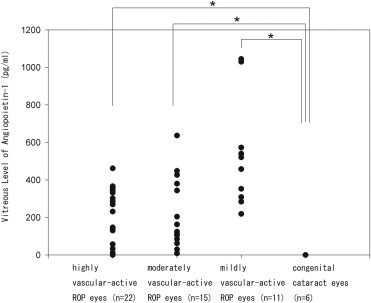
The mean concentrations of Ang-1 and Ang-2 were 201.9 and 7832.1 pg/mL in highly vascular-active ROP eyes, 216.1 and 7731.2 pg/mL in moderately vascular-active ROP eyes, 533.8 and 1685.9 pg/mL in mildly vascular-active ROP eyes, and 0 and 41.5 pg/mL in control eyes. The vitreous Ang-1 level was significantly higher ( P < .05) in highly, moderately, and mildly vascular-active ROP eyes than in control eyes. The vitreous Ang-2 level was significantly higher ( P < .05) in highly and moderately vascular-active ROP eyes than in control eyes. There was a significant negative correlation ( r = −0.406; P = .040) between the Ang-1 and Ang-2 levels in moderately and mildly vascular-active ROP eyes.
Conclusions
The balance of Ang-1 and Ang-2 in the vitreous may be important in the pathogenesis of ROP.
Retinopathy of prematurity (ROP), first reported as retrolental fibroplasia by Terry in 1942, is a retinal vascular disorder that develops in eyes with incomplete blood vessel development at birth. The sprouting of new blood vessels, pathologic neovascularization, induced by hypoxia is the crucial event in the pathology of ROP. In severe cases, the pathologic neovascularization leads to fibrovascular proliferation, vitreoretinal traction, and tractional retinal detachment, which then leads to severe loss of vision. Various factors may be involved in regulating the pathologic neovascularization, and among them vascular endothelial growth factor (VEGF) has been reported to be the dominant factor.
The vitreous level of VEGF has been shown to be higher in stage 4 ROP eyes than in control (congenital cataract) eyes, and the VEGF level was correlated with the vascular activity. In addition, anti-VEGF therapy, for example, intravitreal injection of bevacizumab, a humanized monoclonal antibody against VEGF, can reduce the angiogenic activity in ROP eyes.
The angiopoietins (Angs) are growth factors that modulate the processes of not only physiological angiogenesis but also pathologic neovascularization, particularly associated with VEGF. Ang-1, Ang-2, Ang-3, and Ang-4 are members of the Ang family. Among them, Ang-1 and Ang-2 are ligands of tyrosine kinase receptor Tie2 and have similar binding affinities for Tie2. Ang-1 is expressed by pericytes in vitro and in vivo. It induces autophosphorylation of Tie2 and promotes remodeling, maturation, and stabilization of blood vessels by recruiting surrounding support cells and extracellular matrix. Ang-2, on the other hand, is expressed by endothelial cells in vivo and is an antagonist for the Tie2 receptor by inhibiting its autophosphorylation. Ang-2 induces endothelial destabilization and promotes angiogenesis in the presence of VEGF, and destabilization by Ang-2 in the absence of VEGF leads to the regression of fragile vessels. The production of Ang-2 is upregulated by hypoxia and VEGF.
The purpose of this study was to measure the vitreous levels of Ang-1 and Ang-2 in ROP eyes and to compare the levels to that in eyes with congenital cataracts. We also determined the correlation between the Ang-1 and Ang-2 levels in the vitreous.
Methods
Forty-eight eyes of 36 infants (17 female and 19 male infants) with stage 4 ROP (4A, 36 eyes; 4B, 12 eyes) were studied. The mean gestational age of the infants was 24.2 weeks (range, 22–26 weeks), and the mean birth weight was 640 grams (range, 332–977 grams). All of the infants underwent primary vitreous surgery at the Osaka University Hospital, Osaka, Japan from July 5, 2007 through December 24, 2009. All of the eyes underwent indirect photocoagulation of the avascular peripheral retina before the vitrectomy.
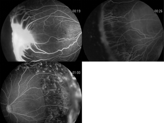
Fundus examinations with a slit lamp and contact lens (Volk Quad Pediatric Lens; Volk Optical Inc, Mentor, Ohio, USA) were performed under general anesthesia. During the examinations, fundus photographs and fluorescein angiograms were taken with a RetCam 120 digital fundus camera (Massie Research Laboratories, Inc, Pleasanton, California, USA). The stage of the ROP was based on the International Classification of Retinopathy of Prematurity. In addition, the stage 4 ROP eyes were classified into 3 groups: highly vascular-active ROP, moderately vascular-active ROP, and mildly vascular-active ROP, as described ( Figure 1 ). Eyes with highly vascular-active ROP initially received 0.5 mg of intravitreal bevacizumab (IVB) and underwent vitrectomy 1 to 7 days after the injection. Vitrectomy was performed on moderately and mildly vascular-active ROP eyes without IVB.
Six eyes of 5 infants with congenital cataract (3 female and 2 male infants), whose ages ranged from 1 month to 4 years, were studied as controls. All of these infants were full-term babies and did not have any other ocular or systemic complications.
Undiluted vitreous samples were collected from eyes with ROP during 3-port closed vitrectomy with a 23-gauge system before the infusion valve was opened. In the eyes with congenital cataract, the 2-port limbal approach was used. After lens aspiration, the aqueous humor in anterior chamber was replaced by viscoelastic material. A posterior continuous curvilinear capsulorrhexis was performed, and undiluted vitreous samples were collected during the anterior vitrectomy.
The vitreous samples were collected in sterile tubes, which were then placed on dry ice and stored at −80°C until the assay. For the protein assay, the vitreous samples were thawed and centrifuged at 15 000 rpm for 10 minutes at 4°C. The supernatants were used to determine the vitreous levels of Ang-1 and Ang-2 by enzyme-linked immunosorbent assay with kits for human anti-Ang-1 and anti-Ang-2 (R & D Systems, Minneapolis, Minnesota, USA). The minimum detectable levels of the tests were 3.45 pg/mL for Ang-1 and 8.29 pg/mL for Ang-2. If the raw data were less than the minimum detectable levels, they were set to 0 for the statistical analyses. Each assay used 30 μL for Ang-1 and 25 μL for Ang-2 of the vitreous sample/well, and the assay was performed twice. The optical density was determined at 450 nm with an absorption spectrophotometer (ARVO MX ; PerkinElmer Japan, Kanagawa, Japan) with the correction wavelength set at 540 nm.
Statistical analyses were performed using SPSS software (Sigma Stat; Systat Software, Inc, San Jose, California, USA). Data are presented as the means and ranges. Kruskal-Wallis 1-way analysis of variance (ANOVA) on ranks was used to compare the vitreous concentrations of Ang-1and Ang-2 among the groups, followed by Dunn’s method to detect significant difference between 2 groups. The correlation between the Ang-1 and Ang-2 levels was determined by the Pearson product moment correlation. A P value less than .05 was considered to be statistically significant.
Results
Among the eyes with stage 4 ROP, 22 eyes were classified as highly vascular-active ROP, 15 eyes as moderately vascular-active ROP, and 11 eyes as mildly vascular-active ROP. The mean vitreous level of Ang-1 was 201.9 pg/mL (range, 0 to 461.7 pg/mL) in the highly vascular-active ROP eyes, 216.1 pg/mL (range, 8.5 to 637.0 pg/mL) in the moderately vascular-active ROP eyes, 533.8 pg/mL (range, 218.9 to 1044.9 pg/mL) in the mildly vascular-active ROP eyes, and 0 pg/mL (range, 0 to 0 pg/mL) in the control eyes. The vitreous levels of Ang-1 were significantly different ( P < .001) among the 4 groups. The levels of Ang-1 in the highly, moderately, and mildly vascular-active ROP eyes were significantly ( P < .05) higher than that in the control eyes ( Figure 2 ).
The vitreous levels of Ang-2 were significantly different ( P < .001) among the 4 groups. The mean vitreous level of Ang-2 in the highly vascular-active ROP eyes that had received IVB was 7832.1 pg/mL (range, 1136.6 to 21 078.1 pg/mL). The mean vitreous level of Ang-2 was 7731.2 pg/mL (range, 599.5 to 17 956.0 pg/mL) in the moderately vascular-active ROP eyes, 1685.9 pg/mL (range, 231.0 to 5055.1 pg/mL) in the mildly vascular-active ROP eyes, and 41.5 pg/mL (range, 0 to 125.5 pg/mL) in the control eyes. The levels of Ang-2 in the highly and moderately vascular-active ROP eyes were significantly ( P < .05) higher than that in control eyes ( Figure 3 ).


