 Vascular Tumors of the Retina and Optic Disc
Retinal Hemangioblastoma (Capillary Hemangioma)
General Considerations
Retinal vascular tumors include hemangioblastoma (capillary hemangioma), cavernous hemangioma, racemose hemangioma, and acquired vasoproliferative tumor. Each has different clinical features, systemic implications, complications, and management.
Retinal hemangioblastoma can be solitary, without systemic disease, or a component of von Hippel-Lindau (VHL) syndrome (1, 2, 3, 4, 5, 6, 7, 8, 9, 10, 11, 12, 13, 14, 15, 16, 17, 18, 19, 20, 21, 22, 23, 24, 25, 26, 27, 28, 29). VHL syndrome is an autosomal dominant condition with various combinations of retinal hemangioblastoma, cerebellar hemangioblastoma, pheochromocytoma, hypernephroma, pancreatic cysts, endolymphatic sac tumor, and several other tumors and cysts (1, 2, 3). The locus for VHL gene is on chromosome 3 (3p25-26) (10), and inactivation of a tumor suppressor gene appears to play a key role, similar to the situation with regard to retinoblastoma. A patient with a retinal hemangioblastoma should be evaluated periodically for VHL syndrome. The incidence of VHL syndrome is about 1 in 40,000 live births, and there is an estimated 7000 cases in the United States (11). The mean age at diagnosis is 18 years for patients with VHL and 36 years for those without VHL (10). Development of new retinal tumors after age 40 years is rare. In a patient with a solitary hemangioblastoma, the risk of developing VHL is 45% if the patient is <10 years of age at diagnosis and 1% if >60 years of age. Hemangioblastoma may not be unique to VHL. A similar tumor has been seen in associated with Marshall-Stickler syndrome (17).
Clinical Features
Retinal hemangioblastoma is typically a reddish-pink tumor in the peripheral retina or on the optic disc. It can occur as an exudative or tractional type. The exudative type has intraretinal and subretinal exudation similar to Coats disease. In contrast to Coats disease, however, it shows one or more distinct red masses with dilated, tortuous, feeding and draining blood vessels. The tractional type of retinal hemangioblastoma can be similar, but is also characterized by retinal gliosis, vitreoretinal traction, vitreous hemorrhage, and tractional retinal detachment. With either type, yellow exudation is often located in the macular area, remote from the peripheral tumor. Spontaneous regression of the tumor rarely occurs (21).
Diagnostic Approaches
The diagnosis of retinal hemangioblastoma is usually made by the typical ophthalmoscopic features described. Fluorescein angiography shows rapid hyperfluorescence of the mass in the arterial phase and late hyperfluorescence, often with leakage of dye into the vitreous. It is important to differentiate the feeding artery and the draining vein because that information is needed in treatment, as discussed later. Indocyanine green angiography and ultrasonography add little to the diagnosis. Optical coherence tomography may identify the mass in the retina and help to delineate associated changes like retinal edema and localized retinal detachment. Computed tomography, magnetic resonance imaging, or other imaging studies should be done to detect the aforementioned systemic lesions associated with VHL.
Genetic testing can be helpful in excluding the diagnosis of VHL syndrome in patients with solitary retinal hemangioblastoma. Of 10 patients with solitary lesions, all tested negative for the VHL gene using Southern blot, conformationsensitive gel electrophoresis, and direct sequence analysis (8).
Pathology
Histopathologically, retinal hemangioblastoma is composed of spindle cells, small blood vessels, and clear stromal cells (1). The stromal cells are believed to be the cells of origin of this tumor. However, their specific nature has not been clarified, and they do not appear to be vascular endothelial cells (20). Hence the term capillary hemangioma may not be accurate. Currently, retinal hemangioblastoma appears to be the preferred term because of its similarity to the cerebellar hemangioblastoma.
Management
Management of retinal hemangioblastoma depends on tumor size, location, complications, and whether the patient has VHL syndrome (3,6). Those associated with VHL syndrome generally appear at an earlier age, are more aggressive, and require active treatment. Juxtapapillary or epipapillary retinal hemangioblastomas may be more difficult to manage because of their locations (13,15).
Some small asymptomatic retinal hemangioblastomas can be cautiously followed and may remain stable or, rarely, show spontaneous regression (21). Tumors with limited retinal exudation or detachment can be managed by laser photocoagulation or cryotherapy, and fairly good control is generally achieved (4,18). More-advanced lesions may require additional retinal detachment surgery. External beam radiotherapy has been used as salvage treatment after other treatments have failed (4). Our group and others have found plaque radiotherapy to be effective in selected cases (16).
Other methods that have had limited use so far include photodynamic therapy (7,22), transpupillary thermotherapy (14), and transretinal feeder vessel ligation combined with vitrectomy and photocoagulation (9).
One of the new treatments beginning to gain some recognition is the use of vascular endothelial growth factor (VEGF) inhibitors (5,29). One case report described reduction in cystoid macular edema and improved vision, but no decrease in tumor size, using the intravenous VEGF inhibitor SU5416 (5). This treatment is generating enthusiasm at the time of this writing, but its true value is not yet established.
Selected References
1. Gass JDM. Retinal and optic disc hemangiomas. In: Gass JDM, ed. Stereoscopic Atlas of Macular Diseases, 2nd ed. St. Louis: CV Mosby;1997:850-859.
2. Shields JA, Shields CL. Vascular tumors of the retina and optic disc. In: Shields JA, Shields CL, eds. Intraocular Tumors. A Text and Atlas. Philadelphia: WB Saunders; 1992:393-419.
3. Shields JA, Shields CL. Systemic hamartomatoses (“phakomatoses”). In: Shields JA, Shields CL, eds. Intraocular Tumors. A Text and Atlas. Philadelphia: WB Saunders, 1992:513-539.
4. Raja D, Benz MS, Murray TG, et al. Salvage external beam radiotherapy of retinal capillary hemangiomas secondary to von Hippel-Lindau disease: visual and anatomic outcomes. Ophthalmology 2004;111:150-153.
5. Girmens JF, Erginay A, Massin P, et al. Treatment of von Hippel-Lindau retinal hemangioblastoma by the vascular endothelial growth factor receptor inhibitor SU5416 is more effective for associated macular edema than for hemangioblastomas. Am J Ophthalmol 2003;136:194-196.
6. Singh AD, Nouri M, Shields CL, et al. Treatment of retinal capillary hemangioma. Ophthalmology 2002;109:1799-1806.
7. Schmidt-Erfurth UM, Kusserow C, Barbazetto IA, et al. Benefits and complications of photodynamic therapy of papillary capillary hemangiomas. Ophthalmology 2002;109:1256-1266.
8. Singh AD, Ahmad NN, Shields CL, et al. Solitary retinal capillary hemangioma: lack of genetic evidence for von Hippel-Lindau disease. Ophthalmic Genet 2002;23:21-27.
9. Farah ME, Uno F, Hofling-Lima AL, et al. Transretinal feeder vessel ligature in von Hippel-Lindau disease. Eur J Ophthalmol 2001;11:386-388.
10. Singh AD, Nouri M, Shields CL, et al. Retinal capillary hemangioma: a comparison of sporadic cases and cases associated with von Hippel-Lindau disease. Ophthalmology 2001;108:1907-1911.
11. Singh AD, Shields CL, Shields JA. von Hippel-Lindau disease. Surv Ophthalmol 2001;46:117-142.
12. Singh A, Shields J, Shields C. Solitary retinal capillary hemangioma: hereditary (von Hippel-Lindau disease) or nonhereditary? Arch Ophthalmol 2001;119:232-234.
13. McCabe CM, Flynn HW Jr, Shields CL, et al. Juxtapapillary capillary hemangiomas. Clinical features and visual acuity outcomes. Ophthalmology 2000;107:2240-2248.
14. Parmar DN, Mireskandari K, McHugh D. Transpupillary thermotherapy for retinal capillary hemangioma in von Hippel-Lindau disease. Ophthalmic Surg Lasers 2000;31:334-336.
15. Garcia-Arumi J, Sararols LH, Cavero L, et al. Therapeutic options for capillary papillary hemangiomas. Ophthalmology 2000;107:48-54.
16. Kreusel KM, Bornfeld N, Lommatzsch A, et al. Ruthenium-106 brachytherapy for peripheral retinal capillary hemangioma. Ophthalmology 1998;105:1386-1392.
17. Shields JA, Shields CL, Deglin E. Retinal capillary hemangioma in Marshall-Stickler syndrome. Am J Ophthalmol 1997;124:120-122.
18. Shields JA. Response of retinal capillary hemangioma to cryotherapy. Arch Ophthalmol 1993;111:551.
19. Majji AB. Paramacular Von Hippel angioma with tractional macular detachment. Ophthalmic Surg Lasers 2002;33:145-147.
20. Chan CC, Vortmeyer AO, Chew EY, et al. VHL gene deletion and enhanced VEGF gene expression detected in the stromal cells of retinal angioma. Arch Ophthalmol 1999;117:625-630.
21. Welch RB. Von Hippel-Lindau disease: the recognition and treatment of early angiomatosis retinae and the use of cryosurgery as an adjunct to therapy. Trans Am Ophthalmol Soc 1970;68:367-424.
22. Atebara NH. Retinal capillary hemangioma treated with verteporfin photodynamic therapy. Am J Ophthalmol 2002;134:788-790.
23. Othmane IS, Shields C, Singh A, et al. Postpartum cerebellar herniation in von Hippel-Lindau syndrome. Am J Ophthalmol 1999;128:387-389.
24. Atebara N, Shields JA. Capillary hemangioma of the optic disc associated with a total retinal detachment. Ophthalmic Surg 1993;24;686-688.
25. Shields CL, Mashayekhi A, Luo CK, et al. Optical coherence tomography in children: analysis of 44 eyes with intraocular tumors and simulating conditions. J Pediatr Ophthalmol Strabismus 2004;41:338-344.
26. Ling H, Cybulla M, Schaefer O, et al. When to look for Von Hippel-Lindau disease in gastroenteropancreatic neuroendocrine tumors? Neuroendocrinology 2004;80:39-46.
27. Dollfus H, Massin P, Taupin P, et al. Retinal hemangioblastoma in von Hippel-Lindau disease: a clinical and molecular study. Invest Ophthalmol Vis Sci 2002;43:3067-3074.
28. Milewski SA. Spontaneous regression of a capillary hemangioma of the optic disc. Arch Ophthalmol 2002;120:1100-1101.
29. Aiello LP, George DJ, Cahill MT, et al. Rapid and durable recovery of visual function in a patient with von Hippel-Lindau syndrome after systemic therapy with vascular endothelial growth factor receptor inhibitor su5416. Ophthalmology 2002;109:1745-1751.
▪ Retinal Hemangioblastoma
Retinal hemangioblastoma is found most often in the sensory retina away from the optic disc. It appears as a reddish-pink intraretinal tumor with a dilated, tortuous feeding retinal artery and a similar draining vein. It can assume an exudative form, a tractional form, or a combination of the two.
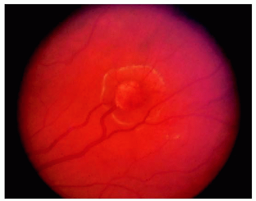 Figure 20.1. Small retinal hemangioblastoma with minimal surrounding exudative retinal detachment. |
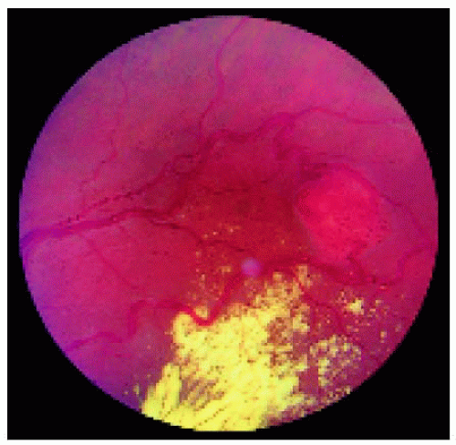 Figure 20.2. Retinal hemangioblastoma with typical intraretinal exudation. The red lesion is almost the same color as the background fundus, but it is better identified by the dilated vessels that feed and drain the mass. In this case, the yellow exudation is mostly inferior to the vascular lesion. |
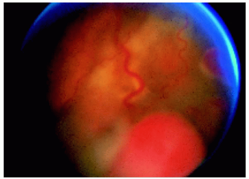 Figure 20.3. Larger, inferiorly located retinal hemangioblastoma with secondary exudative retinal detachment. |
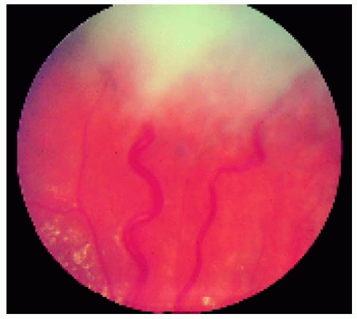 Figure 20.4. Early tractional form of retinal hemangioblastoma. The tumor is obscured by overlying fibrosis in the vitreous, but the dilated blood vessels suggest its presence. In this case, the traction is relatively minor. There is mild scattered retinal exudation more posteriorly. |
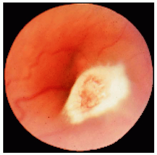 Figure 20.5. “Free-floating” retinal hemangioblastoma. Vitreous traction, usually associated with a posterior vitreous detachment, has blanched the tumor and pulled it into the overlying vitreous cavity anterior to the retina. The retinal feeder vessels are intact here, but they can bleed, leading to vitreous hemorrhage in such cases. |
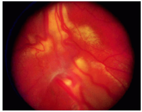 Figure 20.6. Combined exudative and tractional type of retinal hemangioblastoma. Note the traction on the retinal blood vessels and the yellow intraretinal and subretinal exudation. |
▪ Retinal Hemangioblastoma: Wide-Angle Imaging
Wide-angle fundus photography provides a broad view of retinal hemangioblastoma and helps to delineate the extent of exudation, retinal detachment, and vitreous traction.
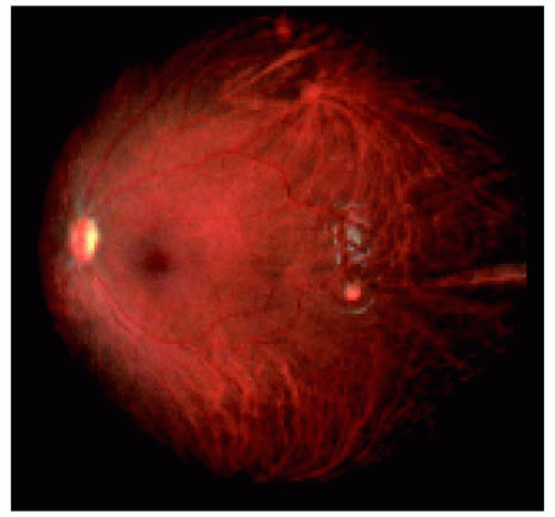 Figure 20.7. Small retinal hemangioblastoma located posterior to the equator temporally in the left eye of a teenaged boy with von Hippel-Lindau syndrome. Note that the small lesion is fed by an artery from the inferior vascular arcade and drained by a vein into the superior arcade. |
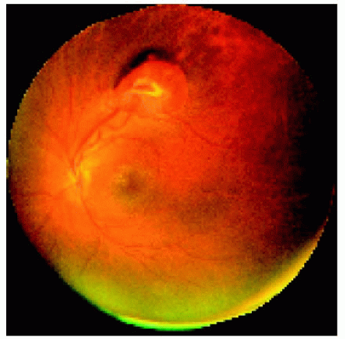 Figure 20.8. Retinal hemangioblastoma in the superior fundus of the left eye in a teenaged girl with von Hippel-Lindau syndrome. Note the dilated retinal artery and vein that supply and drain the mass and the remote yellow exudation in the foveal region. |
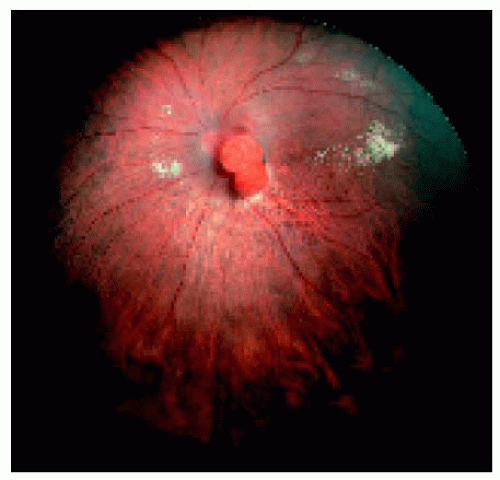 Figure 20.9. Bilobed retinal hemangioblastoma located immediately inferonasal to the optic disc in a 55-year-old man with no findings of von Hippel-Lindau syndrome. The patient has been followed for 2 years with no change in the lesion. He has 20/50 vision due to subretinal fluid and mild cystoid foveal edema. |
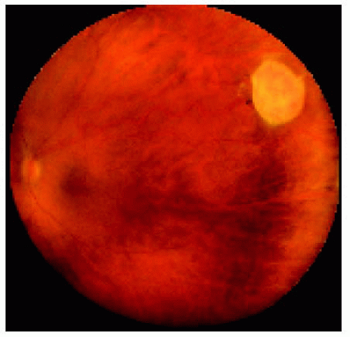 Figure 20.10. Yellow-colored retinal hemangioblastoma located between the equator and the ora serrata superotemporally in the left eye. Note the feeder blood vessels and sparse exudation. There is vitreous traction on the lesion that may account for the yellow color due to blanching of blood vessels in the lesion. |
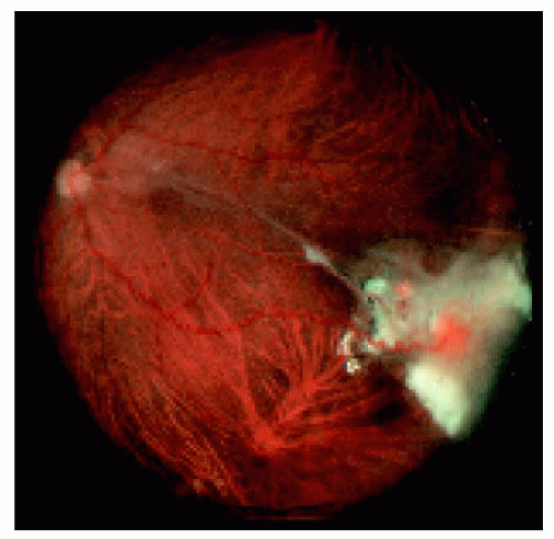 Figure 20.11. Tractional form of retinal hemangioblastoma located near the equator inferotemporally in the left eye. Note the vitreous traction band between the lesion and the posterior pole and the retinal and preretinal fibrosis that partly precludes a view of the tumor. The patient had enucleation of the opposite eye as a young child for complications of retinal hemangioblastoma, and her mother had von Hippel-Lindau syndrome. |
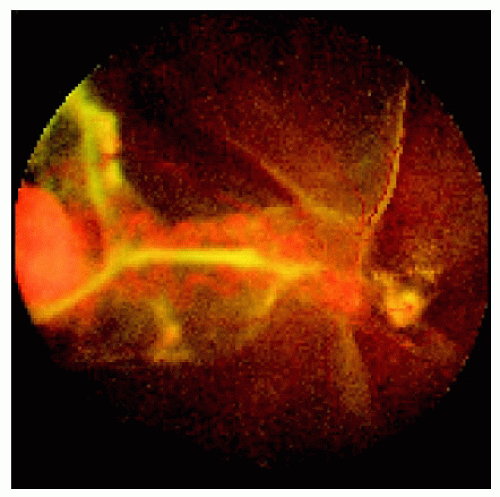 Figure 20.12. Fairly large retinal hemangioblastoma located between the equator and the ora serrata nasal in the left eye. There is a total exudative, nonrhegmatogenous retinal detachment secondary to the tumor. There appears to be a second small hemangioblastoma adjacent to the optic disc in the area of traction. |
▪ Retinal Hemangioblastoma (Nodular) of the Optic Nerve
In some instances, a retinal hemangioblastoma can lie partly or entirely over the optic disc. In such cases, the prominent feeding and draining blood vessels are less apparent. It can assume a nodular or sessile growth pattern. This form has the same relationship to von Hippel-Lindau syndrome as the peripheral type.
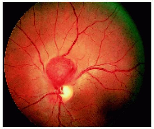 Figure 20.13. Nodular retinal hemangioblastoma overlying the superior margin of the optic disc. (Courtesy of William Hagler, MD.) |
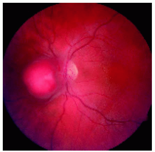 Figure 20.14. Nodular retinal hemangioblastoma overlying the nasal margin of the optic disc in a 33-year-old woman with no evidence of von Hippel-Lindau syndrome. Note the subtle retinal traction and subtle retinal exudation in the papillomacular bundle. |
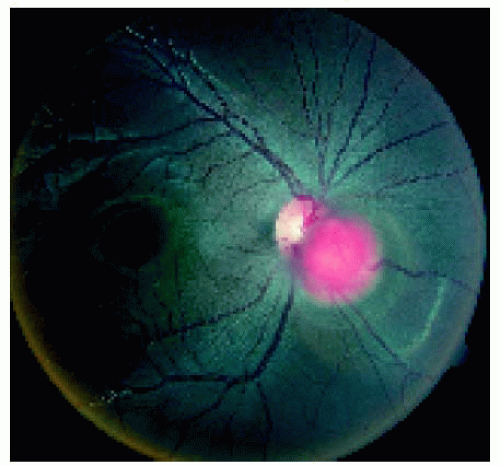 Figure 20.15. Nodular retinal hemangioblastoma overlying the inferonasal margin of the optic disc in an African American teenaged male who had no systemic or genetic evidence of von Hippel-Lindau syndrome. There was no yellow exudation at this time, but optical coherence tomography showed very shallow subretinal fluid in the foveal area, accounting for 20/30 visual acuity. |
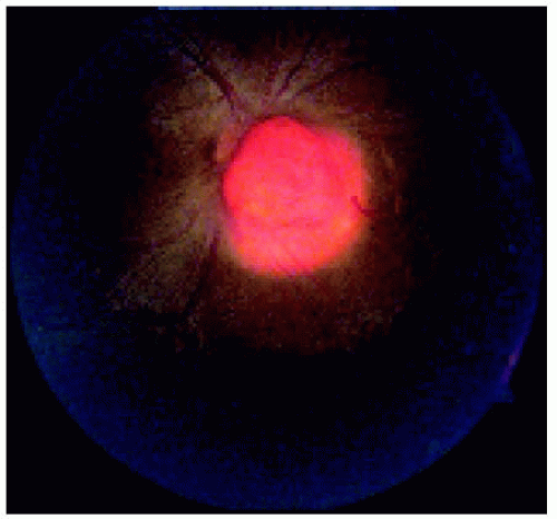 Figure 20.16. The same lesion shown in Figure 20.15, 3 years later. The lesion has shown slow enlargement, but vision is 20/40 at this time. The lesion showed no regression after systemic and periocular corticosteroids, and further treatment had not been decided on at that time. |
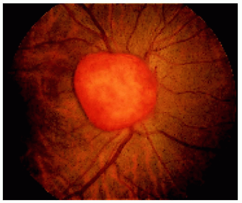 Figure 20.17. Nodular retinal hemangioblastoma overlying the entire optic disc. (Courtesy of Fox Boswell, MD.) |
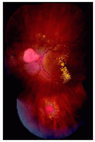 Figure 20.18. Retinal hemangioblastoma on the nasal margin of the optic disc in a patient with von Hippel-Lindau syndrome. Note the second, more peripheral retinal hemangioblastoma inferiorly that has been treated elsewhere with surrounding laser photocoagulation. |
▪ Retinal Hemangioblastoma (Sessile) of the Optic Nerve
Sessile retinal hemangioblastoma may have indistinct margins and be more difficult to recognize than the nodular variant.
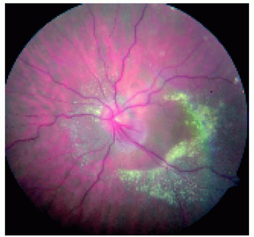 Figure 20.19. Subtle retinal hemangioblastoma on the inferotemporal margin the of optic disc with secondary circinate retinal exudation in a middle-aged woman without von Hippel-Lindau syndrome. Note the yellow circinate exudation peripheral to the lesion and the clear area between the lesion and the exudation. |
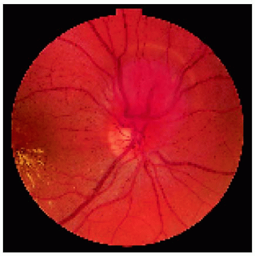 Figure 20.20. Sessile retinal hemangioblastoma over superior margin of optic. Note the larger feeding blood vessel and the intraretinal exudation in the foveal area (to the left). |
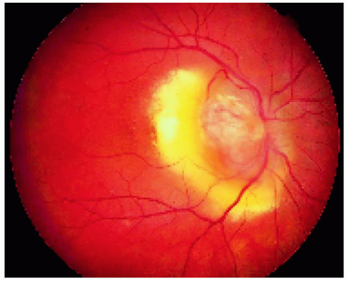 Figure 20.21. Sessile retinal hemangioblastoma covering the optic disc and producing a dense circinate exudation. |
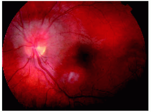 Figure 20.22. Two retinal hemangioblastomas in the posterior pole of the left eye. Note the sessile lesion on the superior margin of the optic disc and a second lesion inferior to the fovea with retinal feeder and draining blood vessels. Two distinct lesions like this are virtually pathognomonic of von Hippel-Lindau syndrome. |
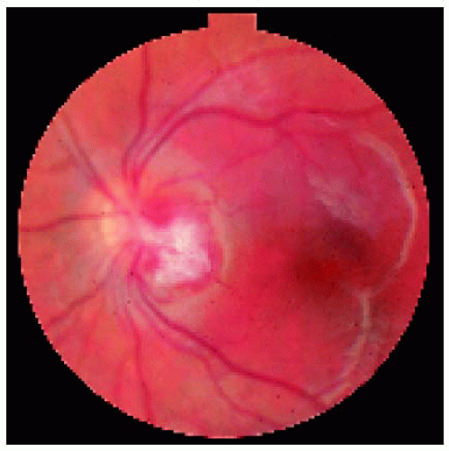 Figure 20.23. Retinal hemangioblastoma of the optic disc in a 5-year-old child with von Hippel-Lindau syndrome. There is subretinal serous fluid in the macular area but no yellow exudation. This patient was seen a number of years ago, and no treatment was given. |
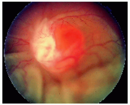 Figure 20.24. Appearance of the lesion shown in Figure 20.23, six years later, when the patient was referred to us. The lesion had enlarged dramatically, and there is a total secondary exudative retinal detachment. Several attempts to reattach the retina were unsuccessful, and enucleation was eventually necessary. |
▪ Retinal Hemangioblastoma: Fluorescein Angiography
Fluorescein angiography of retinal hemangioblastoma has very typical (if not pathognomonic) features.
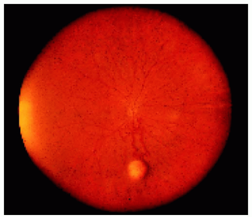 Figure 20.25. Wide-angle fundus photograph of retinal hemangioblastoma at the equator inferiorly. Note the dilated, tortuous retinal blood vessels between the optic disc and the tumor. |
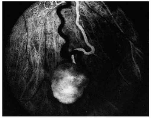 Figure 20.26. The lesion shown in Figure 20.25 in the arterial phase, showing filling of the feeding artery. The draining vien (to the left) appears dark at this time but filled rapidly in the next 2 seconds. |
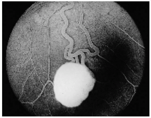 Figure 20.27. The same lesion in the recirculation phase, showing fading fluorescence of the prominent artery and vein and hyperfluorescence of the mass. |
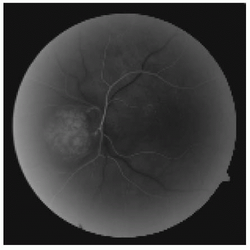 Figure 20.28. Fluorescein angiogram in the early arterial phase of the lesion in Figure 20.26, There is minimal early hyperfluorescence of the lesion at this time. |
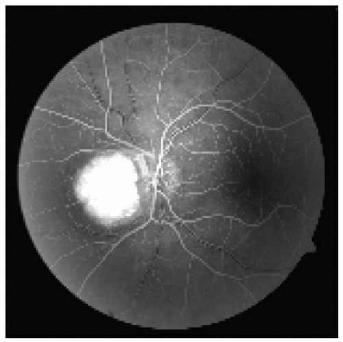 Figure 20.29. The lesion shown in Figure 20.28 in the venous phase. Note the intense hyperfluorescence of the mass. |
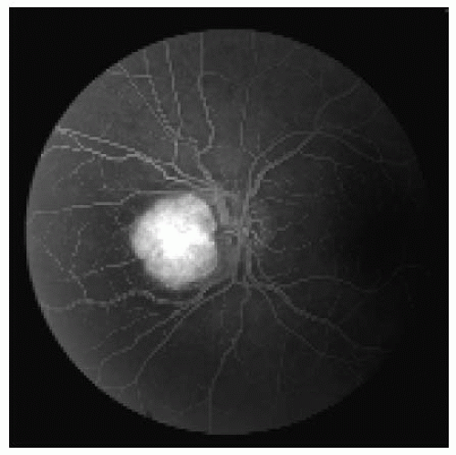 Figure 20.30. The same lesion in the recirculation phase, showing a persistent intense hyperfluorescence of the tumor but not appreciable leakage of dye into the vitreous cavity. Most retinal hemangioblastomas show some leakage into the subretinal space or vitreous. |
▪ Retinal Hemangioblastoma: Association with von Hippel-Lindau Syndrome
The Von Hippel-Lindau (VHL) syndrome is characterized by various combinations of retinal hemangioblastoma, cerebellar and spinal cord hemangioblastoma, pheochromocytoma, hypernephroma, endolymphatic sac tumors, and a variety of other vascular and cystic lesions in various parts of the body. The genetic defect has been localized on the short arm of chromosome 3. Presented here is the case of a young boy with sporadic VHL syndrome who developed endolymphatic sac tumor at age 6 years and multiple bilateral retinal hemangioblastomas at age 12 years, findings that are pathognomonic of VHL syndrome.
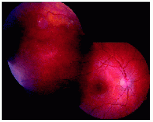 Figure 20.31. Fundus of the right eye, showing two subtle superior retinal hemangioblastomas. These could be overlooked on a cursory examination. |
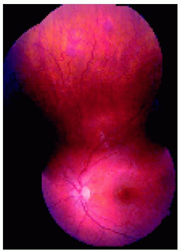 Figure 20.32. Fundus of the left eye, showing two subtle superior retinal hemangioblastomas. These could also be easily overlooked. |
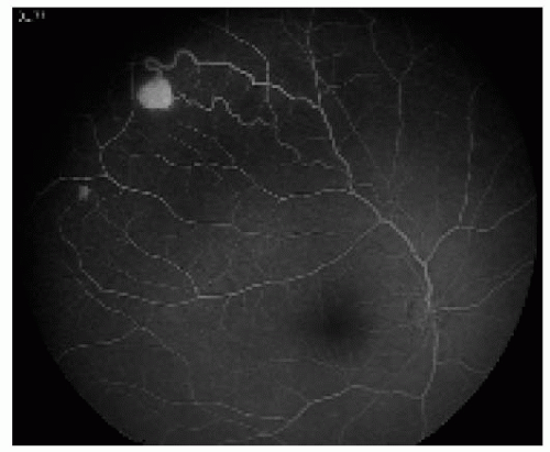 Figure 20.33. Fluorescein angiography of the right eye, showing hyperfluorescence of the two retinal hemangioblastomas. |
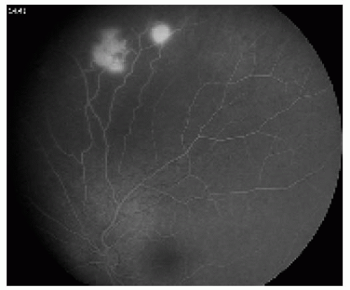 Figure 20.34. Fluorescein angiography of the left eye, showing hyperfluorescence of the two retinal hemangioblastomas. Several other tumors were detected in this eye. |
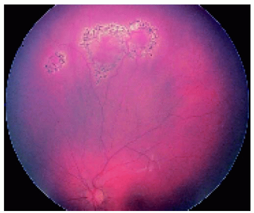 Figure 20.35. Appearance of the tumors in the left eye after laser photocoagulation, showing chorioretinal scars. |
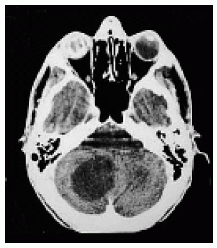 Figure 20.36. Axial computed tomogram of another patient with von Hippel-Lindau syndrome, showing a cystic mass in the cerebellum. Note the opaque, phthisical right globe due to prior complications of longstanding retinal detachment from retinal hemangioblastomas. |
▪ Retinal Hemangioblastoma: Clinicopathologic Correlation
In some cases, aggressive retinal hemangioblastoma may not be controlled, and enucleation of the affected eye may be necessary because of pain, secondary glaucoma, or phthisis bulbi. The fellow eye must be followed closely in all cases.
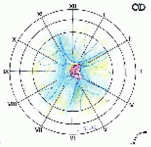 Figure 20.37. Fundus drawing of a red optic disc mass and secondary retinal detachment in a 4-year-old girl with a negative family history for von Hippel-Lindau syndrome. |
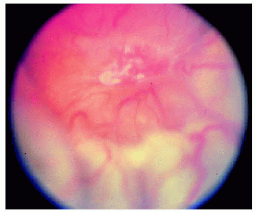 Figure 20.38. Fundus photograph of the ill-defined optic disc mass shown in Figure 20.37. Note the retinal detachment inferiorly. After unsuccessful attempts at retinal detachment repair, the blind, painful eye was enucleated. |
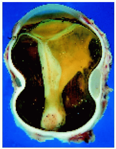 Figure 20.39. Grossly sectioned eye, showing a mass over the optic disc, total retinal detachment, and a silicone encircling band from the retinal detachment surgery. |
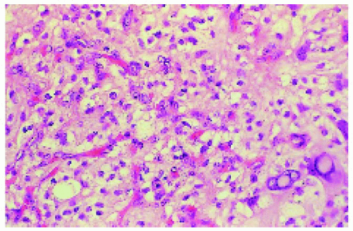 Figure 20.40. Histopathology of the tumor, showing a vascular mass composed of capillary caliber vessels and intervascular stromal cells with foamy cytoplasm. (Hematoxylin-eosin × 150.) |
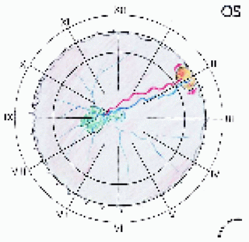 Figure 20.41. The patient’s opposite eye was entirely normal until 3 years after the initial diagnosis, at which time a peripheral retinal hemangioblastoma developed and preretinal macular fibrosis occurred, requiring surgical peeling of the preretinal membrane. The patient continues to have limited vision in the remaining eye. |
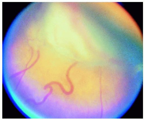 Figure 20.42. Peripheral fundus photograph of the eye shown in Figure 20.41, showing dilated retinal blood vessels and retinovitreal fibrous tissue over the peripheral vascular tumor. |
▪ Retinal Hemangioblastoma in Older Patients and in a Patient with Marshall-Stickler Syndrome
Retinal hemangioblastoma can occur as a sporadic lesion or as a part of von Hippel-Lindau (VHL) syndrome. It is usually diagnosed in the first two decades of life, particularly in patients with the VHL gene. In some cases, it can occur in older individuals with no personal or familial evidence of VHL syndrome. A similar retinal tumor has also been observed in association with Marshall-Stickler syndrome, an autosomal dominant condition characterized by typical facies, arthropathy, cataracts, myopia, and retinal detachment.
Shields JA, Shields CL, Deglin E. Retinal capillary hemangioma in Marshall-Stickler syndrome. Am J Ophthalmol 1997;124:120-122.
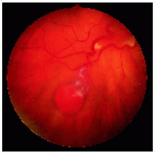 Figure 20.43. Solitary retinal hemangioblastoma in a 65-year-old woman with no von Hippel-Lindau syndrome. |
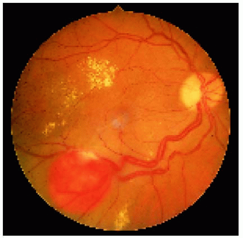 Figure 20.44. Solitary retinal hemangioblastoma in a 62-year-old man with no von Hippel-Lindau syndrome. |
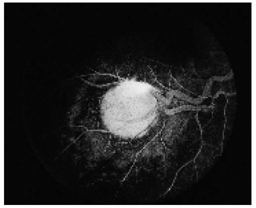 Figure 20.45. Fluorescein angiogram of the lesion shown in Figure 20.44, demonstrating typical features of a retinal hemangioblastoma. |
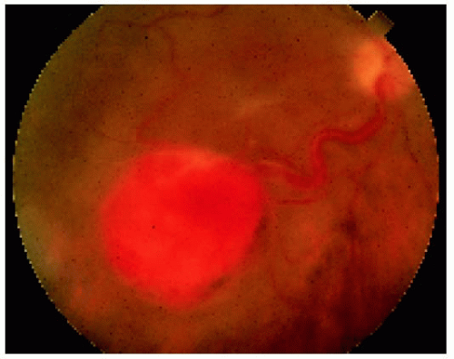 Figure 20.46. Fundus photograph of the lesion shown in Figure 20.44 after 9 years, revealing enlargement of the lesion. It was treated with plaque radiotherapy.
Stay updated, free articles. Join our Telegram channel
Full access? Get Clinical Tree
 Get Clinical Tree app for offline access

|