Chapter 107 The swollen optic disc
The diagnosis of the cause of a swollen optic disc is made by history and the accompanying visual and other ocular signs and symptoms, not just by ophthalmoscopy of the disc, even though that plays an important role (Figs 107.1–107.23). Figure 107.1 shows the importance of this and how one presentation can progress to another. For instance, papilledema (disc swelling due to raised intracranial pressure) starts with normal acuity and color vision and slightly enlarged blind spots but can progress to severe visual loss.

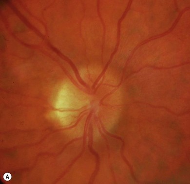
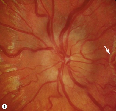
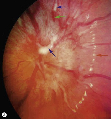
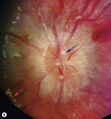
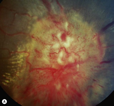
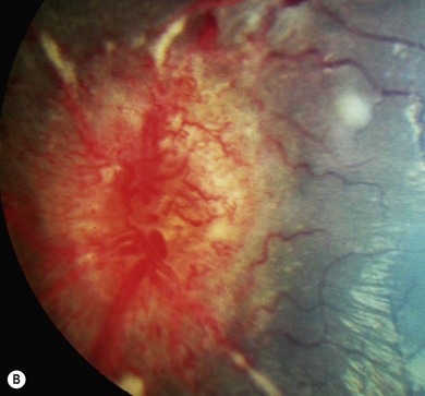
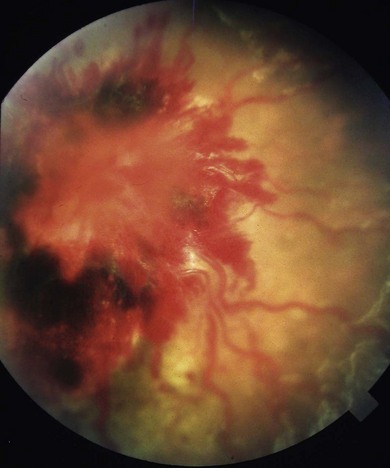
Fig. 107.5 This 7-year-old boy with a brain stem glioma presented with diplopia associated with bilateral sixth nerve palsies (see Chapter 83) that had started gradually about 2 months before and more recent symptoms of raised intracranial pressure. The optic discs are grossly elevated with markedly dilated capillaries and veins, numerous cotton wool spots, and the right eye has a macular star, as a result of protein/lipid accumulation from transudation from disc capillaries. Although the blind spots were very large on Goldmann fields, the visual acuity was 0.1 (6/7.5, 20/25, 0.8) right and 0.0 left.