Chapter 54 The optic chiasm
Introduction
The chiasm is so named because it is shaped like the Greek letter chi.1 Over 2 million nerve fibers pass through it: most are visual but some non-visual fibers project from the optic chiasm to hypothalamic nuclei, forming the retinohypothalamic tract subserving circadian rhythms.2 The ratio of crossed to uncrossed fibers in the human chiasm is about 53 : 47.3
Evolutionary considerations
The chiasm provides the major route for the juxtaposition of corresponding parts of the visual field in each eye. It is important for binocular vision. To establish fusion, it is necessary to have overlapping visual fields, congruence of corresponding retinal elements, and extraocular muscles to maintain alignment of the visual axes.4 In lateral-eyed animals, optic fibers from each eye decussate entirely to the contralateral hemisphere. The percentage of uncrossed fibers increases as the orbits rotate anteriorly and the frontal field of single binocular vision increases.5 The reason that our visual system is crossed is controversial5,6 (Fig. 54.1).
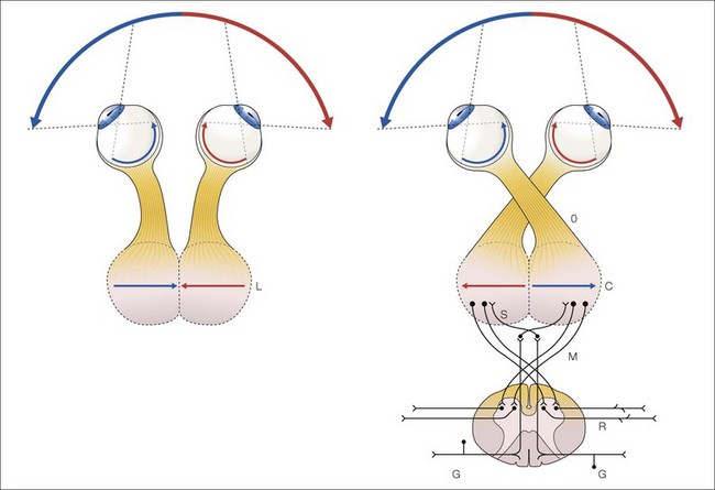
Our foveas are placed where nasal (panoramic) and temporal (binocularity providing) retinas meet.6 This allows vision to subserve its elemental functions:
Anatomy
The optic nerves, chiasm and optic tracts extend posteriorly and upwards 45 degrees from the optic canals in adults and children.7 The chiasm lies in the suprasellar cistern several millimeters above the diaphragma sellae. The anterior cerebral arteries and the anterior communicating arteries lie anteriorly and above the chiasm and optic nerves. The carotid arteries lie laterally with the posterior communicating artery passing underneath the optic tracts. The chiasm lies in the floor of the anterior end of the third ventricle. Expansion of the third ventricle may cause chiasmal compression. Posteriorly lies the hypothalamus and the pituitary stalk, the tuber cinereum, and the mamillary bodies. The optic nerves emerge from the optic canals. The length of the intracranial optic nerve varies so the position of the chiasm in relation to other structures also varies. When the optic nerves are short, the chiasm is “prefixed”, when long it is “postfixed”. Von Willebrand’s knee is an artefact.8
Embryology
The chiasm appears in the first month of life,9 arising from a thickening of the floor of the forebrain. Retinal ganglion cells grow down the optic stalks and enter the floor of the third ventricle where they decussate to form the optic chiasm. The pattern of axon crossing occurs in two phases.10 The first retinal axons meet in the midline at the ventral diencephalon forming an X-shaped chiasm; subsequent axons grow into either the ipsilateral or contralateral optic tract.11
A morphological specialization of the axon called a growth cone allows the axon to sense and respond to signals in the embryonic brain environment.12 Neuronal and glial cells provide guidance information to ingrowing RGC axon. Histologic evidence suggests that uncrossed axons are confined within the lateral portion of the optic nerve and do not approach the chiasmal midline.13 Neurons and radial glial cells in the ventral diencephalon serve a role in retinal ganglion cell axon guidance during chiasm formation. Growth associated proteins in retinal growth cones enable RGC axons to progress and perform path finding tasks. In the mouse, neurons at the site of the future chiasm are required for its formation by retinal ganglion cell axons. A growth associated protein essential for chiasm formation has been identified.14 There is substantial overproduction of neurons which later die back by apoptosis.15 The chiasm reaches its definitive form by the fourth month of gestation.
Transcription factors that pattern the developing chiasm have been identified.16–19 Foxd1 expressed in the developing temporal retina and its downstream effectors (tyrosine kinase membrane proteins) are involved in patterning of the optic chiasm in chicks and mammals.17,18 Foxd1 imprints axons ipsilaterally. Foxd1 is expressed in progenitors of Zic2 positive retina ganglion cells and is the determinant of temporal retinal identity.17,18 Neuropilin molecules, transmembrane proteins, serve as receptors for axonal guidance to regulate axon divergence at the chiasm in mammals.19 Neuropilin 1 (NRP1) is expressed at the chiasm midline and acts on contralateral retinal ganglion cells to provide growth-promoting and chemo-attractive signals for commissural axon crossing at the chiasm.19
Signs and symptoms
Developmental defects and suprasellar tumors are common in children (Box 54.1). Most chiasmal syndromes result from neoplastic disorders, developmental derangements, radiation injury, inflammation, infection, demyelination, infarction, transection, or hypoplasia.20 Dominant optic atrophy may present with a bitemporal hemianopia simulating a chiasmal disorder.20
With optic nerve involvement there is significant color vision defect. Stereoacuity tests and Bagolini striated lens are useful tests in patients with suspected chiasmal compression.4 Decreased stereoacuity is common with chiasmal lesions, even when no visual field abnormalities are detectable.4 Stereopsis can be elicited with complete chiasmal transection by haploscopic stimulation of the intact temporal retinas.21 This shows the sensory capacity for stereopsis is retained in chiasmal transection but the absence of motor fusion precludes stereopsis.22 Bagolini striated lens testing reveals a binocular “mountain” pattern in chiasmal lesions.23 Children with early-onset chiasmal disease may present with nystagmus. The classic form is see-saw nystagmus, but most patients have a compound nystagmus with vertical, horizontal, and rotary components (see Chapter 90).
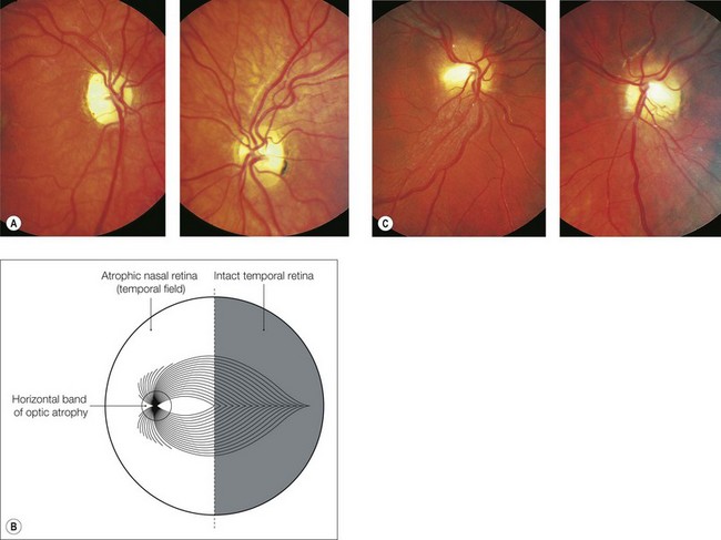
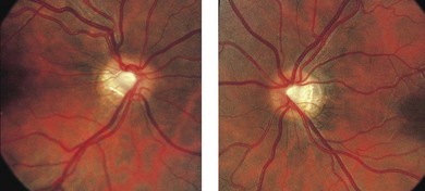
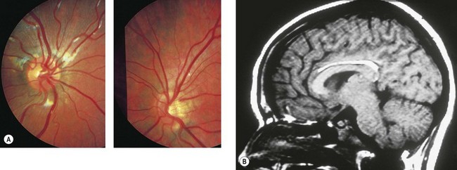
Optic atrophy frequently occurs in chiasmal disease. There may be a generalized loss of neurons, or a pattern of band atrophy due to the loss of the fibers subserving the temporal fields and the preservation of fibers subserving the intact nasal field (Fig. 54.2). In developmental chiasmal defects and tumors there are often optic disc anomalies (Figs 54.3 and 54.4), e.g. hypoplasia.24,25 Congenital suprasellar tumors can produce horizontal “bow-tie” cupping with selective loss of the nasal and temporal nerve fiber layer.26 Papilledema in these optic discs occurs predominantly in the superior and inferior poles (Fig. 54.2C).
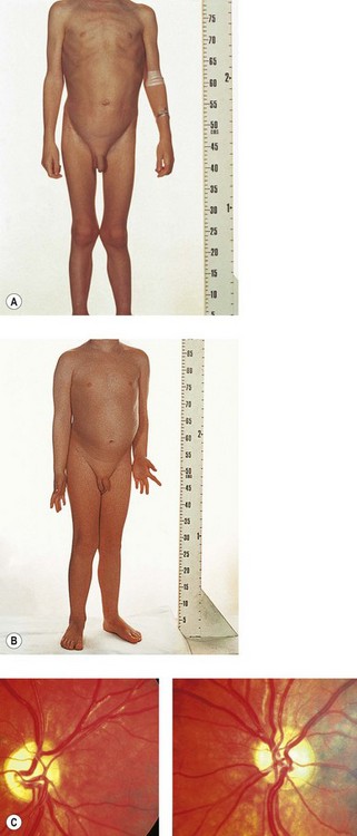
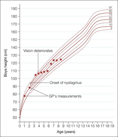
Because of the proximity of the hypothalamus and pituitary gland, endocrine and growth defects may occur. Infants with hypothalamus-involving tumors may develop Russell’s diencephalic syndrome: emaciation with loss of subcutaneous fat (Fig. 54.5), accelerated growth in length relative to weight (Fig. 54.6), and personality changes with euphoria and hyperactivity. One must examine and measure the infant’s general body habitus and inquire about weight gain.
In bitemporal hemianopia the hemifield slide phenomenon may occur (Fig. 54.7). Since only the nasal portion of each visual field is functioning fully, corresponding retinal points between the two eyes no longer exist. Sensory fusion becomes impossible, and motor fusion cannot maintain alignment. A previous heterophoria becomes a manifest deviation. Esodeviations lead to letters or words appearing deleted. Exodeviations produce letters or words appearing duplicated. A vertical hemifield slide causes the child to lose track of which line of text they are reading. These children do not complain of diplopia but rather a duplication of the middle of words or objects. The hemifield slide phenomenon does not require a complete bitemporal hemianopia and can occur as the initial symptom.27a Signs and symptoms of chiasmal disease are summarized in Table 54.1. Rarely, chiasmal tumors can produce photophobia as their presenting symptom.27b
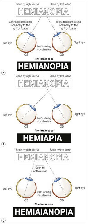
Fig. 54.7 Hemifield slide phenomenon.
(Fritz KJ, Brodsky MC. Elusive neuro-ophthalmic reading impairment. American Orthoptic Journal 1992; 42: 159–164. Reprinted by permission of The University of Wisconsin Press.)
Table 54.1 Chiasmal diseases in children
| Developmental defects | Albinism |
| Achiasmia | |
| Aplasia | |
| Anophthalmia | |
| Tumors | Chiasmal glioma |
| Craniopharyngioma | |
| Pituitary adenoma | |
| Dysgerminoma | |
| Retinoblastoma (“trilateral”) | |
| Trauma | Transection |
| Hematoma | |
| Contusion | |
| Traction | |
| Infiltration | Langerhans’ cell histiocytosis |
| Sarcoidosis | |
| Juvenile xanthogranuloma | |
| Chiasmal neuritis | Postviral |
| Postimmunization | |
| Multiple sclerosis | |
| Optochiasmatic arachnoiditis | Tuberculosis |
| Neurosyphilis | |
| Fungal | |
| Cysticercosis | |
| Vascular anomalies | Arteriovenous malformation |
| Cavernous angioma | |
| Radiation | Acute visual loss months to years after radiation |
| Empty sella | Third ventricular distension secondary to aqueductal stenosis |
| Downward traction on chiasm secondary to surgical scarring on pituitary apoplexy |
Further investigations
Further investigations consist of endocrine studies, neurophysiological evaluation (see Chapter 8) and neuroimaging. Neurophysiological studies may detect a crossover defect (particularly in a preverbal child) and quantitatively and qualitatively assess the visual defect. Magnetic resonance imaging (MRI)28,29 provides neuro-anatomical detail of the chiasm and surrounding structures. Computed tomography (CT) scanning may provide important information about tumor and bony changes involving the parasellar area.
Developmental defects
Developmental derangements of the optic chiasm include:
Albinism (see Chapter 40)
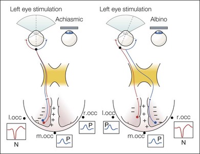
Anomalous decussation of chiasmal projections occurs in albinos.23 Retinogeniculate axons arising from ganglion cells in the portion of the temporal retina within 20 degrees of the vertical meridian decussate abnormally in the optic chiasm to synapse in the contralateral lateral geniculate nucleus.30–32 The human optic chiasm retains a predominance of crossed fibers. MRI shows smaller optic nerves, chiasm, and tracts, with a wider angle between the two optic nerves and the two optic tracts.33 The crossed predominance can be diagnosed electrophysiologically by asymmetrical hemispheric visual evoked potentials34,35 (Fig. 54.8; see Chapters 8 and 40). Pigment around the optic disc plays an important role in axonal guidance, suggesting that loss of retinal epithelial pigment could be the source of chiasmal misrouting in albinism.36 Recently, the role of the transcription factor Zic2 has been implicated in the chiasmal misrouting in albinism.37