Chapter 35 The lens
Embryology
The lens develops as a thickening of the surface ectoderm overlying the optic vesicle. This “lens placoid” appears on day 26–27 in the human embryo. In the chick, a tight extracellular matrix-mediated adhesion occurs between the optic vesicle and the surface ectoderm.1 The mitotically active surface ectoderm is fixed in place, resulting in cell crowding, elongation, and thickening of the placoid. Adhesion of the optic vesicle to the lens placoid ensures eventual alignment of the lens and retina in the visual axis. However, there is no direct cellular contact between the basement membranes of the optic vesicle and surface ectoderm.2 The lens placoid then invaginates to form the lens pit. This becomes the lens vesicle. Lens vesicle detachment is the initial event leading to formation of the anterior segment of the eye (day 33). It is accompanied by migration of epithelial cells via a keratolenticular stalk, cellular necrosis, and basement membrane breakdown.3 Disruption to this process by exogenous or endogenous factors can result in anterior segment developmental abnormalities (see Chapter 32). The detached lens vesicle is lined by a single layer of columnar epithelial cells surrounded by a basal lamina, the future lens capsule. Primary lens fiber formation occurs in the epithelial cells lining the posterior surface of the lens vesicle. This is promoted by the adjacent retinal primordium.4 The lens is thus dependent upon the retinal primordium for cytodifferentiation. The primary lens fibers fill the lumen of the lens vesicle. Elongation of lens cells adjacent to the retina forms the embryonal lens nucleus. The anterior lens cells nearest the corneal primordium remain a cuboidal monolayer and become the lens epithelium. This remains mitotically active for life, providing future lens fiber cells. Epithelial cells differentiate into secondary lens fibers at the lens equator (lens bow). These fibers elongate both anteriorly and posteriorly and insert over the primary lens fibers. They are thickest at the equator. This produces preferential growth of the equatorial diameter of the fetal lens. Secondary lens fibers meet anteriorly and posteriorly at the Y sutures.
Zonular fibers are derived from the non-pigmented ciliary epithelium during the fifth month of gestation. The glycoprotein fibrillin is the main component of the ciliary zonules. There are two isoforms, fibrillin-1 and fibrillin-2. Fibrillin-1 polymers form, without any significant elastin, a structural scaffold of extensible microfibrils. These are arranged in parallel bundles to form the zonular fibers. Disorders affecting the structure or function of these fibrillin-rich microfibrils result in zonular dysfunction, in particular ectopia lentis.5 Fibrillin-1 gene (FBN1) mutations cause Marfan’s syndrome (MFS) possibly some forms of Weill-Marchesani syndrome (WMS) and various other type-1 fibrillinopathy phenotypes in which ectopia lentis is common. Conversely, fibrillin-2 gene (FBN2) mutations can cause Beals’ syndrome which is phenotypically similar to MFS but is rarely associated with ectopia lentis (see below).
The tunica vasculosa lentis (TVL) is a vascular network derived from the hyaloid artery posteriorly and a parallel radial palisade of anastomoses with the annular blood vessel laterally. It envelops the developing lens and nourishes it at a time when aqueous production and formation of the anterior chamber have not yet begun. This intraocular network of vessels begins development in the first month of gestation, is maximal in the second to third month, and begins to regress by the fourth month. It has largely disappeared by birth.6 Persistent TVL is commonly seen in premature neonates and failure of regression of the TVL and hyaloid arteries can cause a range of developmental anomalies collectively termed persistent fetal vasculature (PFV).
Developmental abnormalities of the lens
Congenital aphakia results from failure of induction of embryonic surface ectoderm to form a lens placoid and lens vesicle and is invariably associated with significant anterior and posterior segment developmental abnormalities and poor visual function. Primary congenital aphakia can result from a variety of teratogenic events (e.g. congenital rubella) in the first 4 weeks of embryogenesis and usually results in microphthalmos, severe anterior segment developmental abnormalities, and posterior segment colobomata.7 Secondary congenital aphakia occurs as a result of spontaneous absorption or expulsion of the developing lens. It is associated with less severe ocular anomalies such as PFV.
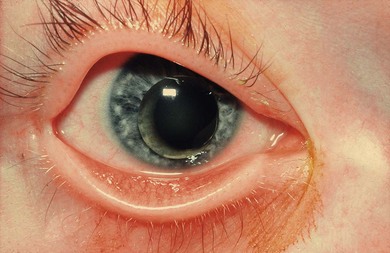
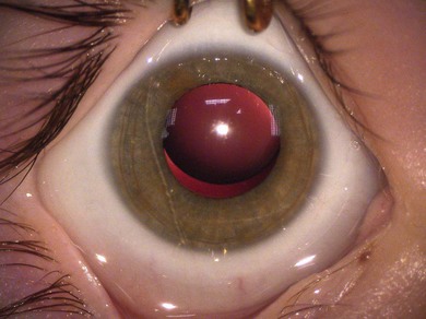
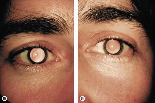
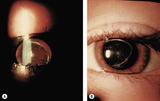
Microspherophakia is a developmental anomaly with a spherically shaped lens of reduced size and diameter (Fig. 35.1). It may result from abnormal or arrested development of secondary lens fibers. It can occur as an isolated autosomal recessive congenital disorder with mutations in the LTBP2 gene identified in one pedigree8 or as part of a hereditary systemic disorder, the most common of which are, autosomal dominant and recessive forms of WMS. It is often accompanied by ectopia lentis (Fig. 35.2).
Duplication of the lens is a very rare anomaly associated with corneal metaplasia, uveal coloboma, and cornea plana.11 It is presumed that metaplastic changes in the surface ectoderm prevent normal lens placoid formation and thus lead to multiple lens vesicles.
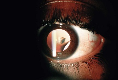
Lens coloboma (Fig. 35.3) occurs in areas where there is a failure of zonular development. Lens indentations or scalloped defects in the lens edge demarcate areas of absent zonules. They may occur unilaterally as an isolated anomaly or bilaterally as part of a uveoretinal coloboma phenotype. Lens colobomas may be seen secondary to zonular damage by the congenital ciliary body tumor medulloepithelioma.9 A localized opacity is often found in the region of the lens coloboma; this is particularly true where it is associated with a ciliary body medulloepithelioma. Most lens colobomas occur inferiorly or inferotemporally.
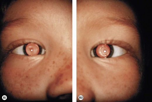
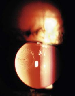
Lenticonus (Figs 35.4−35.6) and lentiglobus are developmental malformations of the anterior or posterior lens surfaces. Lenticonus is a conical defect and lentiglobus a spherical defect. Posterior surface abnormalities are more common than anterior surface anomalies. Both are usually axial. The resulting refractive error through the central lens is often much more myopic and astigmatic than through the peripheral lens. Lentiglobus is more common than lenticonus and usually unilateral.10 Lentiglobus is a progressive, well-circumscribed globular bulging of the posterior capsule of the lens. Most cases (95%) are unilateral and there is little evidence that unilateral lentiglobus is a familial condition. In contrast, bilateral posterior lentiglobus is more likely to be inherited as an X-linked or an autosomal dominant trait.11 It usually presents as the only ocular anomaly; however, it has rarely been associated with microcornea, Duane’s syndrome, and anterior lenticonus. Associated cataract is thought to be caused by mechanical stretching of lens fibers. Lenticonus is more common than previously thought.12 It may be familial with autosomal dominant or X-linked recessive inheritance (Figs 35.4 and 35.5). It may also be seen with Down’s syndrome or in the presence of a persistent hyaloid artery remnant (Fig. 35.6).
Stay updated, free articles. Join our Telegram channel

Full access? Get Clinical Tree


