Substance
Amount
Lutein
6.0 mg
Vitamin C
300.0 mg
Vitamin E
60.0 mg
Vitamin B2
3.0 mg
β-carotene
1,200.0 μg
Niacin
12.0 mg
Zinc
9.0 mg
Selenium
45.0 μg
Copper
0.6 mg
Manganese
1.5 mg
Thirteen patients, seven male patients (mean ± SD age, 69.9 ± 4.3 years) and six female patients (mean ± SD age, 79.8 ± 9.6 years) were included in the study. The informed consents were given according to the tenets of the Declaration of Helsinki. Approval from the institutional human experimentation committee was also granted. All of the patients had the same WHO classification and the same type of lens opacity in both eyes. Patients with ocular complications, such as uveitis and retinopathy, were excluded. Patients with systemic diseases, such as diabetes, were also excluded.
Central pieces of the anterior capsule measuring 5 mm in diameter were collected before and after supplementation as pre- and post-intake samples continuous curvilinear capsulorhexis during cataract surgery. Three tablets (the recommended daily dosage) of Ocuvite + lutein®, the antioxidant supplement, were administered orally every day beginning the day after the first surgery for 6 weeks. Both the pre- and post-intake samples were frozen immediately after collection and stored at −80 °C until measured.
The anterior capsule pieces were homogenized and proteolyzed using MagNa Lyser Green Beads (Roche). The RNA was purified with the MagNa Pure Compact RNA Isolation Kit (Roche). Using a Transcriptor High Fidelity cDNA Synthesis Kit (Roche), cDNA was reverse transcribed by the Gene Amp PCR system 9700 (AB). The cDNA of the control RNA (both positive and negative control RNA included with the kit) was also reverse transcribed at the same time. Using the cDNA, Universal® Probes (Roche) and primers (Nihon Gene Research, Inc.), which are shown in Table 17.2, the mRNAs of G6PDH and 18S were quantified using the Light Cycler® LC480 (Roche) for RT-PCR.
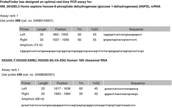
Table 17.2
Probes and primer of the mRNAs of G6PDH and 18S
The expression level of each mRNA was represented as a Cp value, where Cp was defined as the threshold cycle of RT-PCR at which the amplified product was detected. The Cp values were calculated based on a standard curve that was determined from an internal standard and the control RNA for each substance. The mRNA levels of each substance were indicated relative to internal standard and control RNA.
17.4.2 Results
In post-intake samples, the expression levels of G6PDH and 18S were significantly increased (Fig. 17.1). Both genders had the same tendencies. However, a significant difference only existed among the female patients. There were no differences between genders in pre- or post-intake samples. There were no significant correlations between the expression levels of 18S in the pre- and post-intake samples (Fig. 17.2). However, the tendencies toward lower expression of 18S in the pre-intake samples and higher expression of 18S in the post-intake samples were shown in both genders.
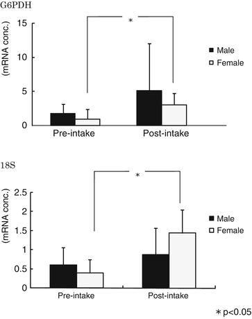
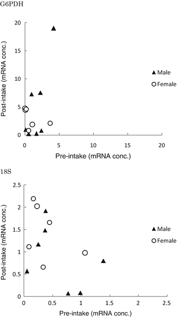
Fig. 17.1
In post-intake samples, the expression levels of G6PDH and 18S were significantly increased. Both genders had the same tendencies. However, a significant difference only existed among the female patients. There were no differences between genders in pre- or post-intake samples
Fig. 17.2
There were no significant correlations between the expression levels in the pre- and post-intake samples. However, the tendencies toward lower expression of 18S in the pre-intake samples and higher expression of 18S in the post-intake samples were shown in both genders
There were significant positive correlations between G6PDH and 18S expression levels in the post-intake samples among male patients (Fig. 17.3). G6PDH expression increased by more than four times and 18S expression almost doubled in the post-intake samples compared with the expression levels in the pre-intake samples.
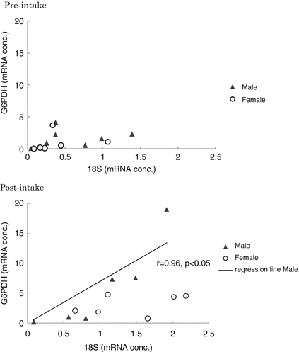
Fig. 17.3
There were significant positive correlations between G6PDH and 18S expression levels in the post-intake samples among male patients. G6PDH expression increased by more than four times and 18S expression almost doubled in the post-intake samples compared with the expression levels in the pre-intake samples
17.4.3 The Effects of Increases in Expression of G6PDH
As the lens is under the anaerobic status, the pentose phosphate cycle is more important than TCA cycles for energy production from glucose in the lens. G6PDH is known as the rate-determining enzyme of the pentose phosphate cycle, thus the increase in gene expression of G6PDH in the post-intake samples may indicate the activation of the pentose phosphate cycle followed by an increase of NAD(P)H in the LECs. Glutathione reductase (GR) is activated due to an increase in the level of coenzyme NAD(P)H, leading to an increase in the level of GSH. Thus, the increasing GSH and GR in turn increase the scavenging of peroxides after supplement intake.
G6PDH showed significantly higher expression levels in the post-intake samples from the female patients more significantly than those from the male patients. Innate antioxidative abilities decrease in elderly female patients due to hormonal imbalance. Estrogens are known to have antioxidant effects [33, 34] due to direct scavenging of free radicals [35] and upregulation of antioxidative enzymes [36]. In postmenopausal female patients, peroxidation increases because of dramatically decreased levels of estrogen. Moreover, NAD(P)H from the pentose phosphate cycle is a coenzyme in the estrogen antioxidation cycle which continuously regenerates the antioxidative potential of estrogens [37]. After supplement intake, an increase in G6PDH is thought to increase NAD(P)H to compensate for the decrease in estrogen among the postmenopausal female patients in this study.
17.4.4 The Effects of Increases in Expression of 18S
The 18S is the active center of protein synthesis in the 40S ribosomal subunit. The increased expression levels of 18S in post-intake samples suggest an increase in the transcription and synthesis of proteins and enzymes, including enzymes involved in the pentose phosphate cycle. Furthermore, the amount a product of the pentose phosphate cycle, ribose-5-phosphate (R5P), may also increase with G6PDH after supplement intake. R5P is necessary for nucleic acid synthesis and is related to the production of the ribosome. Therefore, gene transcription is possibly activated and increased with the increase in G6PDH. This is another possible mechanism for the proportional increase in 18S expression and G6PDH expression after taking the supplement.
17.5 Conclusion
The mRNA expression of G6PDH increased with 18S proportionally in cataractous lenses after intake of the supplement including lutein. It suggests the antioxidative effect of lutein in the lens and that may be effective in preventing the progression of cataract formation.
References
1.
Lin J. The association between copper ions and peroxidative reaction in diabetic cataract. J Japanese Ophthalmol Soc. 1996;100:672–9.
Stay updated, free articles. Join our Telegram channel

Full access? Get Clinical Tree


