Chapter 50 Telescreening for Diabetic Retinopathy
Introduction
The criteria for human diseases amenable to screening approaches were defined by the World Health Organization in 19681 and diabetic retinopathy fulfills all of these. Visual impairment due to diabetic retinopathy is a significant health problem; however, it has a recognizable presymptomatic stage.2 The DCCT and the UKPDS established that intensive diabetes management to obtain near-euglycemic control can prevent and delay the progression of diabetic retinopathy in patients with diabetes.3,4 Timely laser photocoagulation therapy can also prevent loss of vision in a large proportion of patients with sight-threatening diabetic retinopathy.5 Screening for diabetic retinopathy saves vision at a relatively low cost, which has been demonstrated in various studies.6,7 The American Academy of Ophthalmology recommends annual dilated eye examinations beginning at the time of diagnosis for patients with type II diabetes.2 For those with type I diabetes, the recommendation is retinal examination 3–5 years after diagnosis, with annual exams thereafter.2 The barriers for successful screening are numerous and include the high cost of care, poor awareness levels, lack of symptoms in the early stages of disease, socioeconomic factors and poor geographical access to care.8 Current screening programs for diabetic retinopathy are either ophthalmologist-based (with actual presence of the ophthalmologist at the site of screening) or ophthalmologist-led (no ophthalmologist at the site of screening). Table 50.1 summarizes the key differences between the two models. Telemedicine for retinopathy screening is an ophthalmologist-led screening model, which may be a logical potential alternative for patients who have been noncompliant with the traditional face-to-face examination by an ophthalmologist. Telemedicine is the exchange of medical data by electronic telecommunications technology allowing a patient’s medical problems to be evaluated, monitored, and possibly treated while the patient and physician are located at sites physically remote from each other.9 Ophthalmology is uniquely suited for telemedicine as it is a highly visual and image intensive specialty and digital imagery is easily transmitted by electronic means. Remote assessment for diabetic retinopathy provides an ideal model for telehealth screening initiatives and, in fact, has become one of the most common uses for telemedicine in ophthalmology.
Table 50.1 Differences between the ophthalmologist-led and ophthalmologist-based models for screening for diabetic retinopathy
| Ophthalmologist-led model (Telescreening) | Ophthalmologist-based model | |
|---|---|---|
| Brief description | Paramedical staff acquire data/images, which are then transferred for interpretation by ophthalmologist | Screening is performed by ophthalmologist |
| Feasibility | Yes, with less human resources | Needs trained expert |
| Maintenance | Required | Not required |
| Capital expenditure | More | Less |
| Revenue expenditure | Less | More |
| Interobserver bias | Less | More |
| Digital photo archiving | Yes | No |
| Acceptance by community | Yes | Yes |
Guidelines for telescreening program
American Telemedicine Association telehealth practice recommendations for diabetic retinopathy
American Telemedicine Association (ATA), Ocular Telehealth Special Interest Group, and the National Institutes of Standards and Technology Working Group, established the telescreening guidelines for diabetic retinopathy.10 The ATA recommends that telehealth programs for diabetic retinopathy should demonstrate an ability to compare favorably with ETDRS film or digital photography. For screening programs with low thresholds for referral, the International Clinical Diabetic Retinopathy Disease Severity Scale may be used in place of ETDRS scales, however, protocols should state the reference standard used for validation and relevant datasets used for comparison.
Category 1: The program allows identification of patients who have no or minimal diabetic retinopathy and distinguishes them from those who have more than minimal diabetic retinopathy.
Category 2: The program allows identification of patients who do not have sight-threatening diabetic retinopathy and distinguishes them from those who have potentially sight-threatening diabetic retinopathy.
Steps of telescreening
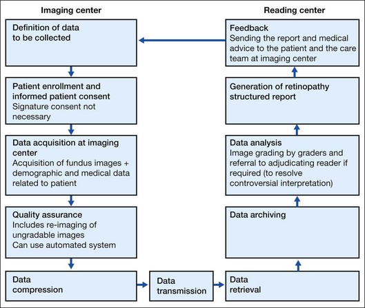
The flow of steps of the telescreening process is diagrammed in Fig. 50.1. In brief, patient enrollment is performed after defining the data to be collected. Since ocular telescreening services for diabetic retinopathy satisfy the criteria of low risk telehealth procedures and are within commonly accepted standards of practice, signature consent may not be required. However, practitioners should provide patients with information about the telescreening program they would reasonably want to know, including differences between care delivered using ocular telehealth approaches versus traditional face-to-face encounters, and a description of what is to be done at the patient’s site and the remote site. The data collected includes fundus images, along with patient examination findings (identification, demographic, and medical information) and some morphological information that is used to make a clinical decision. Fundus images of both eyes of the patient are acquired under a fixed, predetermined imaging protocol. These images are taken by a trained technician using a fundus camera. Due to various factors, the quality of the acquired images may be below the grading standard, thus not providing any meaningful information for examination by the reader. This can be addressed by employing an automatic image quality assessment module. An automatic image quality assessment module will ensure that the images transmitted for diagnosis conform to prescribed gradability standards. During the quality assurance process, the gradable images are selected for compression, whereas the identification of poor quality images can trigger reimaging by the technician. The patient data comprising the clinical data and the fundus images are compressed to make them suitable for low-bandwidth network connectivity. The patient data are transmitted to the servers via the Internet or satellite. At the reading center, the images are graded for presence of retinal lesions and the determination of a diabetic retinopathy level; referred to “next level” graders if necessary; and a retinopathy structured report is generated. Only qualified readers should perform retinal image grading and interpretation. If a reader is not a licensed eye care provider, specific training is required. A licensed, qualified eye care provider with expertise in diabetic retinopathy and familiarity with telescreening program technology should supervise the readers. An adjudicating reader (an ophthalmologist with special qualifications in diabetic retinopathy by training or experience) may resolve discrepant interpretations. Image processing algorithms should undergo rigorous clinical validation. A report comprising the findings, the results and the medical advice given by the expert is made available to the patient and the care team at the remote site through an accessible interface.
Technical considerations
Image acquisition
The gold standard for telescreening is the ETDRS 7 mydriatic standard field 35 mm stereoscopic color fundus photographs.11 However, more practical alternatives, such as digital fundus photography12,13 and nonmydriatic fundus photography,14,15 have been evaluated. Digital imaging has the advantage of faster and easier acquisition, transmission, and storage. Several investigators have reported a high level of correlation between stereoscopic digital imaging and slide film for the identification of most features of diabetic retinopathy.12,13
Regarding nonmydriatic fundus photography, a higher rate of unreadable photographs has been reported through undilated versus dilated pupils.14,15 Diabetic persons often have smaller pupils and a greater incidence of cataracts, which may limit image quality if the procedure is performed through an undilated pupil. Pupil dilation using 0.5% tropicamide is associated with a minimal risk of angle closure glaucoma. Programs using pupil dilation should have a defined protocol to recognize and address this potential complication.
The unsatisfactory performance of nonmydriatic photography has led to the concept of “targeted mydriasis,” offering mydriasis only to a preselected group of patients, in whom undilated photography is known to produce dismal results.15 However, the exact “target” remains to be defined. Based on ROC curve analysis, Raman et al.16 predetermined the cutoff values for “target mydriasis” groups as vision <6/12 (20/40 Snellen equivalent) and age >59 years. Staged mydriasis is another option.17 In this model, a nonmydriatic single digital photograph for screening is taken. If an unsatisfactory nonmydriatic photograph is obtained, the patient undergoes immediate pupillary dilation with 1% tropicamide and the photograph is then repeated. Using this protocol, 75–80% of patients do not require mydriasis.
Compression
Compression may be used if algorithms have undergone clinical validation. Image data can be compressed using a variety of standards, including JPEG, JPEG Lossless, JPEG 2000, and Run-length encoding (RLE). The International Standards Organization (ISO/IEC JTC1/SC2/WG10) has prepared an International Standard, ISO/IS-15444–1 (JPEG 2000 Part 1), for the digital compression and coding of continuous-tone still images. This standard is known as the JPEG 2000 Standard. Digital Imaging and Communication in Medicine (DICOM) recognizes JPEG and JPEG 2000 for lossy compression of medical images.18 ATA recommends that the compression types and ratios should be periodically reviewed to ensure appropriate clinical image quality and diagnostic accuracy. Some studies have attempted to look at the effect of various levels of compression on the quality of the image with both subjective and objective parameters.19,20 The level of acceptable compression ranges from 1 : 28 to 1 : 52.19,20
Data transfer, archiving, and retrieval
The described telemedicine models reported earlier used the Internet to transmit images.21,22 In rural areas and mobile clinics, satellite transmission is a more preferred option because of poor infrastructure. A variety of technologies are available for data communication and transfer. Telescreening programs should determine specifications for transmission technologies best suited to their needs.
Stay updated, free articles. Join our Telegram channel

Full access? Get Clinical Tree


