Most squamous cell carcinomas (SCCs) of the upper aerodigestive tract are
conventional type and as such show, to some degree, a recapitulation of stratified squamous epithelium. Certain SCCs, however, of the upper aerodigestive tract have different growth patterns and histologic features. Not only can these tumors sometimes behave differently from conventional-type SCCs, but they can also present the pathologist with differential diagnoses that range from benign to malignant (
Table 5.1). For example, verrucous carcinoma, a locally aggressive, nonmetastasizing variant of SCC, can be very difficult, if not impossible, to differentiate from benign disease on biopsy.
This chapter discusses the clinicopathologic features of the most common variants of SCC found in the upper aerodigestive tract. Verrucous carcinoma, papillary SCC, basaloid SCC, spindle cell carcinoma, adeno-squamous carcinoma, adenoid SCC, and undifferentiated carcinoma are all discussed along with their histologic mimics and methods for arriving at their correct diagnoses. It especially focuses on difficulties that are faced with small biopsy specimens.
A short discussion of the molecular aspects of these neoplasms is also presented. As discussed in
Chapter 3 on precursor lesions, SCCs of the upper aerodigestive tract arise after a series of genetic events, although these currently appear complex and somewhat variable. It is unclear whether the lack of or acquisition of certain molecular abnormalities leads to the specific phenotypic and behavioral differences between these tumors. Data are currently mixed as to whether these tumors contain specific abnormalities that would allow for their distinction from one another and conventional-type SCC.
1 Variant histologies are seen at some sites
of the tract more often than others and are, in general, more likely to be associated with etiologies other than tobacco and alcohol use (
Table 5.2).
VERRUCOUS CARCINOMA
Verrucous carcinoma was first described and characterized as a distinct entity in 1948 by Lauren Ackerman and is sometimes referred to as “Ackerman’s tumor.”
2 It may develop at different anatomic sites throughout the upper aerodigestive tract and body. When it involves the upper aerodigestive tract, it is most commonly located in the mouth, specifically on the buccal mucosa, gingiva, or the larynx.
3,4,5,6,7,8,9,10,11,12 and
13 These tumors occur in older individuals, usually in the seventh or eighth decade of life, and, for
oral lesions, are often related to the use of oral tobacco. Other etiologic factors include smoked tobacco, alcohol, and poor oral hygiene. The role, if any, that human papillomavirus (HPV) plays in the development of these lesions remains controversial despite extensive studies.
14,15 It does appear that if HPV has a role, it is limited, even in female, nonsmoking patients who develop oral tumors in association with proliferative verrucous leukoplakia. Most studies show that men are much more likely than women to be afflicted with these neoplasms.
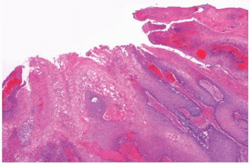
The tumors grossly appear warty and fungating with ulcerations. Histologically, verrucous carcinomas are defined narrowly. They have a wartlike appearance, with abundant keratosis and parakeratosis arising from a folded, thickened squamous epithelium (
Fig. 5.1, e-
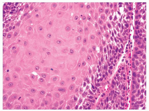
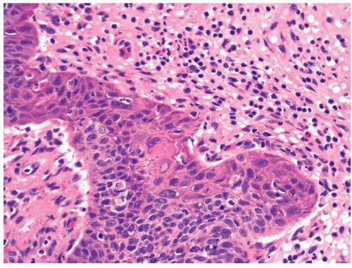
Fig. 5.1). This leads to the noted appearance of “church spires.” The squamous cells mature toward the surface and only minimal cytologic atypia should be present (
Fig. 5.2, e-
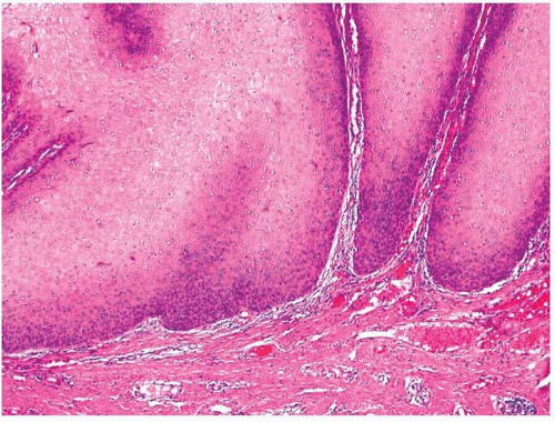
Fig. 5.2). That said, some of the squamous cells may undergo individual keratinization (dyskeratosis), and occasional squamous pearls may be seen within the squamous epithelium. Mitotic figures should be located within or near the basal epithelium. Nuclei have vesicular chromatin with small nucleoli. The bases of the tumors show downgrowths of broad tongues of mature, well-differentiated squamous
epithelium that invade the underlying tissues with a somewhat circumscribed, pushing border (
Fig. 5.3, e-
Fig. 5.3). Infiltrating irregular nests, typical of conventional-type SCC, should not be present. At the base of the tumors, a dense lymphoplasmacytic infiltrate may be present, as may occasional foreign body-type granulomas.
Although many SCCs will have
verrucoid features, it has been noted that the diagnosis of verrucous carcinoma should only be made when tumors meet the strict histologic criteria listed earlier if the diagnosis is
to have clinical meaning. Tumors that are verrucoid, yet do not meet the strict criteria for the diagnosis of verrucous carcinoma, will generally behave in a manner similar to more conventional-type SCCs. True verrucous carcinomas, however, have no metastatic potential. It is estimated that when strict criteria are applied, verrucous carcinomas represent less than 5% of oral and laryngeal tumors.
To diagnose verrucous carcinoma, ample sectioning of an excisional specimen and good sampling of the base of the lesion are needed. For this reason, an unequivocal diagnosis based upon a small biopsy is usually not warranted. Instead, qualified diagnoses such as SCC with verrucoid features or even verrucoid lesion should be considered, especially as benign diagnoses may sometimes feature in the differential diagnosis. Indeed, even today, most verrucous carcinomas are originally diagnosed as benign lesions on biopsy. The surgeon should be made aware of the diagnostic difficulty with these cases, as clinical findings often provide the information needed to guide subsequent therapy. Communication between the pathologist and surgeon is paramount. With final resection specimens, we are loath to use the term “hybrid carcinoma,” which others use for lesions that have features of verrucous carcinoma with focal infiltrating areas. It has been well known since the description of this tumor that some cases of upper aerodigestive tract conventional-type SCCs are deceptively bland and have some but not all of the required histologic features of verrucous carcinomas.
More aggressive yet benign forms of leukoplakia can have histologic features resembling verrucous carcinoma and are discussed in
Chapter 3.
16,17 These lesions have been clinically termed “proliferative
verrucous leukoplakia” and often histologically show
verrucous hyperplasia. They may be differentiated from verrucous carcinoma both clinically and histologically. Clinically, these lesions tend to recur and often develop into invasive SCCs, both verrucous carcinoma and conventional-type SCC; however, they should not form destructive mass lesions. Histologically, verrucous carcinoma will show features of invasion with much more acanthosis and larger tongues of squamous epithelium extending deeply into the stromal tissue. The epithelium of verrucous hyperplasia should not extend more deeply than the adjacent uninvolved epithelium.
18Verruca vulgaris has been reported in the larynx and may also enter into the differential diagnosis.
19 Like verrucous hyperplasia, these lesions should not invade the stroma. It has also been noted that the rete pegs tend to be thinner with verrucae and that verrucae have more prominent keratohyalin granules. Finally, papillary SCC is also mentioned occasionally as a differential diagnostic consideration. The lesions are not very similar histologically and generally should not be confused (see below). Still, there appears to be some histologic overlap in phenotype and some cases can show mixed features.
Immunohistochemistry has shown the accumulation of p53 protein in many of these tumors, and proliferation markers are often noted to be overexpressed when compared with benign epithelium (although not to the degree of more cytologically abnormal cases).
15,20 Retinoblastoma protein expression remains intact and some growth factors, such as c-erbB-3, have been shown to be overexpressed.
21 Loss of heterozygosity (LOH) studies investigating multiple microsatellite markers have shown that these tumors have fewer abnormalities than more poorly differentiated SCC variants such as basaloid SCC or spindle cell carcinoma and have an LOH incidence similar to that of well-differentiated conventional-type SCC.
1 The tumors frequently contain deletions at 9p, a region believed to be involved early in the development of upper aerodigestive tract SCC. Whether these tumors lack any specific genetic abnormalities that would explain their lack of metastatic potential has not conclusively been shown. While some have shown differences of LOH at 4q and 17p between verrucous SCC and conventional-type SCC, these differences were not confirmed in a study that compared verrucous carcinoma with only welldifferentiated, conventional-type SCC. In our experience, verrucous carcinomas are neither immunoreactive with antibodies to p16 nor do they contain high-risk HPV by in situ hybridization.
PAPILLARY SQUAMOUS CELL CARCINOMA
Papillary SCC is an uncommon variant of SCC that may involve the upper aerodigestive tract.
22,23,24,25,26,27,28 and
29 The lesions occur most frequently in older male patients, like nearly all variants of SCC in this region. This variant occurs throughout the upper aerodigestive tract, but most often involves the larynx, oropharynx, and sinonasal tract. Because of its rarity, the
pathogenesis remains unclear. Approximately half of the cases in a recent study were secondary to HPV infection; these tumors most frequently involved the oropharynx.
29 Occasional cases do develop in patients with previously diagnosed benign squamous papillomas.
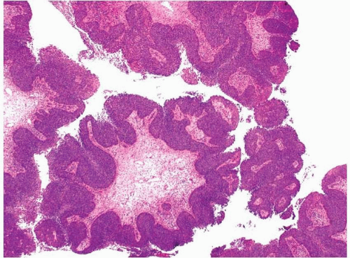
Grossly, these lesions are largely exophytic and appear papillary or even warty, akin to verrucous carcinomas. These lesions are defined histologically and have a low-power appearance similar to sinonasal papillomas (
Fig. 5.4, e-
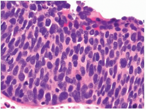
Fig. 5.4). Numerous complex papillary and filiform structures extend in all planes, often rendering the assessment of true tissue invasion extremely difficult. The papillary fronds are covered with a stratified squamous epithelium, which has overt features of malignancy, replete with lack of maturation, increased nuclear to cytoplasmic ratios, nuclear irregularities, and numerous mitotic figures located throughout the entire thickness of the epithelium (
Fig. 5.5, e-
Fig. 5.5). Koilocytic change is often noted (e-
Fig. 5.6). The epithelial component thus appears similar to high-grade squamous intraepithelial lesions of the cervix. Intracellular keratinization or dyskeratosis may be present; however, surface keratosis is often not seen or may be only focal (
Fig. 5.6). The papillary fronds have fibrovascular cores that usually contain some degree of lymphoplasma-cytic infiltrate, which then extends to the base of these lesions where it becomes denser.
The complex nature of these specimens can make the assessment of invasion difficult, especially when the invasive component remains papillary. Often, however, the invasive component will have infiltrating, irregular nests of squamous epithelium morphologically identical to conventional-type SCC. As with verrucous carcinoma, invasion can
almost never be excluded at biopsy, and because of the complex and highly exophytic nature of the noninvasive component of these tumors, definitive invasion can rarely be diagnosed at biopsy. Resection specimens should be carefully assessed for invasion, as invasive lesions tend to behave in a manner similar to that of conventional-type SCC of the upper aerodigestive tract. Lesions that lack invasion can be locally aggressive
and difficult to fully resect, especially when they involve the sinonasal tract. Such lesions often progress to invasive malignancies.
The most common differential to be considered with these lesions includes benign papillary lesions, especially sinonasal or schneiderian papillomas. The degree of epithelial atypia present in these lesions should not generally be seen in other squamous papillary lesions of the upper aerodigestive tract, and the diagnosis is usually not very difficult. Occasional sinonasal or laryngeal papillomas can show inflammation with cytologic atypia or even dysplasia with numerous mitotic figures, however, and methods for definitively distinguishing such lesions from noninvasive papillary carcinomas are lacking histologically. In general, papillary SCC should be a destruction lesion, whereas laryngeal papillomas are not.
Distinguishing these lesions from verrucous carcinomas should not be difficult microscopically, although the two may appear similar grossly (
Table 5.3). Thompson et al.
25 have also argued that the lesions should be differentiated from a more garden variety
exophytic SCC. They point out that true papillary SCC has a better prognosis than exophytic SCC. Histologically, it is more filiform and not as complex. The authors note that papillary SCCs appear more like stalks of celery cut on cross section and should not resemble cauliflower.
Studies investigating genetic abnormalities in these tumors are somewhat limited due to their rarity.
1,26 Overexpression of p53 protein has been shown by immunohistochemistry. LOH studies have shown a similar incidence of genetic abnormalities between these tumors and verrucous carcinoma and well-differentiated, conventional-type SCC. It is interesting to note that these tumors showed an increased LOH for a
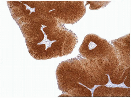
microsatellite marker on the long arm of chromosome 11 when compared with other variants of SCC, although the finding was not statistically significant. Tumors secondary to high-risk HPV infection are diffusely and strongly immunoreactive with antibodies to p16 (
Fig. 5.7).
29
BASALOID SQUAMOUS CELL CARCINOMA
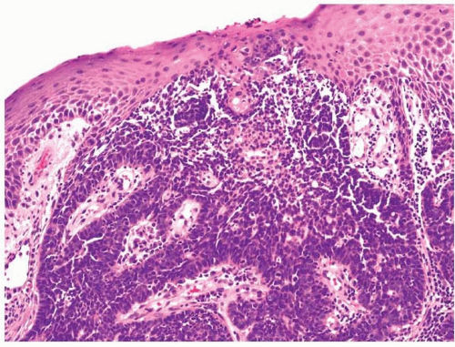
Some SCCs at various sites throughout the body retain a distinctly epidermoid phenotype while lacking cellular maturation and extracellular keratin formation. The cells remain immature in appearance and resemble those of the basal layer of a typical stratified squamous epithelium. For this reason, these tumors are termed “basaloid SCCs,” although a variety of names have been used for these tumors when they occur at different sites in the body (e.g., cloacogenic carcinoma when they arise in the anus).
30,31,32 and
33Basaloid SCCs of the upper aerodigestive tract occur in older individuals, usually in their seventh or eighth decade of life, and occur predominantly in men. First reports of these tumors have described a strong association with tobacco and alcohol use; however, many cases that develop today are secondary to infection by high-risk HPV and occur in younger patients than those first presented.
34,35,36,37 and
38 The tumors show
a predilection for the involvement of the base of tongue, hypopharynx, and supraglottic larynx but have been noted throughout the entire upper aerodigestive tract. Most patients present at a high stage with nodal metastases. The behavior of these tumors is related to HPV status, with tumors harboring infection behaving better stage for stage than conventional SCCs, whereas those not secondary to HPV infection behave the same or worse than other SCCs.
39