Normal Anatomy and Histology
The upper aerodigestive tract and ear are unified by location and function. Because this region does not represent a single organ per se, a discussion of the anatomy and histology will be somewhat complicated. That said, an understanding of the two is essential for surgical pathologists as they struggle to interpret biopsies from these sites. The correct interpretation of lesional histology presupposes an understanding of normal histology. Furthermore, such an understanding also enhances one’s awareness of how certain lesions tend to occur throughout the system (e.g., squamous cell carcinoma and salivary gland-type neoplasms), while others only develop at specific sites (e.g., olfactory neuroblastoma and schneiderian papilloma).
THE NASAL CAVITY
The nasal cavity is bounded anteriorly by the external nares and posteriorly by the nasal choana. It is composed of the left and right nares, which are separated by the nasal septum. The septum is both osseous and cartilaginous and is composed of the vomer (inferior and posterior), the ethmoid bone (superior), and septal cartilage (anterior). Laterally, the “scroll-shaped” superior, middle, and inferior turbinates or conchae are present. Openings to the paranasal sinuses are present laterally and include the openings to the sphenoid sinuses (located behind the superior turbinate), the frontal sinus ostia (located in the anterior region of the middle turbinate), the primary (and accessory) maxillary ostia (located in the hiatus semilunaris), and openings to the anterior, middle, and posterior ethmoid air cells (located in the middle turbinate and behind the superior turbinate). The superior portion of the nasal cavity is bounded by the cribriform plate.
The vast majority of the nasal cavity is lined by ectodermally derived tissue.1 The most anterior mucosa, that of the vestibule, is lined by a
keratinized, stratified squamous epithelium. This epithelium is continuous with that of the external nose and the mucosa includes adnexal structures such as hair follicles and sebaceous and sweat glands (Fig. 1.1). As one progresses posteriorly, this epithelium is replaced by nonkeratinizing squamous cells and ciliated columnar cells, with occasional mucous or goblet cells and intermediate cells (schneiderian mucosa) (Figs. 1.2, 1.3 and 1.4). Seromucinous glands are present beneath the epithelium within a loose stroma. Occasional melanocytes may be present within the surface epithelium or the underlying glands and may even be seen in the lamina propria overlying the septum and turbinates. At the lower, anterior septum, a small tubular sac lined by nonciliated columnar cells (the vestigial vomeronasal organ of Jacobson) may be seen.1 This organ is better developed in the more olfactory-inclined animals. Dense, muscular vascular tissue is present beneath the seromucinous glands and is sometimes noted to resemble erectile tissue (Fig. 1.5). Lymphatic channels, small nerves, and occasional, scattered inflammatory cells can also be present.
keratinized, stratified squamous epithelium. This epithelium is continuous with that of the external nose and the mucosa includes adnexal structures such as hair follicles and sebaceous and sweat glands (Fig. 1.1). As one progresses posteriorly, this epithelium is replaced by nonkeratinizing squamous cells and ciliated columnar cells, with occasional mucous or goblet cells and intermediate cells (schneiderian mucosa) (Figs. 1.2, 1.3 and 1.4). Seromucinous glands are present beneath the epithelium within a loose stroma. Occasional melanocytes may be present within the surface epithelium or the underlying glands and may even be seen in the lamina propria overlying the septum and turbinates. At the lower, anterior septum, a small tubular sac lined by nonciliated columnar cells (the vestigial vomeronasal organ of Jacobson) may be seen.1 This organ is better developed in the more olfactory-inclined animals. Dense, muscular vascular tissue is present beneath the seromucinous glands and is sometimes noted to resemble erectile tissue (Fig. 1.5). Lymphatic channels, small nerves, and occasional, scattered inflammatory cells can also be present.
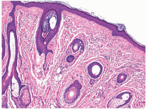 FIGURE 1.1 Squamous mucosa of the nasal vestibule. |
The cribriform plate, the medial superior turbinate, and the superior one-third of the nasal septum all may be lined by olfactory mucosa (Fig. 1.6). This specialized mucosa is composed of tall, eosinophilic sustentacular or supporting cells, elongated, ciliated olfactory neurons, and small basally located cells. The sustentacular cells can sometimes contain lipofuscin, which may give the area a yellow appearance, clinically.2 Beneath the mucosa are serous glands (the glands of Bowman). Focally, olfactory mucosa may be intermixed and replaced by schneiderian mucosa.
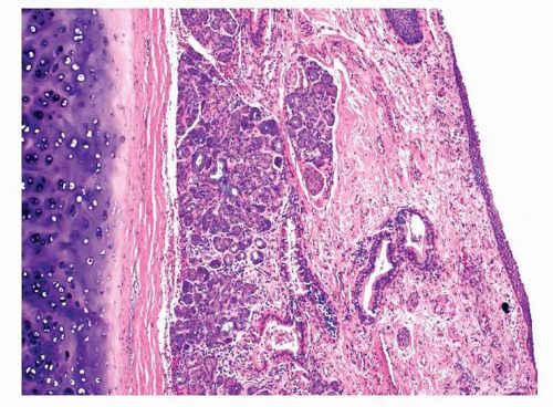 FIGURE 1.2 Schneiderian mucosa with underlying seromucinous glands. |
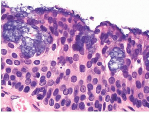 FIGURE 1.3 Schneiderian epithelium with mucous cells. |
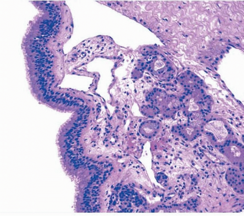 FIGURE 1.4 Proximal schneiderian mucosa. The epithelium is comprised mostly of ciliated, columnar cells. |
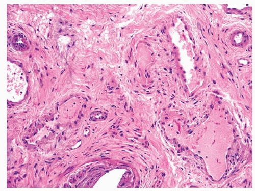 FIGURE 1.5 Fibromuscular vascular tissue. |
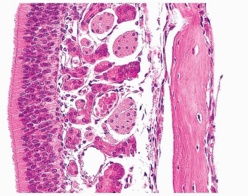 FIGURE 1.6 Olfactory epithelium (Balogh K, Pantanowitz L. Mouth, nose, and paranasa sinuses. In: Mills SE, ed. Histology for Pathologists. 3rd ed. Philadelphia, PA: Lippincott-Williams & Wilkins; 2007:403-430.) |
THE PARANASAL SINUSES
The paranasal sinuses include the frontal, maxillary, sphenoid, and ethmoid sinuses. The ethmoid sinuses are composed of numerous (usually 3-18) ethmoid air sacs. Each of the sinuses is in communication with the nasal cavity and may be considered “diverticula” of the nasal cavity. They grow until adulthood and are usually somewhat asymmetrical in shape and size.
The paranasal sinuses are lined by ciliated columnar cells, with occasional mucinous cells (Fig. 1.7). These cells tend to be less tall than those of the nasal cavity and the mucosa is only half as thick as the nasal mucosa. Between the epithelium and the bone is only a thin layer of fibrous tissue. Seromucinous glands are rare and are present primarily near the ostia to the nasal cavity.
THE ORAL CAVITY
The mouth begins at the vermilion border of the lips. It is encircled by the hard and soft palates (superior), cheeks (lateral), and anterior two-thirds of the tongue and floor of the mouth (inferiorly). The posterior junction with the oropharynx (the fauces) is located superiorly at the posterior edge of the soft palate and uvula, laterally at the tonsillar pillars, and inferiorly at the circumvallate papillae of the tongue (the sulcus terminalis).
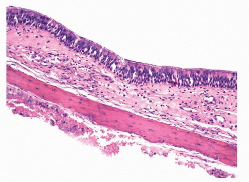 FIGURE 1.7 Mucosa of the paranasal sinus. |
Although there are some regional differences, the majority of the oral cavity is lined by a nonkeratinizing squamous epithelium (Fig. 1.8). Varying degrees of keratinization may be present overlying the gingiva and the hard palate as they are exposed to mastication. Melanocytes and Merkel cells may be present and, although they are difficult to identify, they are generally scattered throughout the oral mucosa. The tongue is lined by filiform, fungiform, foliate, and circumvallate papillae, which
may have taste buds along the sides of their invaginations (Fig. 1.9). These are composed of gustatory, sustentacular, and basal cells akin to olfactory mucosa of the nasal cavity. Immediately beneath the epithelium throughout the mouth is a basal lamina that is composed of type IV collagen, heparan sulfate, laminin, and enactin.2


may have taste buds along the sides of their invaginations (Fig. 1.9). These are composed of gustatory, sustentacular, and basal cells akin to olfactory mucosa of the nasal cavity. Immediately beneath the epithelium throughout the mouth is a basal lamina that is composed of type IV collagen, heparan sulfate, laminin, and enactin.2
Stay updated, free articles. Join our Telegram channel
Full access? Get Clinical Tree
