Benign Squamous Proliferations and Neoplasms
Most neoplasia of the upper aerodigestive tract shows squamous differentiation. Clinically, squamous lesions may be single or multifocal, benign, aggressive, or malignant. Risk factors for the diseases overlap, as do clinicopathologic features, and the definitive diagnosis of some lesions may be extremely difficult, especially with small biopsy specimens. This chapter discusses benign squamous proliferations and neoplasms as they occur throughout the upper aerodigestive tract, with further discussion of some distinct clinicopathologic entities.
PAPILLOMAS
Unifocal and multifocal squamous papillomas occur throughout the upper aerodigestive tract, from the nasal vestibule, to the oral cavity, to the larynx. They can develop at any age, but often occur in children and young to middle-aged adults, possibly reflecting a relationship with exposure to human papillomavirus (HPV). Some lesions may be distinguished as condylomas or verrucae when they are caused by HPV; however, such distinction is usually not necessary, and unequivocal infection is rarely demonstrated. It should be noted that although koilocytic change may be helpful for identifying lesions induced by HPV, the appearance of an HPV-associated viral cytopathic effect is also not entirely specific. True HPV-induced lesions are often multiple and are more common in individuals infected with human immunodeficiency virus (HIV) or who are otherwise immunosuppressed. While most papillomas are not considered to be at risk for malignant transformation and do not show dysplastic changes, lesions in immunocompromised patients may show dysplasia and may be at increased risk for malignant transformation.
Grossly, these lesions are exophytic and may vary considerably in size. A rough, warty appearance may be noted. Histologically, the squamous
epithelium is thickened and covers papillary cores of stromal tissue (Fig. 2.1, e-Fig. 2.1).1 Keratinization may be present and the granular cell layer may appear thickened (Fig. 2.2). The stratified squamous epithelium usually shows normal maturation with limited, if any, atypia. Mitotic figures should be located in or immediately above the basal layer of the epithelium, and some degree of chronic inflammation may be present, especially within the stroma at the base of the lesions (e-Fig. 2.2). The stroma of papillomas of the nasal vestibule will contain skin appendages.2 Many lesions have broader bases with more rounded surfaces and less keratinization and thus appear similar to condylomata of the cervix, replete with focal koilocytic change (Fig. 2.3).
epithelium is thickened and covers papillary cores of stromal tissue (Fig. 2.1, e-Fig. 2.1).1 Keratinization may be present and the granular cell layer may appear thickened (Fig. 2.2). The stratified squamous epithelium usually shows normal maturation with limited, if any, atypia. Mitotic figures should be located in or immediately above the basal layer of the epithelium, and some degree of chronic inflammation may be present, especially within the stroma at the base of the lesions (e-Fig. 2.2). The stroma of papillomas of the nasal vestibule will contain skin appendages.2 Many lesions have broader bases with more rounded surfaces and less keratinization and thus appear similar to condylomata of the cervix, replete with focal koilocytic change (Fig. 2.3).
Benign squamous papillomas must be distinguished from malignant or premalignant diseases such as verrucous carcinoma or verrucous hyperplasia (see Chapters 3 and 4). Patients with verrucous carcinoma or verrucous hyperplasia are older and will usually have a long history of tobacco use. Neither verrucous carcinoma nor verrucous hyperplasia generally appears as discrete polypoid masses, and verrucous carcinoma is a destructive lesion that infiltrates the underlying stroma in a pushing manner. As mentioned, squamous papillomas of the upper aerodigestive tract appear less verrucoid than typical skin lesions and more like condylomata.
A particular lesion of the oral cavity, focal epithelial hyperplasia, arises more commonly in children and young adults and tends to be more common in certain geographic locations and in certain ethnicities.3 These lesions are benign and regress as patients age. Numerous small, slightly
raised, pink papules are seen grossly, which may become confluent. Histologically, there is squamous hyperplasia, usually devoid of papillary architecture. Koilocytes are readily identified and the clumped chromatin may sometimes appear similar to mitotic figures (mitosoid bodies).
raised, pink papules are seen grossly, which may become confluent. Histologically, there is squamous hyperplasia, usually devoid of papillary architecture. Koilocytes are readily identified and the clumped chromatin may sometimes appear similar to mitotic figures (mitosoid bodies).
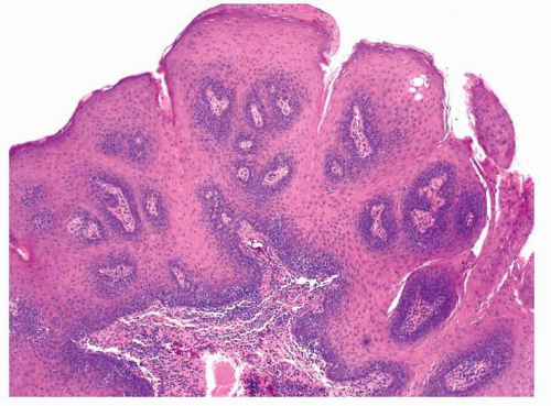 FIGURE 2.1 Squamous papilloma of the palate. |
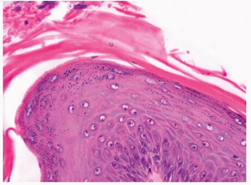 FIGURE 2.2 Keratinization and thickened granular cell layer in a squamous papilloma. |
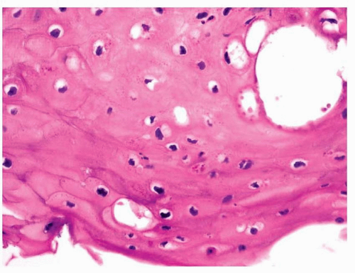 FIGURE 2.3 Koilocytes in a papilloma. |
LARYNGEAL PAPILLOMAS AND PAPILLOMATOSIS
Squamous papillomas of the larynx are categorized according to their number (solitary or multiple) and the age of the patient affected (juvenile or adult).4,5 Most laryngeal papillomatosis occurs in younger patients. The lesions are usually located in the region of the true vocal cords; however, they may be found throughout the larynx and even throughout the oropharynx and trachea.4 They often recur after resection and may cause airway obstruction. The juvenile lesions and some of the adult ones are related to infection of the mucosa with HPV types 6 and 11.4 In cases of juvenile papillomatosis, it is thought that infection occurs at birth, whereas in adults, it is believed that the infection develops at a later age.4 Grossly, the lesions are variably sized but are usually smaller than a centimeter in greatest dimension. Histologically, these lesions are characterized by a branched fibrovascular core, covered with maturing stratified squamous epithelium (Fig. 2.4, e-Fig. 2.3).4 Keratosis is generally not seen. Intraepithelial dysplasia is infrequent but has been noted, and severe or full-thickness dysplasia is very uncommon and should suggest a diagnosis of papillary carcinoma (Fig. 2.5).6 Koilocytic change may be seen. Rare examples of extremely well-differentiated squamous cell carcinoma arising in patients with laryngeal papillomatosis have been recorded.7,8
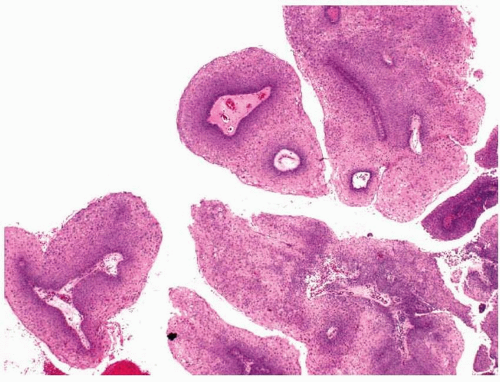 FIGURE 2.4 Laryngeal papillomas. |
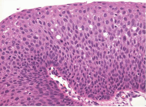 FIGURE 2.5 Mild to moderate squamous dysplasia in a laryngeal papilloma. |
The differential diagnosis should include other squamoproliferative lesions, especially squamous cell carcinoma. Some laryngeal papillomas in older individuals may be very difficult to distinguish from papillary squamous cell carcinomas or verrucous carcinomas. In general, these carcinomas are clinically destructive and will show clear-cut invasion. Unfortunately, helpful clinical history is often not provided, and invasion may be difficult to assess with small biopsy specimens. Papillary squamous cell carcinoma should show full-thickness cytologic atypia; however, as was mentioned, laryngeal papillomas may also show cytologic atypia. For this reason, clinical follow-up and complete excision are recommended for any adult with a solitary papillary lesion that shows any degree of atypia. Verrucous carcinomas will not, by definition, show more than mild cytologic atypia and should show keratinization. They should not be diagnosed if the base of the lesion cannot be assessed on biopsy to verify destructive invasion.
SINONASAL (SCHNEIDERIAN) PAPILLOMAS
Sinonasal or schneiderian papillomas arise almost exclusively within the nasal cavity and paranasal sinuses from the ectodermally derived ciliated columnar epithelium that lines these surfaces; they arise rarely at other sites.9,10 They occur in middle-aged individuals and are at least twice as common in men as in women.10,11 They are classified according to their growth pattern (exophytic/fungiform vs. endophytic/inverted) and by their epithelial cell type (squamous vs. oncocytic/cylindrical cell), although some lesions may show combined features.10,11


Stay updated, free articles. Join our Telegram channel
Full access? Get Clinical Tree
