Major symptoms
Minor symptoms
Facial pain/pressure
Dental pain
Nasal obstruction/congestion
Ear fullness/pain
Nasal discharge
Headache
Hyposmia/anosmia
Halitosis
Cough
Fever
Fatigue
The American Academy of Pediatrics divides sinusitis into 5 different categories based on the duration and frequency of symptoms (see Fig. 7.1) [3]. Acute sinusitis is defined as symptoms of less than 4 weeks duration and strongly suggested by the presence of 2 major symptoms or 1 major and 2 minor symptoms (see Table 7.1). Subacute sinusitis is regarded as when symptoms last anywhere from 4 to 8 weeks. Chronic sinusitis is characterized by symptoms lasting longer than 90 days. Recurrent acute sinusitis, which is, defined as recurrent episodes each completely resolving in less than 2 weeks, separated by asymptomatic periods of at least 10 days. Acute exacerbation of chronic sinusitis is when patients with chronic sinusitis develop new acute respiratory symptoms.
Fig. 7.1
Categories of sinusitis . ABS (acute bacterial sinusitis ); chronic (chronic sinusitis)
Anatomy
The paranasal sinuses consist of air-filled paired cavities in the skull named after the bone in which it is located. The maxillary sinuses are located deep to the cheeks and under the eyes. The frontal sinuses are located above the eyes, the ethmoid sinuses between the eyes and the sphenoid sinuses located at the center of the skull behind the eyes. These cavities are involved in the humidification of air, voice resonance, and skull weight. Each of these sinuses drains through an ostium of approximately 1–3 mm in size into the nasal cavity.
The maxillary and the ethmoid sinuses are present at birth and slowly enlarge to adult size by puberty [4]. The maxillary sinuses have an inverted pyramidal shape and are the largest of the sinuses. The natural opening of the maxillary sinus is positioned superiorly on the medial maxillary wall and opens to the ostiomeatal complex, a confluence of sinus drainage from the frontal, anterior ethmoids and maxillary sinuses located between the middle and inferior turbinates. The ethmoid sinuses consist of a honeycomb of air cells that lie medial to the orbits and are separated from the orbits by the lamina papyracea. The ethmoid sinuses are divided into anterior and posterior cells. Infection of the ethmoids can easily spread to the orbit directly through bony dehiscences or by traversing the neurovascular foramina. The frontal sinuses arise from the anterior ethmoid air cells and start developing between the ages of 5–6 years and continue to develop until late adolescence [5]. The frontal sinuses are funnel-shaped structures with the posterior wall separating the sinus from the anterior cranial fossa. The diploic veins connect the vasculature of the sinus mucosa with the intracranial sinuses and veins, providing a potential route of infectious spread. The sphenoid sinus begins to develop between 3 and 5 years of age and continues to grow until late teenage years. The sphenoid sinus ostium is located on the anterosuperior surface of the sphenoid face, and drains with the posterior ethmoid cells into the superior meatus located between the middle and superior turbinates. The optic nerve and the carotid artery are located on the lateral wall of the sphenoid sinus and the pituitary fossa is located in the posterior aspect.
The sinus cavities are lined with pseudostratified ciliated epithelium, which contain globlet cells, and submucosal glands that produce two different layers of secretions. The deep layer consist of serous secretions permitting normal cilia motility and the superficial layer consist of mucinous/viscous secretions which increase viscosity to capture particles and pathogens. The cilia move mucus and debris from the sinus cavities into the nasopharynx.
Pathophysiology
For the paranasal sinuses to have normal function three main factors need to be present: a normal mucociliary function, patency of the sinus ostia, and thin/clear consistency of nasal secretions. The most common causes to disrupt the normal function of the sinuses in children are viral URTIs and allergic rhinitis. Mucosal edema and inflammation can be caused by viral infections, allergic rhinitis, immune disorders, gastroesophageal reflux disease (GERD), nasal obstruction, and/or anatomical abnormalities and systemic disorders. Prolonged sinus obstruction, variation in the character of nasal secretions and alterations in intranasal pressure can lead to bacterial growth, colonization and subsequently infection of the sinus cavities. Although inflammation in any of the sinuses can lead to obstruction of the sinus ostia, the most commonly involved sinuses in a sinus infection are the maxillary and the anterior ethmoid sinuses [6].
In an URTI, the nasal mucosa responds to the virus by producing mucus and recruiting white blood cells to the lining of the nose, inducing an inflammatory response leading to congestion and swelling of the nasal passages [7]. The resultant ostial obstruction creates sinus cavity hypoxia and mucus retention causing the cilia to function less efficiently, creating an environment for bacterial growth.
Allergy is involved in sinusitis due to an inflammatory obstruction of the sinonasal mucosa and ostia caused by the release of histamine and major basic protein by mast cells and eosinophils, respectively. These substances cause vasodilation, mucous secretion, nerve stimulation and smooth muscle contraction causing damage to the surrounding tissue resulting in rhinorrhea, itching, sneezing and postnasal drip [8]. Non-allergic environmental irritants such as cigarette smoke and household chemicals can directly affect the mucosa of the sinuses. In addition to allergy, immunologic compromise may be an important etiologic factor in patients with chronic, refractory sinusitis. Children with immune deficiencies will likely have a history of recurrent and refractory sinus infections associated with otitis media, bronchitis, and lower respiratory infections.
There is a higher prevalence of GER D in patients with refractory sinusitis, suggesting that reflux may be a contributing factor to the pathogenesis of chronic sinus infection. Acid from the stomach can directly injure the nasal mucosa leading to sinonasal edema and impaired mucociliary clearance. Nasopharyngeal reflux has been documented in children with symptoms of chronic sinusitis that present with coughing paroxysms [9]. Despite the low quality of evidence supporting this relationship, GERD treatment should be considered in patients with chronic sinusitis and reflux symptoms, particularly in those patients not responding to conventional sinusitis treatment [10].
Anatomical variations most commonly associated with nasal obstruction and sinus pathology are nasal septal deviation and concha bullosa, (see Fig. 7.2). Intranasal growths such as nasal polyps , benign or malignant tumors, and adenoid hypertrophy are contributing factors to sinus infections. Both anatomical abnormalities and intranasal growths can cause mechanical obstruction of the sinus ostia predisposing patients to sinusitis [11, 12]. However, studies in children have not found a relationship between sinus disease and anatomical variations, suggesting that other factors play a more essential role in the development of pediatric sinusitis. In addition, the adenoid pad can act as a nidus of bacteria contributing to recurrent or chronic bacterial sinusitis.
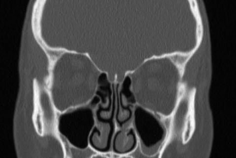
Fig. 7.2
Non-contrast CT sinus showing an area of nasal obstruction . Left septal spur impinging an enlarged inferior turbinate with mild mucosal thickening of the left maxillary sinus
Systemic diseases such as cystic fibrosis and primary ciliary dyskinesia can also alter the normal mucociliary function and cause sinus infections. Cystic fibrosis causes significant alteration in the quality of mucous secreted in the nasal cavities interrupting normal physiology [13]. Primary ciliary dyskinesia affects the cilia morphology and number therefore causing reduced mucous clearance from the respiratory tract and altering the defense mechanisms against environmental particles, bacterial and viral pathogens.
Microbiology
The specific data regarding the contemporary microbiology of sinus disease is limited and much of what is known in children is extrapolated from studies of either nasal colonization or otitis media. Studies of pediatric outpatients with acute otitis media who had middle ear fluid (MEF) and nasopharyngeal (NP) cultures obtained simultaneously have revealed that the middle ear pathogen was also co-isolated in NP culture in 100 % of cases. By contrast however, other pathogens were identified in the NP culture that were not present in the MEF in 47 % of cases [14]. Thus, while NP cultures may provide some information regarding potential otitis and sinus pathogens, the results are hardly definitive. While Streptococcus pneumoniae remains the predominant bacterial respiratory pathogen in children, the microbiology of pediatric respiratory disease has evolved tremendously over the past 14 years. This change is mainly the result of the introduction of the 7-valent followed by the 13-valent pneumococcal conjugate vaccines (PCV7 and PCV13). Following introduction of the PCV7 vaccine, a decline in sinus disease and otitis media due to serotypes of S. pneumoniae included in the vaccine was observed. Studies of children who underwent endoscopic sinus surgery (ESS) following introduction of PCV7, however, revealed an increase in the relative proportion of cases of sinusitis due to S. pneumoniae serotypes not included in the vaccine. Notably the antibiotic-resistant pneumococcal serotype 19A was most common among these non-vaccine serotypes [15]. The prevalence of serotype 19A S. pneumoniae as a sinus pathogen has since declined after widespread use of PCV13 (which includes serotype 19A) [16]. Furthermore, the proportion of cases of orbital abscesses due to S. pneumoniae has declined following introduction of the PCVs [17]. In addition, among isolates causing invasive disease, the percent of pneumococci non-susceptible to penicillin decreased in the post PCV-13 era [18]. Risk factors for infection with pneumococci resistant to penicillin and other antibiotics include age <2 years or >65 years, daycare attendance, medical comorbidities and recent antibiotic use [19, 20]. Studies of invasive pneumococcal disease have shown that among patients with previous antibiotic exposure, proximity to last antibiotic course predicted resistance to that particular antibiotic; such may be the case for pneumococcal sinusitis as well [21].
While the proportion of cases due to S. pneumoniae has declined, other pathogens have increased in frequency. Recent studies of young children with otitis media from Rochester, New York have demonstrated a decline in S. pneumoniae isolation from MEF culture and a relative increase in the prevalence of nontypeable-Haemophilus influenzae (NTHI) . Work from New Zealand has demonstrated that H. influenzae can be identified from MEF through a combination of culture and PCR techniques in up to 60 % of children with otitis in the post-PCV era [22]. Studies in the United States have shown that NTHI may contribute to up to 41 % of cases in children, an increase from 25 % in the pre-PCV era [23]. H. influenzae can present therapeutic challenges in that many strains possess plasmid-encoded β-lactamases. Heilman et al. noted that 26 % of H. influenzae respiratory isolates were β-lactamase-positive [24], however there is tremendous variability around the globe. There has also been noted in recent years outside of the US the emergence of NTHI strains that are resistant to β-lactams through alternative mechanisms other than β-lactamases [25, 26]. The other predominant bacterial pathogen in acute sinusitis to consider in children is Moraxella catarrhalis [27]. M. catarrhalis accounts for up to 20 % of sinusitis pathogens in children [28]. While notably over 90 % of M. catarrhalis produce β-lactamases, susceptibility to other agents remains high [29, 30].
With the rise of community-acquired methicillin-resistant Staphylococcus aureus (MRSA) , much interest has developed in the role that S. aureus may play in respiratory disease. S. aureus has been variably reported as a cause of acute bacterial sinusitis with reports ranging from 0 to >30 % of cases [31]. Many investigators have reported an increased in frequency of MRSA recovered from patients with sinus disease [32]. The exact prevalence of S. aureus in sinus disease is unclear as nasal colonization with S. aureus, in particular MRSA, has increased over time in healthy pediatric and adult controls. Thus, the prevalence of S. aureus as a pathogen of acute sinusitis has likely been over estimated by studies that have relied on NP culture for diagnosis alone. Notably, however, Huang et al. in Taiwan performed cultures of middle meatus drainage in adults and children with maxillary sinusitis and found MRSA in 2.7 % [33]. In contrast to its relatively small role in acute sinusitis, S. aureus can be a prominent cause of chronic sinusitis. In a 3 year review of cases of chronic sinusitis managed with ESS, Whitby et al. identified 56 cases of S. aureus sinusitis of which 21 % were MRSA [34]. Notably, in this study copathogens were isolated in 77 % of cases underscoring that chronic sinusitis is frequently a polymicrobial disease. Among patients with chronic sinusitis, other etiologies to consider include anaerobes, gram-negative bacilli and fungi. Actinomyces has rarely been reported as a cause of sinusitis in adults and children [35], often in the absence of dental caries that are typical for cervicofacial actinomycosis (Fig. 7.3). Allergic Aspergillus sinusitis is occasionally a cause of chronic sinusitis in children with negative cultures who have been refractory to antibiotics; patients typically have a history of atopy, recurrent sinusitis, nasal polyps and the identification of fungi on cultures/smears [36]. Such patients can be managed with thorough debridement and corticosteroids. Among patients with chronic, recurrent or refractory sinusitis consideration should also be given to an immunocompromising condition.
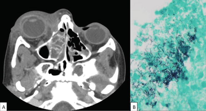
Fig. 7.3
Contrast enhanced CT scan of orbits with maxillary/ethmoid sinusitis due to Actinomyces israelii. (a) Contrast enhanced CT image of an 11-year old previously healthy boy with right-sided ethmoid sinus opacification and abscess in the medial, superior and inferior wall of the right orbit. He was taken to the operating room and histologic examination revealed sulfur granules and anaerobic cultures grew Actinomyces israelii. (b) ×40 magnification of specimen with filamentous branching bacterial organisms identified by MSN staining. Pathologic slide courtesy of Karen Eldin, MD, Dept of Pathology Baylor College of Medicine
Atypical bacterial pathogens are very uncommon causes of sinusitis. Chlamydia (also referred to as Chlamydophila) pneumoniae can rarely be identified as causes of chronic sinusitis in adults or children [37, 38]. Among adults with sinusitis one group of investigators noted that 3.5 % had elevation of complement fixing anti-Mycoplasma antibodies [39]. Given that Mycoplasma antibodies cross react with other pathogens and have a long half-life, relying on this as a diagnostic tool is problematic. Lee et al. performed PCR for Mycoplasma, C. pneumoniae and Legionella from ethmoid sinus samples of 11 adults undergoing endoscopic sinus surgery for chronic sinusitis; none of these patients had molecular evidence of infection due to these pathogens [40]. Conversely, studies of military servicemen with Mycoplasma pneumoniae pneumonia found that nearly two-thirds had radiologic evidence of sinusitis [41]. Thus, for adults or older children with concomitant atypical pneumonia and sinusitis, Mycoplasma could be a potential etiology of disease. While it is believed that viral URTI may create inflammation at sinus ostia, impairing sinus drainage and predisposing to acute sinusitis, the direct role of viruses in infection is unclear. Rises in antibody titers to a number of viruses including influenza, adenovirus and parainfluenza virus have been documented to occur in the setting of acute sinusitis [39]. Other investigators have been able to demonstrate the presence of rhinovirus RNA in maxillary sinus epithelial cells of volunteers with clinically diagnosed sinusitis [42]. Given that viruses can continue to be shed for a period after resolution of an acute respiratory illness, the significance for etiology of the detection of these viruses in patients with sinusitis is unclear.
The diagnosis of sinusitis in a severely immunocompromised child is both a medical and surgical emergency because of the risk of invasive fungal infection. The primary fungi of concern include the Zygomycetes (also referred to as Mucor), Aspergillus spp. and as well as number of less common dematiaceous fungi (such as Curvularia, Bipolaris, Fusarium etc). Such patients are at risk for fungal extension into critical vessels as well as the CNS and aggressive surgical debridement is often necessary (Fig. 7.4). Rarely, acute infection in an otherwise healthy host can extend from the paranasal sinuses into the orbit (Fig. 7.5) or cranial vault. This complication occurs more often in boys (approximately 2:1 gender ratio) and intracranial extension typically occurs in older children (mean age of 11–13 years) [43, 44]. While typical acute sinusitis pathogens can be involved, particularly for intraorbital disease, other organisms predominant in intracranial infection. Common organisms associated with intracranial extension of sinusitis include Streptococcus milleri group, Propionibacterium acnes and S. aureus as well as anaerobes such as Peptostreptococcus spp., Fusobacterium spp. and Prevotella [43, 44]. Notably, both intracranial and intraorbital extension of sinus disease is frequently a polymicrobial infection [43, 45]. An additional rare complication of frontal sinusitis is the development of osteomyelitis of the frontal bone which if a subperiosteal abscess develops may manifest as the so-called Pott’s puffy tumor.
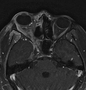
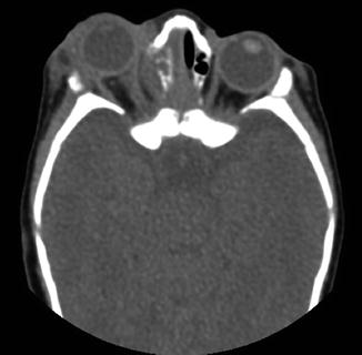
Fig. 7.4
Invasive fungal sinusitis with extension into the right orbit and intracranially. invasive fungal sinusitis in an adolescent with poorly controlled type 2 diabetes. T1-weighted MRI. Right maxillary sinus with extensive disease with extension into the right orbit. This patient presented with severe headaches and facial pain. He underwent debridement and was initiated promptly on voriconazole and amphotericin. Cultures grew Rhizopus. The child and his family denied radical surgical debridement. Despite maximal medical management the child’s disease progressed and he subsequently expired
Fig. 7.5
Maxillary and ethmoid sinusitis with extension into right orbit. Nonenhanced CT of acute sinusitis with extension into the right orbit of a 6 year-old boy. Cultures grew S. pneumoniae
Epidemiology
Understanding the precise clinical epidemiology of sinusitis in children is difficult, in part due to the somewhat subjective nature of clinical diagnosis. Interestingly, sinusitis is not a problem limited to the modern era; there is archaeological evidence of chronic sinusitis in unearthed remains of both adults and children living in medieval Europe [46]. Estimates of the number of cases of sinusitis in adults and children in the United States based on diagnostic codes reach as high as 20 million per year [2]. Some investigators have estimated that acute bacterial sinusitis complicates as many as 8–10 % of viral URTIs in children [2, 47]. Furthermore, sinusitis accounts for 5–10 % of outpatient pediatric visits for which antibiotics are prescribed [48].
Sinusitis is more commonly diagnosed in boys than girls with at least a 1.8:1 gender ratio [49, 50]. The predominant age ranges for pediatric sinusitis vary in the literature from 3 to 12 years of age [49, 50]. In a study specifically examining children less than 3 years of age, Revai et al. noted that the proportion of URTIs complicated by sinusitis peaked in the second year of life [51]. In general, however, sinusitis can occur at any age with the notable caveat that it is an extremely rare diagnosis in infants.
Clinical Manifestations and Diagnosis
Symptoms of an acute pediatric sinus infection are similar to symptoms of a viral URTI, and are strongly suggested by the presence of nasal discharge, nasal obstruction, decreased sense of smell, cough, and/or facial pain/pressure. For patients in whom viral URTI symptoms have not resolved after 7–10 days, acute sinusitis should be suspected. The diagnosis of pediatric sinusitis is mostly clinical and supported by history and findings on physical exam.
Acute bacterial sinusitis is strongly suspected if two major symptoms or one major symptom and two minor symptoms are present (see Table 7.1). Physical examination may be challenging in a pediatric patient but presence of turbinate inflammation and erythema, mucopurulent secretions in the nasal floor, and pooling of secretions from the nasopharynx in the posterior pharynx are highly suggestive of bacterial sinusitis. Endoscopic evaluation can also visualize other abnormalities on exam such as adenoid hypertrophy, nasal polyps as well as to exclude foreign bodies or other causes of nasal obstruction [52]. The presence of nasal polyps on exam should prompt an evaluation for cystic fibrosis.
CBC, ESR, and blood cultures are rarely helpful in cases of uncomplicated acute sinus infection. In cases of recurrent or chronic sinus infections, allergy testing, laboratory evaluation of the immune system to rule out immunodeficiency, mucosal biopsy to rule out primary ciliary dyskinesia and genetic testing for cystic fibrosis should be considered.
A thorough history identifying symptoms for more than 10 days has been shown to significantly correlate with abnormal radiographs and positive cultures, therefore obviating the need for imaging in acute cases. Plain radiographs, including Water and Caldwell-Luc’s views, are often ordered by clinicians to assist in the diagnosis of sinusitis but their roles is limited due to high false-positive rates and are not typically recommended in routine practice [53]. The gold standard for diagnosing bacterial sinusitis is maxillary sinus puncture with aspiration and cultures [54]. However, this procedure is usually done under anesthesia and thus is not practical except in children not responding to broad-spectrum antibiotics, patients who are toxic appearing or in immune deficient patients to target appropriate systemic antimicrobial therapy.
Children have thinner sinus walls and septa, higher bony porosity, open suture lines and larger vascular foramina in their cranial vault making them more susceptible to orbital and intracranial sinus complications than adults. Orbital complications are the most common and severity ranges from eyelid edema to subperiosteal and orbital abscesses (see Fig. 7.5) [55]. Decreased visual acuity, gaze restriction, proptosis, and diminished pupillary reflex are manifestation seen in children with orbital complications. Intracranial complications include meningitis, epidural and subdural abscess, cavernous and sagittal sinus thromboses, and intraparenchymal brain abscesses. Manifestations in pediatric patients with intracranial complications include fever, headaches, lethargy, seizures and neurologic deficits [56]. Otolaryngology and ophthalmology consultations are indicated in a patient with sinusitis and ophthalmologic findings, while otolaryngology and neurosurgery consultations are needed in a patient with sinusitis and neurological findings. Sinus complications are associated with increased morbidity and mortality therefore directed treatment and early management is strongly recommended [57].
CT scan is usually reserved for patients with suspected complications of sinusitis, those who are unresponsive to medical treatment, and patients under consideration for surgical intervention. CT is the gold standard in imaging of the sinuses as it gives better visualization providing higher resolution of bony structures. Axial and coronal views with limited cuts and without contrast usually suffice. Contrast is reserved for suspected suppuration and nasal masses. Mucosal changes, intrasinus collections or growths, and adjacent bone changes can also be visualized. MRI of the sinuses, orbits and brain is usually obtained in the presence of extensive disease and/or when multiple complications are suspected as it better characterize extension of local disease beyond the paranasal sinuses. MRI with contrast gadolinium allows better soft tissue differentiation and high spatial resolution images depicting fine details. A combination of CT and MRI is useful in cases of diagnostic difficulties [53].
Medical Management
While, in general infectious disease problems should be managed with a goal toward culture directed specific antimicrobial therapy, culture data is not typically available for acute sinusitis and therapy must be used empirically. Published guidelines for the management of acute bacterial sinusitis are available from both the Infectious Diseases Society of America (IDSA [58]) and the American Academy of Pediatrics (AAP [59]).
Stay updated, free articles. Join our Telegram channel

Full access? Get Clinical Tree


