Fig. 1.1
Acute otitis externa . Edema and erythema of the pinna and the ear canal can be noted
Treatment
The primary treatment for infectious otitis externa is the application of topical antiseptic or antibiotic agents; however, adjuvant therapies are both important and often overlooked. Careful debridement of the ear canal is important in order to provide access of topical agents to the bacteria or fungi that would otherwise be protected within or behind debris. Furthermore, some topical agents are ototoxic or highly irritating to the middle ear, and thus an assessment of whether the tympanic membrane is perforated or not helps guide the practitioner in the choice of agents. In addition to debridement, maintenance of dry ear precautions promotes resolution of the infection. Affected individuals should be encouraged to use a swimmer’s ear plug or a cotton ball saturated with petroleum jelly to occlude the ear canal when there is any chance of water entering the ear, such as with swimming, bathing, showering, or washing hair. Other measures to avoid the accumulation of moisture within the ear canal should also be taken such as avoiding the use of headphones, hearing aids, occlusive ear buds, or ear plugs other than for brief periods as described above.
Treatment of otitis externa rarely requires systemic antibiotics. Topical treatments fall into three categories: (1) drying and acidifying agents, (2) antimicrobial agents, and (3) steroids [4]. These treatment options are discussed in detail in Chap. 2.
Drying and acidifying agents. A number of acidifying preparations containing acetic acid (combined with isopropyl alcohol, aluminum acetate, or propylene glycol) are available, and can be used for both bacterial and fungal otitis externa. These agents have the potential to be highly irritating to an inflamed or macerated canal epithelium. Furthermore, these agents cannot be used in the presence of a tympanic membrane perforation due to the sensitive nature of the middle ear mucosa. Because of these two reasons, these agents are rarely used to treat acute infection; however they can be used as part of a maintenance routine in individuals with an intact tympanic membrane and a propensity towards otitis externa. The application of such agents (after showering or swimming, for example) can prevent or reduce the frequency of otitis externa.
Antimicrobial agents. Antibiotic drops are the most commonly used therapeutic option for otitis externa. Prior to the introduction of fluoroquinolone topical antibiotics, the mainstay of treatment was a mixture of neomycin, polymyxin, and hydrocortisone (Cortisporin). This preparation had the advantage of being inexpensive as well as helping to reduce inflammation with the presence of steroids. Concerns over the potential ototoxic effects of neomycin (an aminoglycoside) and polymyxin limit the use of these antibiotics to cases without a tympanic membrane perforation [5]. Because general practitioners often initiate therapy in the absence of a clear view of the eardrum, this preparation is seldom first-line therapy.
In cases of fungal otitis externa antifungal agents are required [6]. Miconazole, nystatin, tolnaftate, and ciclopirox olamine are all available in solution form and thus have the advantage of being able to be prescribed by the general practitioner and administered by the patient; however, debridement is an essential aspect of otomycosis therapy.
Steroids. Hydrocortisone and dexamethasone are often included in antibiotic preparations, or may be delivered separately if combination agents are not available. The benefit in symptom resolution provided by the addition of steroids is 0.8 days [5].
Follow Up
Topical treatment of bacterial otitis externa should be carried out for a minimum of 7 days. If a wick was place, it should be removed at the end of the antibiotic course. Dry ear precautions should be maintained until the patient returns for their follow-up appointment, typically in 2–3 weeks. In patients prone to recurrent infections, the application of drying or acidifying drops are used in any maintenance regimen should be avoided until the ear canal is entirely healed and residual signs of inflammation have resolved. Repeat otoscopy must be performed to evaluate for a residual tympanic membrane perforation. Perforation is a rare sequella of otitis externa, except in cases of Aspergillus [7].
Malignant Otitis Externa
Malignant otitis externa , or necrotizing otitis externa , occurs when bacterial or fungal otitis externa spreads beyond the ear canal and affects the temporal bone [8]. In children, this is rare but is seen in immunocompromised individuals. P. aeruginosa and S. aureus are the most common pathogens leading to malignant otitis externa. Clinically, these patients present with longstanding otalgia, which may be out of proportion to the findings on physical exam. Infection may spread from the temporal bone along the skull base and result in cranial nerve paralysis, most commonly of CN VII. In the absence of cranial nerve palsy, the diagnosis of malignant otitis externa requires a high index of suspicion.
Keratosis Obturans and Canal Cholesteatoma
Both of these entities present as an external auditory canal filled with squamous debris. There is often a secondary bacterial infection in both cases resulting in pain or otorrhea; however, on close examination, the two processes represent different etiologies [9]. Keratosis obturans arises from a circumferential accumulation of squamous debris. Debridement will not demonstrate any focal abnormality and the entire canal may be inflamed or irritated. Treatment consists of regular debridement and treatment with topical antibiotics and steroids to reduce inflammation when it occurs. Keratosis obturans is rare in children. Canal cholesteatoma , on the other hand, can be seen in children and typically arises from a focal invasion of the osseous canal by squamous elements. This forms a pocket of keratin debris, which provides a favorable environment for infection. Conservative treatment with topical antibiotics and frequent debridement may allow the canal epithelium to heal, but surgery is often required.
Dermatitis
Allergic, contact, and systemic dermatitis can all affect the external ear. Typically the inflammation associated with these conditions produces a serous otorrhea; however, excoriation of the canal skin by the patient due to itching may sometimes provide a portal of entry for bacteria and see a fulminant bacterial otitis externa as described previously. Treatment of allergic or contact dermatitis is removal of the offending agents. Common offenders are neomycin, shampoo, hairspray, or hearing aid molds. Less commonly steroids can cause hypersensitivity. Systemic dermatitis such as psoriasis or seborrhea can affect the ear and are often treated with topical steroids.
CSF Otorrhea
Spontaneous CSF otorrhea is seldom seen in children, but should be considered in the setting of temporal bone trauma. A ruptured tympanic membrane or tympanostomy tube is required for the fluid to escape the middle ear space. The fluid can be collected and tested for β-transferrin to confirm the nature of the otorrhea. Fluid may be collected at home and kept refrigerated for at least 24 h before being submitted to the laboratory, thus facilitating this testing. Leaks may spontaneously resolve with bed rest and a lumbar drain, but frequently require surgical repair.
Middle Ear
Otorrhea secondary to middle ear pathology is seen when there is a tympanic membrane (TM) perforation. It can have an acute onset in the presence of acute infection or as a complication of an acute process. Longstanding ear discharge results from chronic ear infections.
Acute Otitis Media
Pathophysiology of Otorrhea in AOM
Acute otorrhea in AOM occurs secondary to a perforation of the tympanic membrane. Following transient hearing loss perforation is the second most common intratemporal complication [10]. The purulent fluid within the middle ear is responsible for the TM bulging found in cases of AOM. The non-compliant bony walls of the middle ear associated with acute inflammatory process and mucosal edema creates a high-pressure environment. The compliant tympanic membrane starts to bulge to relieve the pressure. A perforation is seen when permanent pressure over the weakest point of the tympanic membrane creates a breach that will allow the fluid to come out as otorrhea.
Once perforation has happen spontaneous healing may occur and usually the tympanic membrane will close after the suppurative process ends. If the membrane does not heal for longer than 3 months it is considered a chronic perforation. The presence of otorrhea in a chronic perforation is known as chronic otitis media . Suppurative complications of otitis media may happen in the presence of acute or chronic tympanic membrane perforation [11].
Clinical Presentation
The child with AOM often presents with fever, irritability, and ear pain. These symptoms can vary depending on the age of the patient; for example small non-verbal children usually point towards the problematic ear by pulling, tugging or rubbing the affected ear. Gastrointestinal symptoms can also be associated. Older children who are verbal can identify the affected ear.
On physical examination, AOM is diagnosed with moderate to severe bulging of the tympanic membrane or new-onset of otorrhea not due to Acute Otitis Externa. It can also be diagnosed with mild bulging of the TM and recent (less than 48 h) onset of ear pain or intense erythema of the TM with presence of middle ear effusion (Fig. 1.2). Unilateral or bilateral ear compromise should be determined [12]. Interestingly acute otorrhea correlates with a transient relief of otalgia due to sudden decrease of pressure in the middle ear. The presence of otorrhea is easily identified in the ear canal as wetness or abundant discharge from the middle ear through a tympanic membrane perforation (Fig. 1.3).
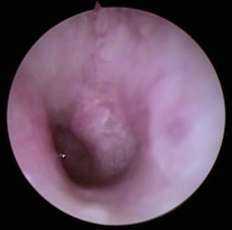
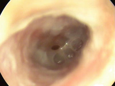
Fig. 1.2
Acute otitis media . TM with severe bulging
Fig. 1.3
Acute suppurative otitis media . Otorrhea through central perforation of the TM, air-fluid level is noted
Severe cases of AOM often presents as a toxic appearing child, with persistent otalgia for more than 48 h with temperature above 39 °C (102.2 °F) in the previous 48 h.
Treatment
Goals of treatment of AOM are to decrease severity and duration of symptoms and to prevent complications. In order to achieve these, patients are treated with analgesics, antibiotics and in cases of recurrent AOM or suppurative complications with tympanostomy tubes.
Initial observation without antibiotics can be offered to children with new onset and uncomplicated mild AOM. Nonetheless, parent/caregiver should be committed to follow-up and be able to begin antibiotics if the child worsens or fails to improve within 48–72 h of AOM onset. In terms of analgesia, it should be used in all cases, whether antibiotics were prescribed or not. Usually oral acetaminophen and ibuprofen are given alone or in combination to treat AOM. Topical analgesic drops are not recommended because there is insufficient evidence about their efficacy in AOM [13].
Stay updated, free articles. Join our Telegram channel

Full access? Get Clinical Tree


