graduated from Ehime University, Japan in 2006 following completion of her residency training in Osaka University Hospital, Japan. From 2007 to 2013, she was at the Division of Ophthalmology, Toyonaka Municipal Hospital, Japan.

3.1 Introduction
Recent advances in micro-incision cataract surgery with foldable intraocular lenses (IOLs) implantation have enabled more effective and comfortable cataract surgeries with sufficient safety [1–3]. As a result, patient expectations have increased regarding the postoperative visual performance.
Various IOL designs generally optimize the quality of vision after cataract surgery [4–8]. However, the selection of new technology IOLs, such as aspherical, toric, and multifocal IOLs, for a particular patient is not easy. In addition, because good optical quality of the cornea is essential for obtaining good visual outcomes after cataract surgery, evaluation of the optical characteristics of the cornea in candidates for cataract surgery is important.
On the other hand, calculating the IOL power for an eye with an irregular cornea such as that which previously underwent refractive surgery presents potential problems for the postoperative refractive error because accurately measuring the corneal power using conventional keratometry is difficult [9–12].
In the current study, we performed corneal topographic analysis of patients scheduled for simple cataract surgery to determine the characteristics of corneal higher-order aberrations.
3.2 Methods
One hundred forty-nine eyes of 124 consecutive cases scheduled for simple cataract surgery at the Osaka University Hospital were included in this retrospective case series. Subjects who required other ocular surgeries such as vitreoretinal surgery or a glaucoma surgery were excluded. The research adhered to the tenets of the Declaration of Helsinki. The institutional review board of Osaka University Hospital approved this study. All participants provided informed consent after the purpose of the study and the procedures were explained.
Corneal topographic analysis was performed using the KR-9000PW (Topcon Corporation, Tokyo, Japan). Two corneal specialists performed visual inspection of color-coded axial power maps with Smolek/Klyce 1.5 diopter (D) power step. Measurements were repeated at least twice to confirm the reproducibility of the topographic maps, and the better map was selected. When appearances of the topographic maps were not reproducible due to poor fixation, head movement, or other reasons, or if the measured area was smaller than 6-mm diameter due to narrow fissure and so on, data was considered as unreliable. Age, gender, and the preoperative distance spectacle-corrected visual acuity (DCVA) of those eyes for which reliable data for the central 6 mm of the cornea were not obtained were compared to those with reliable data.
Four parameters associated with the corneal optical characteristics, i.e., corneal total higher-order aberrations (HOAs), the differences between the central corneal power and simulated K readings (ΔK), the corneal spherical aberration (Z40), and the corneal cylinder, were recorded. Corneal HOA was calculated from the topographic data quantitatively as a set of coefficients of the Zernike polynomials up to the 6th order, and root mean square (RMS, μm) values were used. The total HOAs for the 4-mm corneal diameter were selected to evaluate the clinically relevant corneal irregular astigmatism. Because the blurring effect of 0.5 D defocus corresponds to the RMS value of 0.29 μm for 4-mm diameter, corneal irregular astigmatism was considered to be clinically relevant when the total HOAs exceeded 0.3 μm.
The ΔK was used to determine an abnormal corneal shape that might increase the postoperative refractive error because of inadequate K readings with conventional keratometry when calculating the IOL power [9–12]. A ΔK 0.5 D was considered abnormal value because most of the IOLs were prepared with 0.5 D steps. The Z40 for a 6-mm corneal diameter was chosen to indicate the corneal asphericity. This is because aspherical IOLs show better visual performance than spherical IOLs especially under mesopic conditions and also the asphericity of aspherical IOLs was shown for Z40 for 6-mm diameter [13–15]. A Z40 below 0.1 μm was considered to be an abnormal value because aspherical IOLs with Z40 of −0.20 μm will increase the absolute value of Z40. The optical quality might be reduced due to negative spherical aberration in such cases [16]. Corneal cylinder exceeding 1.5 D was selected as the cutoff value since these eyes were not a good indication for multifocal IOLs but a better indication of surgery for astigmatic correction, such as limbal relaxing incisions (LRIs), touch-up with laser in situ keratomileusis (LASIK), or a toric IOL.
3.3 Results
Reliable data for the central 6 mm of the cornea were not obtained in 33 eyes (22 %) of 33 patients. Reliable data were available for 116 eyes. Although there were no significant differences in age or gender between eyes with and without reliable data, the incidence of eyes with preoperative DCVA worse than 1.0 logMAR unit was significantly higher in eyes with unreliable data than that in eyes with reliable data (Table 3.1, P < 0.001).
Table 3.1
Data of the subjects
Reliable measure | Unreliable measure | p valuea | |
|---|---|---|---|
Number of cases/eyes | 95/116 | 33/33 | |
Gender (men/women) | 39/56 | 15/18 | 0.813 |
Age (years): mean ± SD | 68.6 ± 0.9 | 67.5 ± 1.7 | 0.576 |
DCVA (logMAR) > 1.0 | 4/116 | 10/33 | 0.001 |
The mean ± standard deviation (SD) of the total HOAs was 0.162 ± 0.067 μm. In 5 % of eyes (6 eyes of 116 eyes) with a total HOA exceeding 0.3 μm, two eyes had mild pterygium, two eyes were keratoconus suspect, one eye had undergone LASIK without notice as past history, and one eye had against-the-rule astigmatism with moderate asymmetry (Fig. 3.1).
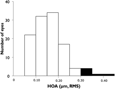
Fig. 3.1
The distribution of corneal total higher-order aberrations for 4-mm diameter. Five percent of patients have clinically relevant irregular astigmatism, which can cause visual impairment
The mean ± SD of the ΔK was 0.16 ± 0.17 D (Fig. 3.2). In 3 % of eyes (4 eyes of 116 eyes), the ΔK exceeded 0.5 D (two eyes were keratoconus suspect, one eye had against-the-rule astigmatism, and one eye had a steep cornea with an extremely prolate shape).
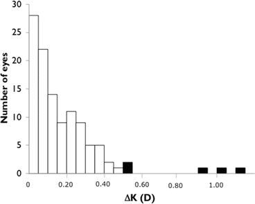
Fig. 3.2
The distribution of the difference between central corneal power and simulated K readings. Three percent of patients have clinically relevant topographic abnormalities. For these cases, corneal topography should be used instead of keratometry to minimize errors in the IOL power
The mean ± SD of the Z40 was 0.247 ± 0.135 μm (Fig. 3.3). In 11 % of eyes, Z40 (13 eyes of 116 eyes) was below 0.1 μm where two eyes were keratoconus suspect, two eyes had high myopia, three eyes had severe myopia with an extremely prolate shape, five eyes had an extremely prolate shape, and one eye had irregular corneal astigmatism.
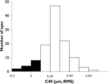
Fig. 3.3


The distribution of corneal spherical aberration for 6-mm diameter. Eleven percent of patients were not good candidates for an aspherical IOL. In these eyes, ocular spherical aberrations can be less negative with implantation of an aspherical IOL
Stay updated, free articles. Join our Telegram channel
Full access? Get Clinical Tree
