1.1 Introduction
Although cataract surgery has been performed for many centuries, technological advances now provide us with the opportunity to have excellent visual outcomes after this surgery. Modern cataract surgery has some aspects of refractive surgery, because the postoperative refractive errors can be reduced by the introduction of the partial coherence laser interferometry and because the surgically induced astigmatism can be minimized by small-incision cataract surgery. Since refractive errors can lead to both a decrease in uncorrected visual acuity and deterioration of quality of vision, reducing possible refractive errors and acquiring good visual outcomes are two essentials to the minimization of spectacle dependence and to the maximization of subsequent patient satisfaction. At present, most cataract surgeons merely consider that it is essential to reduce the possible refractive errors as much as possible for cataract surgery. However, it is known that pupil size also plays an important role in the visual outcomes of the surgical procedure [1–5]. In this chapter, we will look at the rationale for the clinical assessment of the preoperative pupil size in order to predict and maximize the postoperative visual performance in cataract surgery.
1.2 Role of Pupil Size on Postoperative Visual Function
The human visual system is known to suffer from aberrations, diffraction, scatter, finite receptor size, and noise in the neural pathways. The smaller pupil size may have some advantages in its superiority for image formation, such as an increasing depth of focus, a decrease in higher-order aberrations [6, 7], and a decrease in light scatter [8], all of which may, to some extent, offset the deleterious effects of reduced luminance [9] and diffraction. In order to better understand the quality of vision in pseudophakic patients, it is necessary to know what changes in pupil size result from cataract surgery using modern phacoemulsification techniques.
1.3 Background for the Rationale for the Preoperative Assessment of Pupil Size to Predict Visual Outcomes
It has been demonstrated that the postoperative pupil size cannot be predicted from the preoperative size with sufficient consistency, possibly because the pupil is substantially impaired by previous cataract surgery [10–12]. However, modern cataract surgery using newer phacoemulsification techniques has been reported to induce a transient decrease in pupil size immediately after surgery, probably because of the traumatic release of miotic neuropeptides [13–15], but soon recovers to the preoperative levels. Accordingly, the postoperative pupil size can be predicted from the preoperative measurements. Hayashi and Hayashi [16] demonstrated, in a study of 386 eyes undergoing uncomplicated phacoemulsification with IOL implantation, that there was a strong positive correlation between preoperative and postoperative pupil size obtained with the Colvard pupillometer. Sobaci et al. [17] showed no significant differences in pupil diameter or shift between pseudophakic and cataractous eyes using the NIDEK OPD-Scan, under low mesopic or photopic conditions, suggesting that uncomplicated in-the-bag IOL implantation had no significant adverse effect on pupillary mechanics. Therefore, we may successfully predict the postoperative pupil size, affecting the refractive outcomes and subsequent patient satisfaction after cataract surgery, if we can accurately and reproducibly determine the preoperative pupil size. This is the background for the rationale for performing “pupil-customized cataract surgery (PCCS).”
1.3.1 Summary for the Clinician
Pupil-customized cataract surgery (PCCS) means predicting and maximizing the postoperative visual performance and subsequent patient satisfaction by the preoperative assessment of pupil size in cataract patients.
1.4 Why Is the Consideration of Pupil Size Important to Cataract Patients?
Figure 1.1 shows the relationship between pupil size and patient age in 304 normal eyes, demonstrating a significant decrease in pupil size as a function of age [18]. It is quite reasonable that there was a significant negative correlation between pupil size and subject age. However, it should be noted that there was a greater variability in pupil size even in elderly patients, as evidenced by the high coefficient of variation (0.30) in eyes aged 60 years or more. These findings imply that all elderly patients do not necessarily have smaller pupils, suggesting the clinical significance of the individual assessment of pupil size even in elderly patients undergoing cataract surgery.
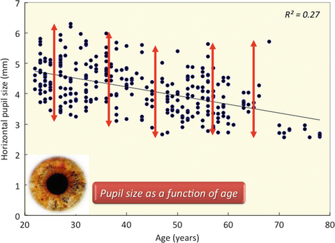
Fig. 1.1
A graph showing a significant decrease in pupil size as a function of age. Interestingly, a greater variability in pupil size was found even in the elderly population
1.4.1 Measurement Technique of Pupil Size
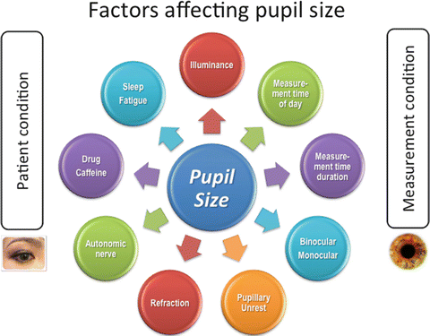
As shown in Fig. 1.2, it is known that pupil size can be influenced not only by the patient background, for example, by age [19–25], manifest refraction [26], and the accommodative state of the eye [27, 28], and by various sensory and emotional conditions [29] but also by measurement conditions affecting the level of retinal illuminance [30, 31]. Accordingly, this measurement does not necessarily offer high reproducibility. It has been demonstrated that dynamic pupil measurement offered a higher repeatability than static pupil measurement and that monocular pupil size was larger than binocular size. Therefore, the binocular dynamic pupillometer is necessary in order to accurately determine pupil size under binocular conditions [32, 33]. Moreover, the pupil diameter should be evaluated dynamically with the open-view type of binocular digital infrared pupillometer, offering high reproducibility, under natural-viewing conditions. The measurements should be performed for several seconds at the same time of day under the same conditions as those in which the patients were resting in order to reduce the individual changes in pupil size. Moreover, the horizontal pupil diameter should be assessed for determining the accurate pupil size, since the sagittal diameter measurements are not very reliable due to the presence of the eye lid especially in elderly patients.
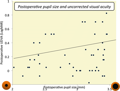
Fig. 1.2
Factors influencing pupil size. Pupil size can be affected not only by the patient background but also by measurement conditions
1.4.2 Summary for the Clinician
Pupil size should be assessed dynamically with the open-view type of binocular digital infrared pupillometer, offering high reproducibility, under natural-viewing conditions.
1.5 Pupil Size in Monofocal IOL-Implanted Eyes
As previously mentioned, modern cataract surgery has been reported to induce no significant change in pupil size [16, 17], although there was a transient decrease in pupil size immediately after surgery. Therefore, we may successfully predict the postoperative pupil size, at least to some extent, if we can accurately and reproducibly measure the preoperative pupil size. Figure 1.3 shows the relationship between the postoperative pupil size and uncorrected distance visual acuity (logMAR) 3 months after uncomplicated monofocal IOL implantation, when the targeted refraction is emmetropia and when no other disease was observed with CDVA of 20/20 or more, demonstrating that there was a significant positive correlation between them. These findings indicate that the postoperative visual function was significantly affected by the postoperative pupil size even in monofocal IOL-implanted eyes. Figure 1.4 shows the relationship between the preoperative pupil size and uncorrected visual acuity after monofocal IOL implantation in the same study population, also demonstrating that there was a significant positive correlation between them. We confirm that the postoperative visual function was also significantly affected by the preoperative pupil size in monofocal IOL-implanted eyes and that we can predict, to some extent, the postoperative visual performance of cataract patients in a clinical setting.
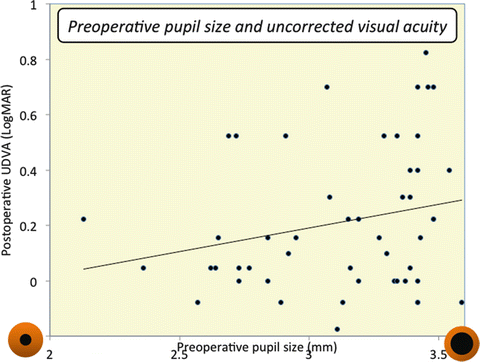
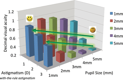
Fig. 1.3
A graph showing a significant positive correlation between the postoperative pupil size and uncorrected distance visual acuity (UDVA) 3 months after monofocal IOL implantation. It is suggested that the postoperative visual function was significantly affected by the postoperative pupil size in monofocal IOL-implanted eyes
Fig. 1.4
A graph showing a significant positive correlation between the preoperative pupil size and uncorrected distance visual acuity (UDVA) 3 months after monofocal IOL implantation. It is suggested that the postoperative visual function was also significantly affected by the preoperative pupil size in monofocal IOL-implanted eyes
1.6 Pupil Size in Multifocal IOL-Implanted Eyes
It is no doubt whether pupil size plays a vital role in visual performance after multifocal IOL implantation, especially after refractive multifocal IOL implantation. Nakamura et al. [34] demonstrated that fewer than 50 % of subjects over 60 years of age had pupil sizes conducive to the near zone of refractive multifocal IOLs. Hayashi et al. [2] found that a smaller pupil correlated significantly with lower near visual acuity in Array refractive multifocal IOL-implanted eyes. Kawamorita and Uozato [35] investigated the relationship between pupil size and the modulation transfer function (MTF) of the refractive multifocal IOL in vitro and suggested that the near MTF increases at the expense of the far MTF with a large pupil. In principle, the basic optical characteristics of diffractive multifocal IOLs are not affected by the changes in pupil size, because distance and near correction are simultaneously present across the full area of the pupil according to their design. However, at present, most so-called diffractive IOLs are in essence a hybrid combination of refractive and diffractive lenses. For example, the ReSTOR IOL has an optic that consists of a diffractive zone in the central 3.6 mm region and a refractive zone in the outer region of diffractive zone. The refractive zone is for improving far vision under mesopic condition and requires a pupil larger than 3.6 mm to work. We should keep in mind that visual performance in both refractive and so-called diffractive multifocal IOL-implanted eyes were influenced by pupil size, when we consider the indications for different types of multifocal IOLs.
1.7 Pupil Size in Toric IOL-Implanted Eyes
At present, we often consider the amount of astigmatism as well as the axis of astigmatism for toric IOL implantation. However, pupil size also plays an essential role in postoperative visual performance even in astigmatic eyes requiring toric IOL implantation. We previously showed that uncorrected visual acuity was better in eyes with smaller pupil sizes in both with-the-rule and against-the-rule astigmatic eyes (Fig. 1.5) [36]. For example, uncorrected visual acuity of eyes with 1 D of astigmatism for a 5-mm pupil was almost equivalent to that of eyes with 2 D of astigmatism for a 2-mm pupil. Watanabe et al. [37] also reported, in a study of 36 monofocal IOL-implanted eyes, that pupil size may have an impact on postoperative uncorrected visual acuity in eyes having against-the-rule astigmatism and in eyes with a large pupil diameter and with-the-rule astigmatism. Especially in eyes with larger pupils, it may be necessary to correct the preexisting astigmatism in order to acquire excellent visual outcomes after toric IOL implantation. These findings, although simple, are clinically meaningful because most surgeons neglect the preoperative pupil size for determining the indication for toric IOL implantation in a clinical setting. We should take the three major components (the amount of astigmatism, the axis of astigmatism, and pupil size) into consideration for the surgical correction of astigmatism, such as toric IOL implantation.