graduated from Chiba University in 1975 and received his doctoral degree from Tokyo Institute of Technology (T.I.T.) in 1981. He joined Toppan Printing Company in 1982 and the faculty of engineering of Chiba University in 1990. Dr. Ohnuma is currently an associate professor of medical engineering at Chiba University in Chiba.

graduated from Kagoshima University Faculty of Medicine in Kagoshima, Japan in 2008. He took residency in ophthalmology at Keio University in 2010. He works at Kawasaki Municipal Hospital in Kanagawa, Japan.

graduated from Hamamatsu University School of Medicine in 1986. Dr. Noda had been a part-time professor of Tokyo Women’s University School of Medicine from 1991 to 2012 and is currently a chief in the Department of Ophthalmology at National Hospital Organization Tokyo Medical Center and also a clinical professor of graduate school of nursing studies at Tokyo Healthcare University.

14.1 Introduction
Modern intraocular lens (IOL) implantation usually provides good visual results. Several studies have reported on the optical performance of different types of monofocal and multifocal IOLs using optical bench testing [1–5] and ray tracing [6], and several clinical studies [7–9] have reported on visual performance, mainly, visual acuity (VA) and contrast sensitivity, in patients in whom IOLs have been implanted under various conditions. However, the image quality of the IOLs measured by optical bench testing outside the eye and the results of ray tracing are difficult to extrapolate to conditions in pseudophakic eyes in vivo. However, clinical (psychophysical) tests may be affected by nonoptical factors in the patients’ visual system, so that the tests may not reflect the optical performance of the IOL. Therefore, there is no appropriate method for evaluating the optical performance of an IOL objectively under various conditions. This indicates the need for a new method of evaluating the retinal image quality in eyes with IOLs under conditions that closely resemble clinical cases. We previously reported systems used for visual simulation of retinal images in eyes with an IOL under conditions similar to clinical cases and reported the differences in retinal image quality with monofocal and multifocal IOLs [1]. In this chapter, we describe the results of visual simulation of retinal images in eyes that had been implanted with one of three different multifocal IOLs using a visual simulation system to evaluate the optical performance and characteristics of the IOLs.
14.2 Methods
14.2.1 Visual Simulation System
Our visual simulation system consisted of lenses with an artificial pupil (model eye) and a charge-coupled device (CCD) camera (EO-1312CCD, Edmund Optics Inc., Barrington, NJ) (Fig. 14.1). The model eye had the dimension and refractive index of each surface that we reported previously [10]. The IOL was set in a black IOL holder with a central circular hole that served as the iris pupil. The IOL holder was inserted into the glass cell with the anterior surface of the IOL facing the artificial cornea. The glass cell was filled with pure water. The corneal spherical aberration of the model eye was set at 0.22 μm. The best focus of the model eye with the IOL was set at 5 m. The chart, which consisted of various sizes of Landolt rings, was set at various distances from 30 cm to 5 m, and the simulated retinal images were obtained using this system through 3.0- and 4.5-mm apertures for the ReSTOR IOL (Alcon Inc., Hunenberg, Switzerland) and the Tecnis Multifocal IOL (Abbott Medical Optics Inc., Santa Ana, CA) and through 3.5- and 4.5-mm apertures for the Mplus IOL (Oculentis GmbH, Berlin, Germany).
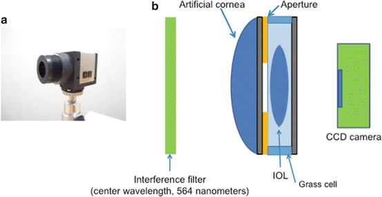
Fig. 14.1
Visual simulation system. (a) The eye model combines lenses with an artificial pupil and a CCD camera. (b) Schema of the visual simulation system
14.2.2 IOLs
The three multifocal IOLs are shown in Fig. 14.2. The AcrySof ReSTOR (SN6AD1) is a multifocal IOL that uses apodization, diffraction, and refraction. The apodized diffractive region is found in the inner 3.6 mm of the optic. The refractive portion is on the outer 2.4 mm of the lens. Apodization is the unique key differentiating feature of this IOL. The diffractive step heights decrease (apodization), which allows control of light energy distribution and directs increasingly more light energy to the distance focal point. This means that more energy is available for reading in patients with smaller pupils; at night most of the energy is directed to the distance focus for larger pupils. A diffractive lens uses only about 82 % of the energy for the two primary images; however, for larger pupils, the outer region is solely refractive, and the total energy use increases to over 90 % for larger pupils with this IOL. The ReSTOR offers different add powers ranging from 0.75 diopter (D) to 4.0 D on the IOL plane. In this study, we used +3.0 D of add power on the IOL plane (SN6AD1).
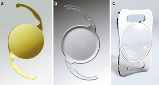
Fig. 14.2
Photograph of the IOLs used in this study. (a) The ReSTOR IOL (SN6AD1). (b) The Tecnis Multifocal (ZMB00). (c) The Lentis Mplus (LS-313 MF30)
The Tecnis Multifocal IOL (ZMB00) is a full optic diffractive, multifocal IOL in which the anterior prolate surface of the well-known Tecnis monofocal optic is combined with a posterior surface with diffractive optic. In this full optic diffractive lens, about 41 % of the light goes to each of the two primary lens powers, and the balance is independent of the pupillary diameter. The add power is +4.0 D on the IOL plane.
The Lentis Mplus IOL (LS-313 MF30) is an inferior segmental, near add, multifocal IOL containing an aspheric distance-vision zone combined with a 3.00-D posterior sector-shaped near-vision zone, allowing seamless transition between the zones. Theoretically, light hitting the transition area of the embedded sector is reflected away from the optical axis to prevent superposition of interference or diffraction. The IOL has a biconvex design with a 6.0-mm optic size and an overall length of 12.0 mm. It is made of an acrylic material, the HydroSmart copolymer (Oculentis GmbH), which is comprised of acrylates and a hydrophobic surface with ultraviolet-absorbing components. Another important feature of the Lentis Mplus is its 360-degree continuous square optic and haptic edge.
Stay updated, free articles. Join our Telegram channel

Full access? Get Clinical Tree


