graduated from Kagoshima University, School of Medicine, Japan in 1995, and specialized in ophthalmology in the Faculty of Medicine, Kyushu University in 1995. He undertook his residency and fellowship in ophthalmology, Kyushu University Hospital, from 1995. From 2004 to present, he is working at Hayashi Eye Hospital, Fukuoka, Japan.

graduated from Hokkaido University, School of Medicine, Japan in 1993, and specialized in ophthalmology in the Faculty of Medicine, Kyoto University, Japan in 1993. He received his doctoral degree from Kyoto University in 2003. From 2000 to 2002, he undertook a research fellowship at Burnham Institute in San Diego, California. From 2007 to present, he is working at Hayashi Eye Hospital, Fukuoka, Japan.

2.1 Introduction
For some pathologic conditions, extraction of a clear lens is an effective treatment option. In eyes with primary angle-closure glaucoma, extraction of a large lens is effective in preventing acute pupillary block [1–3]. During vitrectomy, extraction of a lens facilitates thorough removal of the peripheral vitreous. In eyes that are to undergo penetrating keratoplasty, a slight cataract should be removed simultaneously when subsequent cataract surgery may be difficult. However, because visual function with a young crystalline lens is superior to that with an intraocular lens (IOL), surgeons usually hesitate to perform clear lens extraction in young or middle-aged patients.
The loss of accommodation is a major disadvantage when a young crystalline lens is removed.
However, pseudophakic eyes with a monofocal IOL can achieve relatively good visual acuity in a certain range of distance; this is known as apparent accommodation [4, 5]. On the other hand, the amplitude of ocular accommodation in phakic eyes decreases with aging from middle age on [6, 7]. Although the amplitude of apparent accommodation in pseudophakic eyes with a monofocal IOL has also been shown to decrease from middle age on [8], the decrease is more pronounced in phakic eyes than in pseudophakic eyes. Accordingly, it is assumed that the amplitude of apparent accommodation in pseudophakic eyes becomes equivalent to that of ocular accommodation in phakic eyes of older patients.
Furthermore, previous studies have shown that contrast sensitivity in phakic eyes, specifically in young phakic eyes, is better than that in pseudophakic eyes with a conventional spherical IOL [9–11], although controversy remains [12]. However, more recent studies showed that contrast sensitivity in phakic eyes deteriorates gradually from middle age on [13, 14], especially under scotopic conditions [15, 16]. This has been attributed to an increase in spherical aberration of the lens due to aging [17, 18]. Therefore, because implantation of an aspheric IOL precludes a marked increase in higher-order ocular aberration [19, 20], contrast sensitivity in pseudophakic eyes with an aspheric IOL may be comparable to that in phakic eyes of middle and advanced ages.
The purpose of this study was to compare visual function, including accommodative amplitude, region of accommodation, and contrast sensitivity, between phakic eyes with a clear lens and pseudophakic eyes with a monofocal IOL at 40 years of age and older. In ages at which these visual functions become comparable in phakic and pseudophakic eyes, clear lens extraction may be justified for eyes that show even slight pathology.
2.2 Methods
2.2.1 Patients
All patients scheduled for phacoemulsification surgery were screened for enrollment. The original plan was to recruit phakic subjects and pseudophakic patients in each of five age groups: 40s, 50s, 60s, 70s, and 80s. However, because patients in their 80s who met the inclusion criteria were rare, patients of this age could not be enrolled for measurement of region of accommodation and contrast sensitivity. The inclusion criterion in the phakic group was that corrected visual acuity (CVA) be 20/25 or better with no or little cataract. The exclusion criteria were severe pathology of the optic nerve, macula, or cornea, severe opaque media other than cataract, previous history of ocular inflammation or surgery, an irregularly shaped pupil, and difficulty in examination or follow-up.
2.2.2 Measurements
The accommodative amplitude of pseudophakic and phakic eyes was examined using an accommodometer (HS-9E, Kowa). The amplitude was measured using the same method described previously [8]. Briefly, the spherical and cylindrical powers in diopters (D) for best distance visual acuity (distance refraction) were first examined using an autorefractometer (KR-7100, Topcon). The refractive status for best near visual acuity at 0.40 m (near refraction) was examined using a near vision tester (Lumichart, HOYA). Then, the near point of accommodation of each eye was determined in the following manner. With the spherical lens equivalent to near refraction added, the patients were asked to read a Landolt visual target that corresponds in size to near visual acuity of 20/29. The visual target was moved from about 1 m up to the near point at which the patients noted blurring of the target. The target was then moved back until it became clear. Measurements were repeated, and the average distance at which blurring and refocusing occurred was recorded as the near point. The accommodative amplitude was calculated by subtracting the diopters of spherical equivalent of best distance and near visual acuity from refractive power of the near point.
CVA was measured from far to near distances using the all-distance vision tester (AS-15, Kowa). This device measures equivalent visual acuity from far to near distances by placement of a spherical lens and various sized visual targets at appropriate distances along the visual axis. For example, the visual acuity at ∞ m is measured by placing a spherical lens (focal distance = 250 mm) at 250 mm from the patient’s eye and a visual target at 500 mm. The region of accommodation in phakic eyes and in pseudophakic eyes at which the patient achieved 20/29 or 20/40 were examined and converted to the diopteric range.
Contrast visual acuity (contrast VA) and that in the presence of glare (glare VA) under photopic and mesopic conditions were examined using the Contrast Sensitivity Accurate Tester (CAT-2000, Menicon) [21]. This device measures the logarithm of the minimal angle of resolution (logMAR) VA using 5 contrast percentages of visual targets (100, 25, 10, 5, and 2.5 %) under photopic and mesopic conditions. Measurement under the photopic lighting condition was made with chart lighting of 100 cd/mm2, while chart lighting under the mesopic condition was 2 cd/mm2. A glare source of 200 lux was located in the periphery at 20° around the visual axis.
2.2.3 Statistical Analysis
Decimal VA was converted to the logMAR scale for statistical analyses. Differences in accommodative amplitude, all-distance VA, and contrast VA and glare VA between the age groups was compared using the Kruskal–Wallis test. Differences in the continuous variables between the phakic and pseudophakic groups were compared using the Mann–Whitney U test, and categorical variables were compared using the Fisher’s exact test or the chi-square test. Any differences with a P value of less than 0.05 were considered to be statistically significant.
2.3 Results
The mean amplitude of ocular accommodation decreased in proportion to the age group, which was a highly significant difference (P < 0.0001; Fig. 2.1), while the difference in the amplitude of apparent accommodation was marginally significant (P = 0.0380). The amplitude of apparent accommodation in the group in their 40s and younger was greater than that in the group in their 70s or 80s. Furthermore, the amplitude of ocular accommodation was correlated strongly with the actual age of each patient (r = −0.758, P < 0.0001), while only a weak correlation was found between the amplitude of apparent accommodation and the actual age (r = −0.191, P = 0.0292).
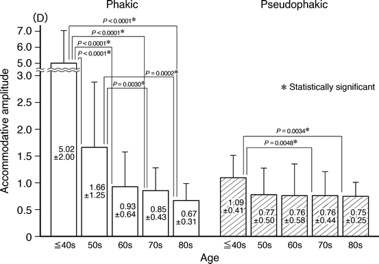
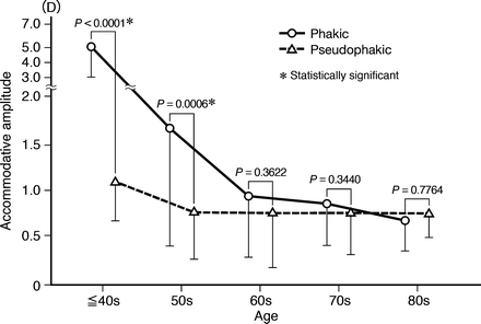
Fig. 2.1
Changes in the mean amplitude of normal accommodation in phakic eyes and of apparent accommodation in the pseudophakic eyes in the five age groups. The mean amplitude of ocular accommodation decreased in proportion to the age, which was significantly different (P < 0.0001). The mean amplitude of apparent accommodation also decreased with age, but the change was marginally significant (P = 0.0380). P values indicate the difference between each two groups [22]
When comparing the mean accommodative amplitude between the pseudophakic and phakic eyes (Fig. 2.2), the mean amplitude of apparent accommodation was significantly less than that of ocular accommodation in the groups in their 40s and younger and in the 50s. However, in the groups in the 60s, 70s, and 80s, no significant difference between eyes was found in the amplitude. The incidence of the patients in whom the amplitude of apparent accommodation in pseudophakic eyes was more than that of ocular accommodation in phakic eyes was significantly different between the five groups (P < 0.0001). The incidence of the patients in their 40s and younger was significantly less than that in the groups in their 60s, 70s, and 80s, and that in the group in their 50s was significantly smaller than that in the group in their 80s.