 Retinoblastoma: Diagnostic Approaches
Retinoblastoma: Diagnostic Approaches
The diagnosis of retinoblastoma is best made by the recognition of its typical clinical features using indirect ophthalmoscopy and slit lamp biomicroscopy (1). In many cases, however, ancillary studies like fluorescein angiography, ultrasonography, computed tomography, and magnetic resonance imaging can provide valuable diagnostic information, especially in cases that are atypical (1, 2, 3, 4, 5, 6, 7, 8, 9, 10, 11, 12, 13, 14, 15, 16, 17, 18, 19, 20, 21, 22, 23, 24, 25, 26, 27, 28, 29, 30, 31, 32, 33).
Fluorescein Angiography
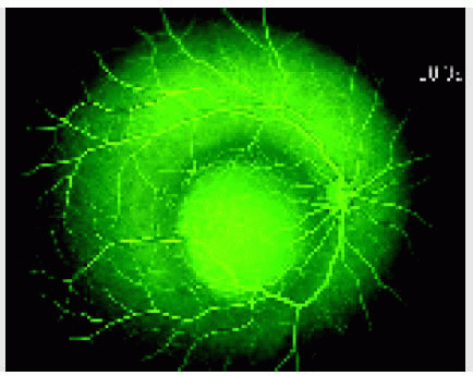
Fluorescein angiography shows a vascular tumor that fills rapidly with fluorescein and shows later hyperfluorescence. During the vascular filing phase, the feeding arteries fill rapidly, and fine reticular blood vessels typically are often seen in the superficial portions of the tumor (2,3). Leakage into the vitreous and subretinal space is seen in more-advanced tumors. After successful treatment of the tumor, the blood supply is diminished or absent and the lesion is more hypofluorescent. Spontaneously arrested or regressed retinoblastomas also show variable degrees of hyperfluorescence even though they are clinically inactive.
Ultrasonography
Ultrasonography is used routinely to assist in the diagnosis and to measure tumor size before and after treatment (4,5). With B-scan ultrasonography, retinoblastoma usually appears as an acoustically solid mass with highly reflective foci representing calcification. A-scan ultrasonography shows high internal reflectivity. Ultrasound is especially useful for exophytic tumors with overlying retinal detachment when the tumor cannot be seen ophthalmoscopically. In such cases, Coats disease is often a diagnostic consideration, but ultrasonography shows a mass in retinoblastoma but usually no mass with Coats disease or other nontumor causes of retinal detachment (6,7). Diffuse infiltrating retinoblastoma is often an exception because it frequently shows no distinct mass and no calcification.
Computed Tomography
Stay updated, free articles. Join our Telegram channel

Full access? Get Clinical Tree


