 Posterior Uveal Melanoma: Pathology
Pathology of Posterior Uveal Melanoma
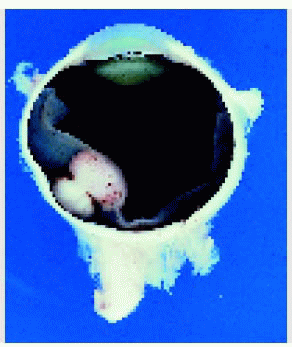
The pathology of uveal melanoma is briefly discussed with regard to routine gross and light microscopy and some more recent developments related to prognosis (1, 2, 3, 4, 5, 6, 7, 8, 9, 10, 11, 12, 13, 14, 15, 16, 17, 18). Posterior uveal melanoma can have characteristic features on both gross and microscopic examination (1, 2, 3). Observations of growth patterns on grossly sectioned eyes can be helpful to the clinician in understanding the clinical behavior of the tumors and in predicting prognosis. Gross examination of an enucleated eye can reveal signs of scleral melanocytosis, nodules of extrascleral extension, tumor growth pattern, extent of secondary retinal detachment, and other important features. Grossly, a choroidal or ciliary body melanoma can be dome-shaped, mushroom-shaped, or diffuse. The mushroom shape occurs from rupture of Bruch’s membrane secondary to tumor growth. Melanoma can be deeply pigmented, partly pigmented, or nonpigmented. It can produce a secondary retinal detachment, subluxation of the lens, or secondary cataract. It can invade the anterior chamber, producing secondary glaucoma, or it can extend through the sclera. A diffuse melanoma can assume a ring configuration in the ciliary body region or a flat, slightly elevated appearance in the choroid.
Low-magnification study of uveal melanomas can provide similar information regarding growth patterns, invasion of adjacent structures such as sclera, vortex veins, optic nerve, retinal pigment epithelium (RPE), sensory retina, and vitreous. It can reveal areas of tumor necrosis. It can demonstrate lipofuscin-laden macrophages at the level of the RPE that correlate with the orange pigment seen clinically on the tumor surface.
In 1931, Callender proposed the widely used cytologic classification of uveal melanoma, dividing melanoma cells into spindle A, spindle B, fascicular, mixed, epithelioid, and necrotic cell types. Later, McLean and associates, from the Armed Forces Institute of Pathology modified the Callender classification and dropped the fascicular and necrotic cell types. Mixed cell type is a combination of spindle and epithelioid cells. This Mclean modification is used by many eye pathologists today (4,5).
It has long been recognized that the cell type of uveal melanoma is related to prognosis. Patients with tumors composed of pure spindle cells have a more favorable prognosis, and those with a component of epithelioid cells (mixed or epithelioid cell types) have a worse prognosis (1,3,8). Melanomas with a low mitotic activity are associated with a better prognosis, whereas those with a greater mitotic activity carry a worse prognosis (1,3,8). There is increased mortality in patients with extrascleral extension of the tumor. More recently, it has been found that the histopathologic presence of networks of closed vascular loops and other abnormal vascular patterns are associated with a less favorable prognosis (10,11). Some uveal melanomas have demonstrated evidence of infiltration of lymphocytes in the tumor. This is also believed to be associated with decreased survival (6,10,12).
Stay updated, free articles. Join our Telegram channel

Full access? Get Clinical Tree


