CHAPTER 148 Otologic Manifestations of Systemic Disease
Systemic diseases that may involve the ear include granulomatous and infectious processes, tumors, bone disorders, storage diseases, collagen vascular and autoimmune diseases, and immunodeficiency disorders (Table 148-1). In some of these diseases, the initial clinical symptoms may occur in the temporal bone and be confused with other diseases that are limited to the ear.
Table 148-1 Systemic Diseases That Affect the Ear
Granulomatous and Infectious Diseases
Langerhans Cell Histiocytosis
LCH (formerly called histiocytosis X) refers to a group of disorders that are characterized by a proliferation of cytologically benign histiocytes. The term histiocytosis X was proposed by Lichtenstein,1 who considered eosinophilic granuloma, Hand-Schüller-Christian disease, and Letterer-Siwe disease to be related disorders because of the similarity of their pathologic lesions. The severity of these diseases and prognosis and treatment differ greatly, however. Histopathologically, the primary lesion of LCH is formed by collections of pathologic Langerhans cells with variable numbers of eosinophils, macrophages, and lymphocytes. Diagnostic characteristics of the pathologic Langerhans cells include nuclei that are deeply indented and elongated on light microscopy, cytoplasm that is pale and abundant, Birbeck granules on electron microscopy, expression of CD1 on the cell surface, and positive immunostaining for S100 protein and for the so-called Langerin protein.2,3 The etiology and pathogenesis of LCH remain unknown, although more recent research has begun to shed some light on these.2,4,5 Current thinking favors the notion that LCH results from immunologic dysfunction, resulting in unchecked proliferation of pathologic Langerhans cells.5
Unifocal eosinophilic granuloma, which occurs in children and young adults, shows a male predominance. It appears as a solitary osteolytic lesion in the femora, pelvis, scapulae, vertebrae, ribs, mandible, maxilla, or skull (including the temporal bone). The lesion may be asymptomatic, or it may cause pain, local swelling, or pathologic fracture. There are no systemic manifestations. The clinical course is typically benign, the prognosis is excellent, and spontaneous regression may occur. Local curettage with or without low-dose irradiation (approximately 60 Gy)6 is usually curative. Temporary splinting or casting may be necessary for weight-bearing bones. Follow-up examination with a radiographic skeletal survey should be performed to detect lesions at other sites; such lesions are almost always found within 1 year.
Hand-Schüller-Christian disease may be best understood as a multifocal form of LCH. It usually occurs in children younger than 5 years old, and it is characterized by multifocal osteolytic lesions with limited extraskeletal involvement of skin, lymph nodes, and viscera. Multiple lesions are evident at diagnosis or develop within 6 months after a unifocal lesion appears. Systemic manifestations include fever, anorexia, recurrent upper respiratory infections, anterior cervical lymphadenopathy, otitis media, and hepatosplenomegaly. The classic triad of osteolytic skull lesions, exophthalmos as a result of orbital bone involvement, and diabetes insipidus secondary to pituitary disease may be seen in 25% of patients.7 Chest radiograph may show diffuse pulmonary infiltration, particularly in central and perihilar areas. Hilar lymphadenopathy is rare. Diagnosis requires biopsy of an accessible lesion. Spontaneous regression may occur, but the disease is typically chronic, and low-dose chemotherapy may be required to control systemic manifestations.
Letterer-Siwe disease is a disseminated form of LCH that occurs in children younger than 3 years old and manifests with the diffuse involvement of multiple organs. Manifestations include fever, seborrheic or eczema-like rash, oral lesions, lymphadenopathy, hepatosplenomegaly, multiple bony lesions, diffuse replacement of marrow with resulting blood dyscrasias, and pulmonary infiltration with respiratory failure. The disease is virulent, with a poor prognosis and a high mortality rate. Treatment consists of varying combinations of corticosteroids and cytotoxic drugs such as methotrexate, mercaptopurine, vincristine, vinblastine, chlorambucil, cyclophosphamide, and etoposide. High-dose chemotherapy and radiation followed by bone marrow transplantation or hematopoietic stem cell transplantation has been reported to be successful in a few advanced refractory cases of LCH.8
Otologic Manifestations
The mastoid is a common site of involvement in LCH. When small, the lesion is asymptomatic. As it expands, the lesion may manifest in several ways: by erosion of the posterior bony canal wall; by erosion through the cortex of the mastoid, zygomatic, or squamosal portions; or by secondary infection.9 The otic capsule and facial nerve are relatively resistant. Although sensorineural hearing loss (SNHL), vertigo, and facial nerve paralysis can occur, they are infrequent. Similarly, extension beyond the temporal bone involving the jugular fossa and skull base is rare.
The reported incidence of otologic manifestations in patients with LCH is 15% to 61%,7 and they may be the initial presentation of the disease. The most common symptom is otorrhea, followed by postauricular swelling, hearing loss, and vertigo.10,11 The most common sign is granulation tissue or aural polyps in the external auditory canal. The disease may manifest with perforation of the tympanic membrane, otitis media, otitis externa, fistula between the mastoid and the external canal, or nontender postauricular swelling. Occasionally, inner ear symptoms and a positive fistula test are found in the presence of an intact tympanic membrane. The disease often mimics chronic otitis media, and mastoid surgery is frequently performed before the diagnosis is made.9
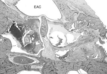
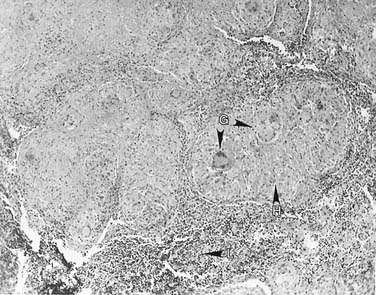
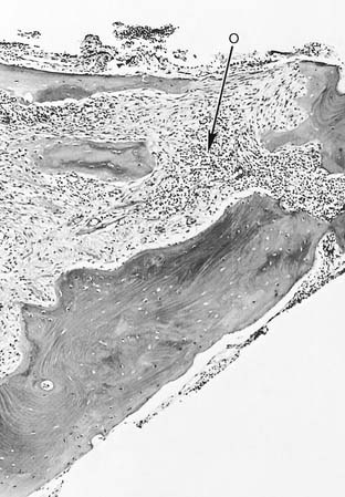
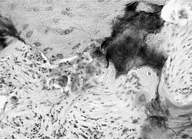
Diagnosis of LCH is suggested by an inflammatory disorder of the middle ear and mastoid that does not respond to routine antibiotic therapy, bilateral destructive ear disease, an elevated erythrocyte sedimentation rate in the absence of acute infection, exuberant granulation tissue after mastoid surgery with a persistently draining cavity, and associated skin and systemic lesions. Radiographs show destructive lesions in the mastoid and temporal bones (Figs. 148-1 and 148-2).9,10,12 The diagnosis is established by biopsy; because the surface of the granulation tissue often shows infection, necrosis, and fibrosis, tissue should be acquired from deeper parts of the lesion.13 Microscopic findings include sheets of histiocytes with a variable number of eosinophils, plasma cells, polymorphonuclear leukocytes, and multinucleated cells (Figs. 148-3, 148-4, and 148-5). Areas of hemorrhage and necrosis are common. A definitive diagnosis of LCH is usually made on the basis of immunostaining and electron microscopic studies.2,4
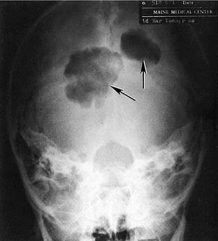
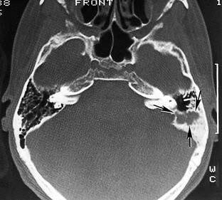
Figure 148-2. Irregular lytic lesion in the mastoid bone (arrows) of a 13-year-old boy with unifocal eosinophilic granuloma.
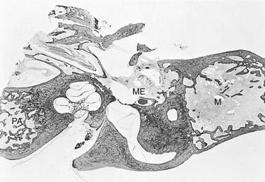
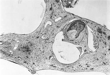
Figure 148-4. A higher power view of part of the middle ear area seen in Figure 148-3. The infiltrate consists of histiocytes, eosinophils, and multinucleated cells. The incus (I) has been partially resorbed (×45).
Tuberculosis
The incidence of tuberculous otitis media has decreased dramatically. During the early part of the 20th century, 1.3% to 18.6% of all cases of chronic otitis media were reportedly caused by tuberculosis, whereas more recent studies report tuberculous otitis media rates of 0.05% to 0.9%.14 The last 20 years have witnessed an increase in incidence of tuberculosis, however, which is caused in part by the aggressive nature of tuberculosis in individuals infected with human immunodeficiency virus (HIV), and by an emerging resistance to antituberculosis drugs.15 Mycobacterium tuberculosis is the infective organism in most cases; occasionally, atypical mycobacteria (e.g., Mycobacterium avium and Mycobacterium fortuitum) are responsible.16
During the early stages of tuberculous otitis media, the tympanic membrane becomes thickened, and the otoscopic landmarks become obliterated. There is a conductive hearing loss as a result of middle ear effusion, a thickening of the tympanic membrane and middle ear mucosa, and a destruction of the ossicles (Figs. 148-6 and 148-7). There is no characteristic pain or tenderness, but lymphadenopathy in the high jugular chain occurs early. Multiple small perforations of the tympanic membrane may occur, with seropurulent drainage. The perforations quickly coalesce to cause loss of the tympanic membrane. Likewise, a myringotomy site in an intact tympanic membrane may enlarge quickly. The middle ear mucosa appears to be hyperemic with polypoid granulation. Osseous involvement results in the sequestration of bone and the destruction of the inner ear, the facial nerve, or both. Destruction of the mastoid tip may result in an asymptomatic, nontender Bezold’s abscess (i.e., a “cold” abscess). Rarely, tuberculosis can also manifest as primary tuberculous petrositis,17 or as SNHL caused by chronic labyrinthitis and tuberculous meningitis.18
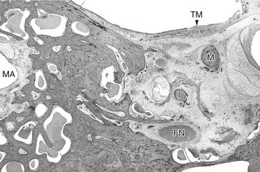
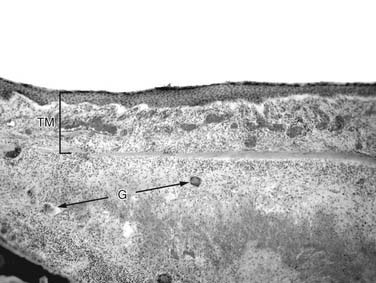
Figure 148-7. A higher power view of the tympanic membrane (TM) seen in Figure 148-6. The TM is intact, but greatly thickened by tuberculous granulation tissue containing the typical epithelioid cells, round cells, and multinucleated giant cells (G) (×100).
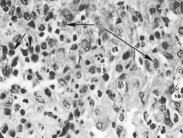
The diagnosis of tuberculous involvement of the middle ear is usually delayed. The characteristic signs and symptoms include multiple perforations of the tympanic membrane that quickly coalesce to form total tympanic membrane loss, nontender cervical lymphadenopathy, intractable otitis media with polypoid granulation, and bony sequestration; these should alert the physician to this possible diagnosis. Otic capsule involvement with resultant loss of auditory and vestibular function may be the first symptom, which indicates a process that differs from the more typical chronic otitis media. Definitive diagnosis is made by histopathologic examination of tissue from the middle ear or mastoid showing a granulomatous process with multinucleated giant cells (Langhans cells) (see Figs. 148-6 and 148-7) and histologic demonstration of acid-fast organisms of tuberculosis. The accurate identification of the mycobacterial species and drug-resistant isolates by culture is still the gold standard. Culture takes several weeks, however, and is time-consuming. The process is facilitated by the use of several different types of nucleic acid amplification tests that have been approved by the U.S. Food and Drug Administration for rapid identification of M. tuberculosis.15,19
The mainstay of management of tuberculosis of the middle ear and mastoid is the systemic use of standard antituberculous chemotherapy.14 Mastoid surgery may be required to remove sequestrated bone. Reconstructive surgery of the ossicles and tympanic membrane is feasible after the infection has been controlled.
Wegener’s Granulomatosis
WG is a granulomatous inflammatory process with necrotizing vasculitis. It primarily affects the upper and lower respiratory tracts and kidneys, but WG can involve essentially any organ in the body. The disease occurs equally in men and women, and the mean age of onset is 40 years. Common presenting symptoms include headache, sinusitis, rhinorrhea, otitis media, fever, and arthralgia.20,21 Upper airway and sinus involvement occurs in 75% to 90% of cases. Pulmonary manifestations include cough, pleuritic chest pain, hemoptysis, and nodular or cavitary infiltrates on chest radiography. Pulmonary manifestations occur in 65% to 85% of cases. Other manifestations include glomerulonephritis (60% to 75% of cases); eye involvement in the form of conjunctivitis, iritis, scleritis, or proptosis (15% to 50% of cases); and dermatologic findings of necrotic ulcerations, vesicles, or petechiae.
Laboratory findings in WG may include normochromic, normocytic anemia; thrombocytosis; positive rheumatoid factor; and hyperglobulinemia, particularly of IgA. The erythrocyte sedimentation rate is almost always elevated. The discovery in 1985 of autoantibodies directed against the cytoplasmic constituents of neutrophils (ANCA, c-ANCA) in patients with WG was a major advance in the diagnosis and understanding of WG.22 A positive ANCA test, especially proteinase 3–specific c-ANCA, is very helpful in establishing or supporting a diagnosis of WG. The specificity of positive c-ANCA testing in WG is greater than 95%; the sensitivity is variable and depends on the phase and type of WG. c-ANCA is positive in more than 90% of patients with systemic and active WG, whereas in limited WG (e.g., ear or head and neck only or inactive disease), the sensitivity of c-ANCA is only 65% to 70%. In addition, the antibody titer parallels the activity of the disease, although there have been conflicting data about the value in using the titer as a guide for immunosuppressive therapy.
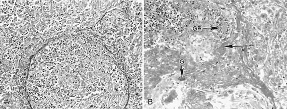
The diagnosis of WG is made histologically by the presence of necrosis, granulomatous inflammation with multinucleated giant cells, vasculitis, and microabscess formation (Figs. 148-8 and 148-9). Small biopsy specimens (e.g., from the ear or upper airways) may lack all of the various diagnostic features, however. In such cases, a diagnosis of WG can be made on the basis of the clinical presentation, ANCA testing, and repeat biopsy specimens taken from the same or related sites.
The etiology and pathogenesis of WG are unknown. Infectious, genetic, and environmental risk factors and combinations thereof have been proposed. The evidence to date suggests that WG is a complex, immune-mediated disorder in which tissue injury results from the interplay of an initiating inflammatory event and a highly specific immune response.23 Part of this response consists of the production of ANCA, directed against antigens present within the primary granules of neutrophils and monocytes; these antibodies produce tissue damage by interacting with primed neutrophils and endothelial cells.
The prognosis of WG has dramatically improved from a mortality rate of 82% before the era of immunosuppressive therapy to the current remission rate of more than 75% with appropriate medical therapy.24 Induction of remission is usually achieved with high doses of corticosteroids, cyclophosphamide, or methotrexate given for 3 to 6 months. Maintenance of remission is achieved with lower doses of corticosteroids and less toxic alternatives to cyclophosphamide, such as azathioprine, methotrexate, trimethoprim-sulfamethoxazole, or other drug combinations. Other therapies that have been used include leflunomide, mycophenolate mofetil, and the tumor necrosis factor inhibitors etanercept and infliximab.
Otologic Manifestations
The middle ear and mastoid are the most common sites within the temporal bone that are involved in patients with WG.20,25 WG may cause obstruction of the eustachian tube with resultant serous otitis media and conductive hearing loss (see Fig. 148-8). Some patients also have purulent otitis media, and others may have frank granulomatous involvement of the middle ear and mastoid. The process can extend to involve the facial nerve and the inner ear, which is usually manifested as rapidly progressive SNHL and loss of vestibular function. Otologic disease may be the sole and initial presenting feature in some patients with WG.
Sarcoidosis
Sarcoidosis is a chronic multisystem disorder of unknown etiology that is characterized by the presence of noncaseating granulomas. It most frequently affects the lungs, although almost all body parts can be affected. The disease shows a female predominance, and is 10 times more common in blacks than in whites. The onset of disease is usually during the third to fourth decade of life. Common presenting manifestations include bilateral hilar adenopathy on chest radiography, cough, and granulomatous skin rash. Other manifestations include iridocyclitis, keratoconjunctivitis, peripheral lymphadenopathy, hepatosplenomegaly, cardiac failure, myalgia, and arthralgia. Neurologic involvement includes central and peripheral manifestations. The facial and optic nerves are the most commonly affected cranial nerves. Either peripheral mononeuritis or polyneuritis may be seen. Laboratory findings may include hilar adenopathy on chest radiography (Fig. 148-10), hypercalcemia, and elevated serum angiotensin-converting enzyme. The histopathologic feature of the sarcoid lesion is noncaseating epithelioid granulomas (Fig. 148-11).
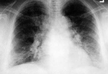
Figure 148-10. Bilateral hilar adenopathy and linear parenchymal densities in a 56-year-old woman with pulmonary sarcoidosis.
The etiology and pathogenesis of sarcoidosis are unknown. The initial pulmonary lesion is an alveolitis characterized by the accumulation of CD4+ T cells, followed by development of noncaseating granulomas. The antigenic stimulus that initiates the disease process remains elusive. Several possible associations have been investigated, including mycobacteria, certain HLA complexes, abnormalities of T cells and the T-cell antigen receptor, and involvement of several different cytokines.26
Spontaneous resolution occurs in many patients. Corticosteroids are beneficial for patients with progressive symptoms or with ocular, cardiac, or central nervous system involvement. For patients who cannot tolerate or do not respond to corticosteroids, alternative drugs include methotrexate, cyclophosphamide, azathioprine, chlorambucil, cyclosporine, chloroquine, pentoxifylline, and antagonists of human tumor necrosis factor-α such as etanercept (Enbrel) and infliximab (Remicade).27
Otologic Manifestations
Otologic manifestations of sarcoidosis include SNHL, vestibular dysfunction, and facial nerve paralysis, and occasionally granulomatous disease of the external or middle ear and mastoid.28–31 The facial nerve is the most commonly affected cranial nerve and is usually involved as part of the triad of uveoparotid fever (Heerfordt’s syndrome) that is characterized by parotitis, uveitis, facial nerve paralysis, and mild pyrexia. The facial paralysis may be sudden in onset, it is often bilateral, and it may resolve spontaneously. Histopathology of the temporal bone in sarcoidosis has been reported in only one case; the main findings were perivascular lymphocytic infiltration and granulomatous inflammation involving the cochlear, vestibular, and facial nerves within the internal auditory canal.32
Syphilis
Congenital and acquired syphilis may affect the middle ear in the late latent and tertiary forms. In the late latent form, the middle ear and mastoid may be affected by a rarefying osteitis with leukocytic infiltration of the ossicles and mastoid bone (Figs. 148-12 and 148-13). A similar but larger lesion of tertiary syphilis, the gumma, shows obliterative arteritis and central necrosis. A gumma of the ear canal or middle ear may result in perforation of the tympanic membrane and a granulomatous appearance of the middle ear mucosa. Definitive diagnosis of syphilis of the middle ear requires a positive serologic test and a histologic demonstration of Treponema pallidum. Syphilitic involvement of the tympanic membrane and middle ear may mimic tuberculosis, and superinfection may result in chronic otitis media.
Inner ear involvement may occur in the absence of macroscopic changes in the tympanic membrane or middle ear. Hennebert’s sign, which is the induction of ocular deviation with positive or negative pressure in the external auditory canal, was believed to indicate a true fistula between the middle and inner ear because of rarefying osteitis of the otic capsule. The more probable cause is fibrous adhesions, however, between the stapes footplate and the membranous labyrinth as a result of endolymphatic hydrops.33 Combined antibiotic and corticosteroid therapy is beneficial for the management of SNHL.34
Lyme Disease
Infection is usually acquired in the summer, and individuals of all ages and both sexes can be affected. Three clinical stages that are similar to the stages seen with cases of syphilis are recognized.35 The first stage (early, localized infection) begins 3 to 33 days after a tick bite with a characteristic skin lesion (erythema migrans). This lesion occurs in 60% to 80% of patients and may be accompanied by minor constitutional symptoms. The second stage (early, disseminated infection) occurs within days or weeks after inoculation and begins with hematogenous dissemination of the organism from the site of inoculation. The symptoms, which mimic a systemic viral illness, include fever, migratory arthralgia, myalgia, headache, meningismus, generalized lymphadenopathy, malaise, fatigue, and secondary annular skin lesions.
After hematogenous spread, B. burgdorferi seems to be able to sequester itself in certain niches and cause localized inflammation in the nervous system, heart, or joints. Neurologic involvement is manifested by meningitis, encephalitis, cerebrospinal fluid lymphocytosis, peripheral neuropathy, myelitis, or cranial neuropathy (including facial nerve paralysis). Cardiac manifestations include atrioventricular block or other arrhythmias, myocarditis, and pericarditis. Joint disease manifests as brief attacks of asymmetrical oligoarticular arthritis, primarily in large joints (especially the knee). The third stage (late or persistent infection) occurs more than 1 year after onset, and can result in chronic, prolonged arthritis; chronic encephalomyelitis; chronic axonal peripheral polyradiculopathy; keratitis (similar to syphilis); acrodermatitis chronica atrophicans; and localized scleroderma-like lesions. A patient may experience one or all of the stages and the infection may not become symptomatic until the second or third stage. Affected tissues show infiltration by lymphocytes and plasma cells. Mild vasculitis and hypercellular vascular occlusion can occur. There is generally no tissue necrosis. In contrast to the findings in patients with syphilis, granulomas, gummas, multinucleated giant cells, and fibrinoid necrosis have not been observed as yet.36
The diagnosis of Lyme disease is usually based on the recognition of the characteristic clinical features, a history of exposure in an area where the disease is endemic, and detection of a specific antibody to B. burgdorferi by enzyme-linked immunosorbent assay and Western blotting.35 The antibody test must be interpreted properly using the criteria of the Centers for Disease Control and Prevention because false-negative and false-positive results can occur. In addition, B. burgdorferi may cause asymptomatic infection, in which case the symptoms caused by another disease may be wrongly attributed to Lyme borreliosis.
The spirochete is highly sensitive to doxycycline, but only moderately so to penicillin. Other effective antibiotics include amoxicillin, erythromycin, cefuroxime, ceftriaxone, and imipenem. Steroids have been used for severe carditis and arthritis. Among patients managed early in the course of the disease, the specific antibody response usually disappears within months, and patients may become reinfected in the future. Among patients with late manifestations such as arthritis, titers decline after successful management, but patients remain seropositive. Although two vaccines were developed and found to be effective in clinical trials, they are not currently being marketed.37
Otologic Manifestations
Facial nerve paralysis is the most common otologic manifestation, with a reported incidence of 3% to 11%38,39; it may be bilateral in 25% of cases. This type of paralysis is seen in the second stage of the disease, and it affects patients of all ages and both sexes. The onset is acute, the duration is weeks to a few months, and the return of function is spontaneous and is usually complete. There may be a history of preceding otalgia, ipsilateral facial pain, or paresthesias.40 Although other neurologic features of the second stage may be seen, facial nerve paralysis can occur as the sole neurologic abnormality (including normal cerebrospinal fluid).
Antibiotics or steroids do not seem to influence the duration or outcome of facial paralysis,38 but they are recommended to treat concurrent symptoms and to prevent the more serious late complications. There is no role for surgery. The precise cause and histopathology of facial nerve palsy have not been elucidated. Histology from other involved peripheral nerves shows perineural and perivascular infiltration by lymphocytes and plasma cells. In chronic and severe neuropathies, demyelination and loss of nerve fibers similar to wallerian degeneration occur.36 It is unclear whether neural lesions result from an inflammatory response to the spirochete or an immune-mediated epiphenomenon.
A well-documented otologic manifestation is an unusual skin lesion called a lymphocytoma, in which intensely red and violet nodules occur on the earlobe during the second stage of disease.35 The lesion consists of benign but hyperplastic lymphocytic follicles in the dermis.36
Auditory and vestibular manifestations, such as SNHL, sudden hearing loss, positional vertigo, and Meniere’s-like symptoms have been described.41–44 More clinical data and temporal bone studies are needed to substantiate these preliminary observations.
Mycotic Diseases
Otologic Manifestations
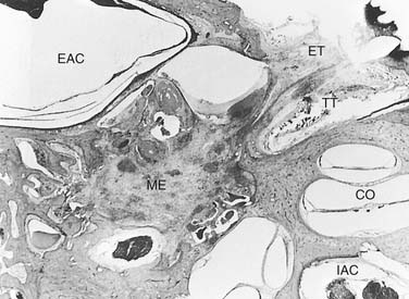
The middle ear and mastoid can be involved as a result of ascending infection along the eustachian tube and tensor tympani (often seen in mucormycosis) (Fig. 148-14) or through superinfection of existing chronic otitis media.45 Destruction of the middle ear cleft ensues, often with extension to the surrounding structures, including thrombosis or rupture of the internal carotid artery.46 Other routes of infection include hematogenous embolic dissemination, which can result in multiple granulomas throughout the temporal bone, and through cryptococcal involvement of the central nervous system, which can cause invasion and degeneration of the nerve trunks in the internal auditory canal.47