 Orbital Secondary Tumors
Orbital Secondary Tumors
General Considerations
By traditional definition, an orbital secondary tumor is a neoplasm that has invaded the orbital tissues to form a neoplastic site in adjacent structures such as eyelid, conjunctiva, intraocular structures, sinuses, nasopharynx, and brain (1,2,3,4,5,6,7,8,9,10,11,12,13,14,15,16,17,18,19,20,21,22,23,24,25,26,27,28,29). This is to be differentiated from a metastatic orbital tumor in which the lesion reaches the orbit from a distant cancer by hematogenous or neural routes. A more detailed breakdown of the specific lesions that can secondarily invade the orbit is provided elsewhere (1); this chapter provides an overview of the subject, followed by selected examples of specific lesions that invade the orbit.
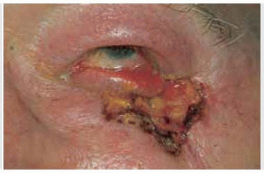
The main eyelid tumors that can involve the orbit secondarily include basal cell carcinoma, sebaceous gland carcinoma, squamous cell carcinoma, cutaneous melanoma, and Merkel cell carcinoma. Conjunctival tumors include melanoma and squamous cell carcinoma, particularly the mucoepidermoid and spindle cell variants. Intraocular tumors include uveal melanoma and retinoblastoma, and, rarely, medulloepithelioma and acquired epithelial tumors of the ciliary body. Sinus tumors that can secondarily invade the orbit include carcinoma of the ethmoid or maxillary sinus and, rarely, rhabdomyosarcoma. Nasopharyngeal tumors include carcinoma, juvenile angiofibroma, and esthesioneuroblastoma. Intracranial tumors include sphenoid wing meningioma and, rarely, glioblastoma. Most of the tumors that can secondarily invade the orbit have been discussed in other chapters and the other atlases. In the authors’ clinical series, the 142 secondary orbital tumors accounted for 11% of the 1,264 orbital lesions. That series was somewhat biased because of the large number of patients with uveal melanoma and retinoblastoma seen by the authors and the fewer cases of primary sinus carcinoma referred to the authors (4).
Clinical Features
The clinical features of a secondary orbital tumor necessarily vary with the type and location of the primary neoplasm. In many instances, there is a prior history of excision or other management of a periorbital lesion. This is particularly true of eyelid, conjunctival, lacrimal sac, and intraocular tumors. In cases of sinus or nasopharyngeal tumors and sphenoid wing meningioma, the orbital manifestations are commonly the initial features of the disease.
Diagnostic Approaches
In cases of suspected secondary orbital tumor, it is important to take a detailed medical history and to perform an ocular and systemic evaluation. External ocular examination, slit-lamp biomicroscopy, and fundus examination can reveal the primary tumor. Imaging studies, particularly computed tomography (CT) and magnetic resonance imaging (MRI), are mandatory to determine the extent of orbital involvement and to assess the status of a primary lesion in the paranasal sinuses, nasopharynx, or brain.
Pathology
The histopathology of a secondary orbital tumor necessarily varies with the type of neoplasm. The pathology of these tumors is covered in textbooks and elsewhere in the current atlases.
Management
The management of orbital extension of the various periorbital tumors can vary greatly from case to case; each case must be individualized. In general, wide surgical resection often followed by irradiation or chemotherapy are treatment options. Massive orbital extension by primary eyelid and conjunctival tumors (basal cell carcinoma, squamous cell carcinoma, melanoma, etc.) are usually managed initially by wide surgical excision, which includes orbital exenteration (5). Massive orbital extension of uveal melanoma (18) or retinoblastoma may also require orbital exenteration, but there are various alternatives for mild degrees of orbital involvement, including techniques of irradiation and chemotherapy. Mild degrees of secondary orbital invasion by lymphoma, metastatic carcinoma, rhabdomyosarcoma, and esthesioneuroblastoma can usually be managed by irradiation. Orbital extension of sphenoid wing meningiomas can be managed by surgical excision, irradiation, or both, depending on the clinical situation. There are many other situations where treatment depends on the entire clinical situation.
Selected References
These sources cite many other references on specific tumors that secondarily involve the orbit.
1. Shields JA. Secondary orbital tumors. In: Shields JA, ed. Diagnosis and Management of Orbital Tumors. Philadelphia: WB Saunders; 1989: 341-377.
2. Rootman J. Neoplasia. Secondary tumors of the orbit. In: Rootman J, ed. Diseases of the Orbit. 2nd ed. Philadelphia: Lippincott, Williams and Wilkins; 2003:280-282.
3. Henderson JW. Secondary epithelial neoplasms. In: Henderson JW, ed. Orbital Tumors. 3rd Ed. New York: Raven; 1994:343-360.
4. Shields JA, Shields CL, Scartozzi R. Survey of 1264 patients with orbital tumors and simulating lesions: the 2002 Montgomery Lecture, part 1. Ophthalmology 2004;111:997-1008.
5. Shields JA, Shields CL, Suvarnamani C, et al. Orbital exenteration with eyelid sparing: indications, technique and results. Ophthalmic Surg 1991;22;292-297.
6. Demirci H, Shields CL, Shields JA, et al. Orbital tumors in the older adult population. Ophthalmology 2002;109:243-248.
7. Glover AT, Grove AS Jr. Orbital invasion by malignant eyelid tumors. Ophthal Plast Reconstr Surg 1989;5:1-12.
8. Shields JA, Shields CL, De Potter. Surgical approach to conjunctival tumors. The 1994 Lynn B. McMahan Lecture. Arch Ophthalmol 1997;115:808-815.
9. Shields JA, Demirci H, Marr BP, et al. Sebaceous carcinoma of the ocular region. Surv Ophthalmol 2005;50:103-122.
10. Shields JA, Demirci H, Marr BP, et al. Sebaceous carcinoma of the eyelids. Personal experience with 60 cases. Ophthalmology 2004;111:2151-2157.
11. Rao NA, Hidayat AA, McLean IW, et al. Sebaceous gland carcinoma of the ocular adnexa: A clinicopathologic study of 104 cases with five year follow-up data. Hum Pathol 1982;13:113-122.
12. Shields JA, Elder D, Arbizo V, et al. Orbital involvement with desmoplastic melanoma. Br J Ophthalmol 1987;71:279-285.
13. Dithmar S, Meldrum ML, Murray DR, et al. Desmoplastic spindle-cell melanoma of the eyelid with orbital invasion. Ophthal Plast Reconstr Surg 1999;15:134-136.
14. Polito E, Leccisotti A. Primary and secondary orbital melanomas: a clinical and prognostic study. Ophthal Plast Reconstr Surg 1995;11:169-181.
15. Crawford JB. Conjunctival melanomas: prognostic factors. A review and analysis of a series. Trans Am Ophthalmol Soc 1980;78:467-502.
Stay updated, free articles. Join our Telegram channel

Full access? Get Clinical Tree


