Ophthalmic Disorders Associated with Selected Primary and Acquired Immunodeficiency Diseases
Ralph D. Levinson
James P. Dunn
Gary N. Holland
Immunodeficiency diseases are complex and increasingly important problems in modern medicine,1,2,3,4 and a broad spectrum of ophthalmic disorders may be among their protean manifestations.5,6,7,8,9 Immunodeficiency can be primary, or it can be acquired as a consequence of other disease states or iatrogenic immunosuppression. In this chapter, selected primary and acquired immunodeficiency diseases are reviewed, with emphasis on their associated ophthalmic findings. The ophthalmic manifestations of the acquired immunodeficiency syndrome (AIDS) are highlighted.
A review of the topic at this length cannot be exhaustive; the subject of immunodeficiency diseases is far too broad and complex. Instead, the reader is provided with a general overview of the topic, on which a rational approach to the evaluation and management of ophthalmic disorders in immunodeficient patients can be based.
PRIMARY IMMUNODEFICIENCY DISEASES
Immunodeficiency can result from a defect in any component in the immune system: lymphocytes, phagocytic cells (mononuclear cells and polymorphonuclear leukocytes), or the complement system. The World Health Organization (WHO) has classified the primary immunodeficiency diseases into the following categories: (1) combined immunodeficiencies (with defects in both T- and B-lymphocyte function); (2) predominantly antibody deficiencies (abnormal B-lymphocyte development or defects or failure of B lymphocytes to respond to T-lymphocyte signals); (3) predominantly T-lymphocyte defects; (4) immunodeficiencies associated with other major defects; (5) complement deficiencies; and (6) congenital defects of phagocytic cell number, function, or both2 (Table 1). B-lymphocyte disorders probably account for half the primary immunodeficiency diseases; the remaining half, in descending order of frequency, are attributed to combined disorders (20%), phagocytic disorders (18%), predominantly T-lymphocyte disorders (10%), and complement disorders (2%).4 Immunodeficiency diseases, however, do not always fall into these broad functional categories. Although classically pure antibody disorders can occur, the interactive nature of the multiple arms of the immune system can result in overlap between these classifications.
Table 1. Primary Immunodeficiency Disorders | ||
|---|---|---|
|
Our knowledge of the abnormal genes and altered biochemical pathways that result in primary immunodeficiency diseases has increased dramatically in the last decade. More than 60 genetic loci have been identified that are associated with specific defects in the immune system.10 Abnormalities in the production of cell surface receptor molecules, cytoskeletal proteins, enzymes involved in purine and oxidative metabolism, immunoglobulins, complement components, and cytokines have been described; in addition, defects in intracellular signaling pathways and T-lymphocyte cell receptors, as well as defective rearrangements of immunoglobulin genes, have been identified. Understanding the genetics and pathophysiology of these abnormal states has provided new insights into the functioning of the immune system in health and disease.
Distinct and disparate immunologic diseases such as X-linked agammaglobulinemia, C3 deficiency, and neutropenia can produce similar clinical features, even though they are caused by unrelated pathogenic mechanisms affecting different components of the immune system. With selective defects of immune function, other intact immune mechanisms may partially compensate for the deficiency and further blur any clinical distinction between disorders. A similar phenomenon has been found in gene knock-out mice, strains that lack very specific effectors of immune function; they reveal redundancies in the immune system that, although confusing in research models, can be critical for maintaining function. Some diseases, however, do have unique clinical features that aid in establishing a diagnosis.
It has been estimated that in the general population approximately 2500 persons per million (0.25%) have primary immunodeficiencies, most of whom are male.11 Selective immunoglobulin A (IgA) deficiency is believed to be the most common primary immunodeficiency, with a reported prevalence of 1:700 in whites.2 It is estimated that agammaglobulinemia occurs in 1 of 50,000 live births and severe combined immunodeficiency in 1 of 100,000 to 1 of 500,000 live births.3
LYMPHOCYTE DISORDERS
Predominantly Antibody Defects
Defects of humoral immunity may involve all or only selected types of antibodies. Antibody deficiency alters several protective immunologic mechanisms, thus increasing the frequency, chronicity, and severity of infections caused by various bacterial, viral, and protozoan pathogens. Lack of secretory antibodies, which usually accompanies a deficiency of serum antibodies, increases host susceptibility to attachment of bacteria, viruses, and parasites to mucosal surfaces.4 Gastrointestinal (GI) and respiratory tract infections therefore predominate, and the ocular surface may be at increased risk. Systemic seeding of pathogens and violation of the blood-brain barrier may occur more readily because levels of antibodies that function in neutralization, cytotoxicity, or antibody-dependent cellular cytotoxicity reactions are low. Failure to form antibody-antigen complexes limits the actions of the innate, nonspecific immune defense mechanisms normally called into play, such as activation of the complement system with subsequent chemotaxis of phagocytic cells.
Bacterial pathogens commonly associated with antibody deficiencies include many encapsulated organisms, such as Streptococcus pneumoniae, Haemophilus influenzae, Staphylococcus aureus, and Neisseria meningitidis. Common viral agents include enteroviruses and rotaviruses. Giardia lamblia and Cryptosporidium species are considered the most common protozoa that cause disease in these patients. Infections with Pneumocystis carinii, (previously classified as a protozoan parasite but now considered to be more closely related to the fungi12), as well as infections with cryptosporidium, occur in patients with X-linked hyper-IgM syndrome.13,14
In addition to recurrent pneumonia and chronic diarrhea, infectious disorders associated with antibody deficiencies include sinusitis, otitis, viral hepatitis, paralytic poliomyelitis, arthritis related to Mycoplasma sp., cystitis, and urethritis. Meningitis and viral encephalitis are the most important complications. Ocular disorders usually involve the eyelid, conjunctiva, or cornea. In most cases, they are attributed to recurrent bacterial infection.
Sjögren’s syndrome and other autoimmune diseases have been associated with immunoglobulin deficiencies,15,16,17 most commonly with IgA deficiency and common variable immunodeficiency. Those with primary Sjögren’s syndrome may be deficient in the IgG2 subclass as well.16 Infiltration of the salivary glands by lymphocytes is observed. It is possible that xerophthalmia will be more commonly recognized as experience with immunodeficient patients increases.
Described subsequently are several immunodeficiency disorders for which there have been specific references to associated ophthalmic disorders. There is no evidence, however, that there are unique ocular changes in any of the specific antibody deficiency syndromes.
X-LINKED AGAMMAGLOBULINEMIA
X-linked agammaglobulinemia (Bruton’s agammaglobulinemia) is associated with a deficiency of a tyrosine kinase that is required for B-lymphocyte maturation; the gene for this enzyme is found on the X-chromosome. Patients with X-linked agammaglobulinemia have repeated bouts of pyogenic infections. Keratoconjunctivitis has been reported in these patients,18 but in most cases, the eyes remain unaffected.19,20 Eczema is common in this condition,21 and involvement of the eyelids might be expected.
SELECTIVE DEFICIENCY OF IMMUNOGLOBULIN ISOTYPES
Some patients with IgG4 deficiency have multiple hordeola and chalazia, marginal corneal infiltrates, and vascularization of the limbus.22 Other cases, however, have no reported ocular findings.23
Little information is available about ophthalmic involvement with other selective antibody disorders. Selective IgM deficiency results in severe eczema, verrucae vulgaris, and recurrent bacterial infections of the skin.21 Periorbital cellulitis has been reported with this condition.3 Patients who have immunodeficiency with hyper-IgM develop severe verrucae vulgaris,21 which could involve the eyelids.
SELECTIVE IGA DEFICIENCY
IgA deficiency is found in 1:700 whites but in only 1:18,500 Japanese.2 Ocular infection with IgA deficiency is believed to be rare.9,24 Ocular disease is more common when other immunoglobulin classes are also grossly affected.18 In cases in which low serum levels of IgA do not reflect a deficit in secretory IgA, the ocular surface may be at no increased risk for infection. It may be dangerous to attempt IgA replacement; patients with IgA deficiency may have anaphylactic reactions to IgA-containing fluids.25 Even if IgA replacement in blood were possible, it would not protect the ocular surface in most cases; IgA cannot be transported into tears without a secretory piece.
Hemorrhagic retinitis was associated with IgA deficiency in one patient, but it may actually have been the result of systemic lupus erythematosus in that situation.26 Retinal vasculitis has been described in patients with common variable immunodeficiency, which may be a related syndrome, because it is associated with low levels of serum IgA in some people.27 IgA levels in those patients were not reported, however.
An infant with an unusual phacomatosis that included findings of oculocutaneous pigmentation, neurologic abnormalities, and cutaneous vascular abnormalities also had an IgA deficiency.28 He had unilateral glaucoma and abnormal intraocular vessels, described as being similar to those found in Sturge-Weber syndrome.
COMMON VARIABLE IMMUNODEFICIENCY
Common variable immunodeficiency is associated with decreased levels of serum IgA and IgG (and in some cases IgM). Approximately 25%; of cases are familial. The genetic defect is not known. There is an equal gender distribution. There are two peaks of disease onset, at ages 1 to 5 years and 16 to 20 years. Patients may have been immunologically normal before onset of IgA deficiency. Patients with IgA deficiency may progress to common variable immunodeficiency. This disorder resembles X-linked hypogammaglobulinemia clinically; the spectrum of associated disorders is similar, although infections are usually less severe and have later onset.3 In some cases, medications (e.g., sulfasalazine, hydantoin, and carbamazepine) have been implicated as causes.29
Franklin and associates18 reported on five patients, four of whom had conjunctivitis or keratoconjunctivitis. H. influenzae was isolated from three of these patients, one of whom also was found to be infected with S. aureus; Staphylococcus epidermidis was isolated from the fourth patient. Ocular findings are believed to be rare in this syndrome, however. Among approximately 500 cases in the United Kingdom, one patient was reported to have acute unilateral uveitis30 and another acute annular detachment of the choroid and retina with panuveitis (William J. Dinning, unpublished data). These findings may have been incidental; they could not be attributed specifically to the immunodeficiency. A patient with common variable immunodeficiency has been reported with severe corneal thinning that was thought to be sterile in both eyes.31 The problem progressed to corneal perforation in his right eye and a secondary H. influenzae endophthalmitis developed, with loss of the eye. The fellow eye was treated with a lamellar corneal graft, and the patient had 20/25 vision in that eye until his death 2 years later.
Autoimmune diseases, including Sjögren’s syndrome, systemic lupus erythematosus, and rheumatoid arthritis have been associated with IgA deficiency and common variable immunodeficiency. Retinal vasculitis, presumed to be autoimmune in origin, has been reported in three patients with common variable immunodeficiency.27 Associated findings included macular edema, vitreitis, optic disc edema, and retinal neovascularization. No evidence of infection was found. Therapy with periocular injections of corticosteroids, oral prednisone, and cyclosporine appeared to be responsible for maintaining 20/40 or better vision in two patients, but visual acuity dropped to 5/200 in both eyes of the third patient.
Combined Immunodeficiencies
SEVERE COMBINED IMMUNODEFICIENCY DISEASE (SCID)
The combined immunodeficiencies are a group of disorders in which both T- and B- lymphocyte function is abnormal. The abnormalities that have been found appear to be primarily in T lymphocytes, highlighting the importance of these cells for normal B-lymphocyte function, as well as in cell-mediated immunity. Defects in the common γ chain of several cytokine receptors, in intracellular signal transduction, in Class II major histocompatibility complex (MHC) expression, in enzymes involved in purine metabolism, and in T-lymphocyte receptor gene rearrangement have all been described in patients with combined T- and B-lymphocyte immunodeficiencies.10
SCID is a designation that refers to potentially fatal, profound defects in both cellular and humoral immunity. Cases may be X-linked, autosomal recessive, or autosomal dominant. Because of the severe lack of normal immune function, patients with these disorders are at risk for overwhelming infection by multiple types of pathogens: viruses, bacteria, fungi, and protozoa. Patients usually succumb to infection during the first year of life, although bone marrow transplantation (BMT) may result in reconstitution of the immune system in some cases. Patients are particularly susceptible to normally mild viral respiratory pathogens (parainfluenza, adenovirus, respiratory syncytitial virus).4 Reports of fatal, disseminated adenoviral infection in these children have not mentioned the cornea or conjunctiva among the sites of disease, however. Perhaps because of the rapidly fatal nature of SCID, morbidity from opportunistic infections of the eye has not been among the commonly reported features of these syndromes. Infection with herpes simplex virus (HSV), type 1, including ocular involvement, has been reported in an infant with SCID, but no details were given.32 Cytomegalovirus (CMV) retinitis has been reported in three children with SCID33,34,35; two patients had been treated by BMT, however.33,35 Severe, disseminated CMV infections occur frequently in patients undergoing this procedure; the relationship between the retinal infection and the immunologic defects of SCID in these patients is therefore uncertain. The third patient was a 5-month-old girl who presented with meningismus and lethargy and was found to have leukocoria.34 SCID was diagnosed during the evaluation of the patient’s systemic illness. White vitreous humor masses were found in both eyes, as well as retinal necrosis and retinal detachment. A fine needle biopsy of the mass in one eye revealed cells with cytoplasmic inclusion bodies consistent with CMV infection. Serologic test results were consistent with recent CMV infection in the patient and her mother. Despite treatment with intravenous (IV) ganciclovir, she died 1 month later from pneumonia; CMV and P. carinii were found in her lungs.
Nezelof’s syndrome refers to the presence, in infants, of cell-mediated immune defects and an embryonic thymus, but normal circulating immunoglobulins; as such, it is a variant of SCID. There has been no consistency in the literature in the use of this term, and, therefore, it is difficult to compare reports of its manifestations. Among five patients with disorders labeled Nezelof’s syndrome, Friedlander and associates36 identified S. aureus blepharitis in one and Pseudomonas aeruginosa-related blepharoconjunctivitis in one. The other patients were without apparent ophthalmic disease. A patient with Nezelof’s syndrome and tuberculous meningitis did have optic atrophy among her constellation of findings.37
OTHER DISORDERS OF T AND B LYMPHOCYTES
Bare lymphocyte syndrome consists of a group of defects that lead to defective expression of Class I or Class II MHC molecules. Candidal chorioretinitis has been reported in a patient with this disorder, but the patient had other, well-established risk factors for disseminated candidiasis, including indwelling catheters and systemic use of broad-spectrum antibiotics.38 The infection cleared with appropriate antifungal therapy; therefore, the effect, if any, of the specific immune defect on the infection cannot be assessed.
Immunodeficiencies Associated With Other Major Defects
WISKOTT-ALDRICH SYNDROME
Wiskott-Aldrich syndrome is a sex-linked congenital immunodeficiency disease with both humoral and cellular disorders. Patients are also thrombocytopenic at birth. The involved gene, designated WASP (for Wiskott-Aldrich syndrome protein), is expressed in hematopoietic cells and is associated with the cytoskeletal reorganization of activated platelets and lymphocytes. The syndrome is characterized by deficiencies in IgM and elevations in IgA and IgE. Patients develop eczema; thrombocytopenia; and recurrent infections, including laryngitis, bronchitis, gastroenteritis, pyoderma, sinusitis, and otitis media. Infection is caused most often by pyogenic bacteria, but patients with this syndrome are also susceptible to various fungi, viruses, and protozoa. Kaposi varicelliform eruption, a disseminated form of cutaneous HSV infection, may occur. Such infections become apparent by 6 months of age, and susceptibility to infection increases with age. Death often occurs in the first decade of life because of bleeding or infection.
Several ophthalmic disorders are associated with Wiskott-Aldrich syndrome.39,40 Patients may develop ocular surface and adnexal disease, including eczema on the eyelids; blepharoconjunctivitis and pannus formation associated with molluscum contagiosum virus (MCV) lesions; and recurrent, sometimes bilateral, HSV keratitis. Recurrent periocular HSV infections have been reported.41 In many reported cases, the specific pathogens responsible for conjunctivitis are not mentioned. Marginal infiltrates and episcleritis also have been reported; these findings have been attributed to an altered immune response to infectious agents. Papilledema and oculomotor disorders have been attributed to intracranial lesions. Hemorrhage, both intraocularly and on the ocular surface, may occur. In a review of reported cases, Podos and associates39 found that ophthalmic disease (excluding eczema of the eyelids) had been mentioned in 18 of 80 cases. Fifteen patients had infections and five had bleeding of the conjunctiva, periorbital tissues, retina, or into the vitreous humor. Excision of MCV lesions by expression and curettage should be performed with caution in these patients because of the increased risk of hemorrhage.
ATAXIA-TELANGIECTASIA
Ataxia-telangiectasia is a rare, autosomal recessive disorder characterized by neurologic abnormalities, including cerebellar ataxia, vascular changes of the conjunctiva and skin, recurrent respiratory infections, and malignancies, especially lymphomas. The gene responsible for ataxia-telangiectasia, termed ataxia-telangiectasia muted (ATM), has been mapped to chromosome 11 (11q). Its gene product is a protein kinase involved in cell cycle regulation and deoxyribonucleic acid (DNA) repair.42 Defects in DNA repair, cell cycle regulation, and chromosomal instability may play a role in the observed increase in lymphoid malignancies, as well as in abnormal development of the thymus.
Patients have alterations in both cellular and humoral immune functions; markedly decreased IgA and IgE levels are often present. The disease is progressive, with gradual loss of immunologic and neurologic function. Patients may become mentally retarded. They usually die as a result of this disease before 20 years of age.
Conjunctival vascular changes and eye movement disorders constitute the ophthalmic manifestations of this syndrome.43,44,45 Dilated venules and irregular vascular segments in the horizontal exposure area give the eye an injected appearance. These findings appear in most people with ataxia-telangiectasia between the ages of 2 and 7 years. These vascular changes have no functional importance. Similar vascular changes occur on the nose, ears, and extremities.
The disorder is associated with various eye movement disorders involving systems that stabilize images on the retina.46 Abnormalities include difficulties in fixation, pursuit, and convergence; nystagmus; an increased delay time for initiation of voluntary horizontal and vertical saccades; hypometria of voluntary saccades (an abnormality in the initiation of the fast component of involuntary saccades induced by optokinetic or vestibular stimuli, with deviation of the eyes in the direction of the slow component, rather than in the direction of the fast component, as in normal study subjects); and decreased slow component velocity of optokinetic nystagmus.44,46 Those affected may have head thrusts when making horizontal refixations, in an attempt to redirect the eyes for fixation on objects of interest; such thrusts are identical to those seen in people with congenital oculomotor apraxia. Visual acuity, pupillary reaction, and fundi are normal.
Other Lymphocyte Disorders
There are many other incompletely characterized disease states that include immunodeficiency among their findings. Those diseases with defects in cell-mediated immunity make the host susceptible to a broad array of infectious diseases caused by bacteria, viruses, fungi, and protozoa. They include opportunistic infections with organisms that generally are commensal to nonimmunocompromised hosts, such as P. carinii and Candida albicans. Devastating, disseminated viral infections by members of the herpesvirus family, adenovirus, rotavirus, enteroviruses, parainfluenza virus, and respiratory syncytial virus can occur. Atypical mycobacterial infections are common and may be widely disseminated.
CHRONIC MUCOCUTANEOUS CANDIDIASIS
Persons with chronic mucocutaneous candidiasis may have subtle defects both in cell-mediated immunity and in specific antibody responses to Candida sp.47,48 These individuals have severe skin and mucous membrane infections, which can include ulcerative candidal blepharitis and candidal keratoconjunctivitis.49 Photophobia from keratopathy may be the first symptom of disease, occurring at an early age. Findings may include fine punctate erosions of the upper cornea, superficial pannus, and superficial stromal infiltration with scarring. Easty9 believes that these findings are due to bacterial, rather than fungal, infections, but organisms are rarely identified.
These immunologic defects rarely result in candidemia or systemic candidiasis, however. Although people with this disorder have not been reported to develop endogenous candidal chorioretinitis or endophthalmitis in the absence of other predisposing factors, deep tissue infection, including central nervous system (CNS) involvement, has been reported.50,51,52
Chronic mucocutaneous candidiasis may occur as an isolated disorder, or it may be one of multiple disorders in several different syndromes. Endocrine disorders are common, being seen in perhaps half of patients.53 In 1962, Gass54 reported that keratoconjunctivitis was a frequent initial finding in young children with multiple endocrine deficiency, autoimmune disease, and candidiasis syndrome, a disorder characterized by idiopathic hypoparathyroidism and adrenal insufficiency. Organisms were isolated from the conjunctiva in only one of these cases; it was suggested that the conjunctival disease was an immunologic reaction to fungal antigen. Wagman and associates55 reported a series of 16 patients with the syndrome, 4 of whom had keratitis, anterior stromal vascularization, and scarring. A report of two siblings with this disorder mentioned the additional findings of eyelash and eyebrow loss in both, and small diffuse keratic precipitates in one.56 Wong and Kirkpatrick57 reported clinical improvement in one patient with this syndrome, who was treated with allogeneic lymphocytes and administration of transfer factor and amphotericin B.
Chronic mucocutaneous candidiasis is also part of a syndrome that includes keratitis, ichthyosis, and deafness.58 The precise immunologic defect has not been identified, and evidence of chronic candidiasis has been found in only approximately 20% of cases. Corneal vascularization apparently occurs in most patients with this syndrome, and loss of eyelashes and eyebrows is common. Cowan and associates59 described a family in which three siblings had defects in lymphocyte function and antibody production that they attributed to a defect in biotin metabolism. Clinical disorders included CNS dysfunction and severe mucocutaneous candidiasis, with blepharoconjunctivitis and corneal ulceration. Although candidal infections usually are associated with defects in cellular immunity, antibody defects, such as occurred in these patients, may play a role in their development as well.49
PRIMARY PHAGOCYTIC DYSFUNCTION
Neutrophils, monocytes, and eosinophils comprise the nonspecific phagocyte system, providing immunologic defense against pyogenic bacteria, such as S. aureus, and fungi, such as Aspergillus sp. To accomplish this role, phagocytes are capable of many sequential functions, including response to chemotaxis, adhesion, opsonization, ingestion, and intracellular microbial lysis. Defects in one or more of these functions can result in chronic or recurrent infections that are refractory to antibiotic therapy. Targets of infection include skin, mucous membranes, lung, bone, lymph node, and components of the reticuloendothelial system.
Disorders of phagocytic function can occur in combination with other immune defects. Wheeland and associates60 described a father and son with chronic purulent blepharitis and secondary corneal ulceration who had abnormal neutrophil function and cellular immunodeficiency. Their disease did not fit other well-established syndromes.
Chronic Granulomatous Disease
Chronic granulomatous disease (CGD) is an inherited disorder characterized biochemically by an inability of phagocytes to use molecular oxygen for generation of important microbicidal byproducts, such as hydrogen peroxide, hydroxyl radicals, and superoxide anions, thus making those affected people susceptible to recurrent life-threatening infections. X-linked recessive and autosomal recessive inheritances are seen. The genetic heterogeneity of the disease primarily reflects different mutations in the genes for the four subunits of reduced nicotinamide adenine dinucleotide phosphate (NADPH) oxidase. The diagnosis can be pursued by in vitro testing of phagocyte function, including measurements of the respiratory burst by chemiluminescence or by measuring the ability of an individual’s phagocytes to kill catalase-positive bacteria in vitro.
CGD is characterized by recurrent and often chronic infections involving the lungs, lymph nodes, skin and soft tissues, liver, GI tract, bone, and brain. The pathogens involved most often in the disease are S. aureus, Aspergillus sp., Chromobacterium violaceum, Pseudomonas cepacia, and Nocardia sp. Most of the pathogens are catalase-positive microorganisms. Catalase-positive microbes destroy the hydrogen peroxide they generate. CGD phagocytes are defective in hydrogen peroxide generation and are deprived of an alternative source of hydrogen peroxide by which their antimicrobial function could be maintained. Infections with catalase-negative microbes, such as S. pneumoniae, are rare in people with CGD.
The ocular signs of CGD fall into two groups: recurrent blepharokeratoconjunctivitis and chorioretinal lesions61,62,63,64 (Fig. 1). Focal, pigmented chorioretinal lesions appear to be a common finding of CGD. Goldblatt and associates64 examined 38 patients with CGD and 36 of their family members. They found that nine of 38 affected children had chorioretinal lesions, as did three asymptomatic carriers of the gene.64 The lesions are usually perivascular or peripapillary in distribution and can progress to large areas of chorioretinal atrophy.62,63,64 They are most often described as punched-out chorioretinal scars. Goldblatt and associates64 described small white foveal lesions similar to drusen in some patients, but the typical chorioretinal lesions have not involved the central macula. Visual field defects may occur, corresponding to the location of chorioretinal scars.63 Histopathologic studies of the chorioretinal lesions show foci of granulomatous inflammation of the choroid and sclera.65 The granulomatous inflammation has been seen to extend through Bruch’s membrane,65 and areas of Bruch’s membrane may be absent.66 Pigment-filled macrophages in the retina and choroid are similar to those found elsewhere in the body.66 Chorioretinal scars with glial proliferation and areas of retinal pigment epithelium (RPE) hypertrophy and hyperplasia surrounded by atrophy of the RPE and the overlying retina are also found.66 Special stains and cultures of the reported ocular pathology and vitrectomy specimens have not revealed any infectious agents.65,66,67
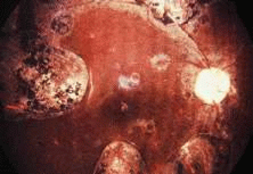 Fig. 1. Typical fundus lesions of chronic granulomatous disease. (Photograph provided by Scott M. Whitcup. From Nussenblatt RB, Whitcup SM, Palestine AG. Uveitis: Fundamentals and Clinical Practice. St Louis: Mosby-Year Book, 1996.) |
In an isolated case report,65 a 3-year-old child with CGD, who had been documented to have chorioretinal scars previously, was found to have chorioretinal lesions that appeared to be actively inflamed with an associated mild vitreitis. The lesions remained stable for several months, but after 7 months, his left eye developed a severe anterior uveitis, vitreitis, and exudative retinal detachment. A lensectomy and vitrectomy was performed. Cultures and stains of vitreous humor were negative. The presumed endophthalmitis did not respond to IV ceftazidime, nafcillin, and amphotericin B administered for 4 weeks. The eye was ultimately enucleated; cultures and stains of the enucleated eye did not reveal any infectious agents. It is not clear, however, whether there might have been an abnormal response to an infectious agent that had been eliminated by the therapy he received. Most people with CGD do not have any evidence of active inflammation or infection, however.
Chronic blepharoconjunctivitis and marginal or punctate keratitis have been reported in patients with CGD, often accompanied by both pannus formation and perilimbal infiltrates.63 These findings are believed to represent an immune reaction to staphylococcal antigens or toxins, rather than a direct invasion of the cornea by organisms. Because it is caused by a catalase-positive bacterium, staphylococcal blepharitis is often a chronic condition in patients with CGD. Regular eyelid hygiene and the selective use of topical antibiotics and corticosteroids has been used to prevent corneal neovascularization and scarring.63
Aggressive and prompt medical and surgical therapy of infections has been important for improving the prognosis of patients with CGD. Granulomata do respond to corticosteroids. The neutrophils of some patients with CGD do respond to interferon (IFN)-γ with a respiratory burst, which may depend on the production of nitric oxide rather than superoxide anions.68 Treatment of CGD with IFN-γ is associated with augmented production of nitric oxide by neutrophils.68 IFN-γ has also been used as a prophylactic agent.69 Although there are reports of cures with BMT, there have been problems with failures to engraft.70
Chédiak-Higashi Syndrome
Chédiak-Higashi syndrome (CHS) is a rare, autosomal recessive disorder, usually occurring in the children of consanguineous parents. It is characterized by partial oculocutaneous albinism, increased susceptibility to both viral and bacterial infections (with both catalase-positive and catalase-negative organisms), anemia, leukopenia, thrombocytopenia, cutaneous ulcers, and peripheral and central neurologic changes, including cerebral atrophy.71 CHS often leads to death in early childhood from infections or, less commonly, from hemorrhage.
Neutrophil abnormalities associated with CHS include neutropenia, reduced bone marrow neutrophil reserves, and reduced neutrophil chemotaxis. Abnormal granules in neutrophils and eosinophils can be seen on routine peripheral blood smears and also are present in other granule-containing cells, such as hepatocytes, renal tubular cells, melanocytes, Schwann’s cells, and thyroid cells.72 Ingestion of particulate organisms occurs normally, but the rate of bacterial killing by neutrophils following ingestion is abnormal because of slow release of the abnormally large peroxidase-positive granules, leading to the delayed appearance of myeloperoxidase within phagocytic vacuoles. In addition to the deficient microbicidal activity of lysosomal degradation, natural killer cell activity also has been demonstrated to be abnormal in some cases.73 Heterozygous carriers of CHS have been identified by studies of degranulation in leukocytes.72
Children surviving past early childhood may be afflicted later with a so-called accelerated phase of the disease, characterized by lymphohistiocytic infiltration of the liver, spleen, nerves, and other tissues. Neoplastic transformation of these cells may occur. In addition to antimicrobial chemotherapy for infections, antineoplastic drugs and corticosteroids have been used to treat this accelerated phase.
Oculocutaneous albinism may be a prominent feature of CHS; an ultrastructural defect of melanosomes distinguishes this condition from true forms of albinism. Giant abnormal melanosomes, believed to be result from fusion of smaller abnormal organelles, have been reported. Hair color ranges from blonde to light brown, but most often has been reported as silver or gray.71,74 The iris is translucent with variable color. The amount of pigment in the uveal tract is variable and may be within normal limits. Histologic examination of the eyes also has revealed decreased pigmentation of the RPE, ciliary body, and choroid. These abnormal granules have been observed in monocytes and neutrophils present in the corneal limbus, iris, and choroid.71,75,76,77,78,79 Ultrastructural study of a conjunctival biopsy specimen from a patient with CHS revealed giant intracytoplasmic lysosomal granules in stromal fibroblasts, which are considered to be pathognomonic for CHS; thus, such a biopsy has been suggested as an ancillary diagnostic test in suspected cases of this syndrome.71
COMPLEMENT DEFICIENCIES
The complement system consists of approximately 20 serum proteins that are critical mediators of antigen-antibody reactions. Sequential activation of the complement proteins (complement cascade) is caused by either antibody-antigen interaction, through the so-called classic pathway, or by various stimulants, such as lipopolysaccharide, aggregated immunoglobulin, cobra venom, thrombin, or proteases through the so-called alternate pathway. Specific complement components and complexes of more than one complement protein serve different functions. C3b, for example, promotes immune adherence, phagocytosis, lymphokine production, and antibody-dependent cell-mediated cytotoxicity. C1,4 and C1,4,2,3 complexes aid in neutralization of viruses to which antibodies have attached. The completed complement cascade leads to lysis of viruses, mycoplasma, protozoa, and spirochetes by creating holes in membranes, capsules, or envelopes.
Deficiencies have been reported for each of the proteins that comprise the complement system. Complement deficiencies can result in increased susceptibilities to infections, connective tissue disorders (systemic lupus erythematosus and other autoimmune diseases), or angioedema, depending on which components are involved. For example, C2-deficient patients generally do not have problems with recurrent infections, unless other components, acting later in the cascade, are deficient as well. In contrast, deficiency of C3, which plays an integral role in both the classic and alternate pathways, results in repeated infections. Deficiencies of the later acting components (C5, C6, C7, or C8) have been linked with serious and sometimes fatal infections with N. meningitidis and Neisseria gonorrhoeae. The association between complement deficiency and autoimmune diseases, such as systemic lupus erythematosus, may relate to the host’s inability to clear circulating immune complexes.
Although complement has been found in tears,80 cornea,81 aqueous humor,82 and sclera,83 ocular infections have not been reported to be prominent features of complement disorders.9 One should assume, however, that patients will be at risk for ocular infections with the same pathogens seen in nonocular sites. Experimental evidence in animal models shows that the complement system may play a role in protecting the eye against corneal infection84 and endophthalmitis85 caused by P. aeruginosa, an organism whose endotoxins activate the alternate pathway. Complement levels are increased in the vitreous humor of inflamed eyes.86 Patients with complement deficiencies who develop intraocular infections for some other reason may, therefore, have more severe disease than their immunocompetent counterparts.
ACQUIRED IMMUNODEFICIENCY DISEASES
Acquired immune dysfunction is more common than the primary immunodeficiency syndromes. An important cause of acquired immunodeficiency that ophthalmologists throughout the world will encounter is AIDS. Nevertheless, there are various conditions, such as malnutrition, other infections (including measles, syphilis, tuberculosis, leprosy), cancer (including Hodgkin’s disease), immunosuppressive drug therapy, trauma, burns, collagen vascular diseases, diabetes, alcoholic cirrhosis, sarcoidosis, and aging, that can result in acquired immunodeficiency and may have associated ophthalmic disorders. The spectrum of disorders resulting from acquired humoral and cellular immunodeficiencies are similar to those of the primary diseases.
In many acquired immunodeficiency states, immune dysfunction does not contribute substantially to clinical manifestations. Reviewed here are several disorders in which altered host defenses lead to prominent clinical problems.
ACQUIRED IMMUNODEFICIENCY SYNDROME
AIDS was first reported in the early 1980s, and its ocular complications were described soon thereafter.87,88,89 It is the most severe in a spectrum of immunodeficiency states caused by infection with the human immunodeficiency virus (HIV).
Infection with HIV is characterized predominantly by T-lymphocyte deficiency. The initial step in HIV infection is binding of the envelope protein of HIV to the receptor on the host CD4+ T lymphocyte; chemokine receptors, such as CCR5 or CXCR4, act as coreceptors and are also necessary for binding. After fusion with the host cell membrane has occurred, the RNA of the virus undergoes reverse transcription, forming DNA, which enters the host cell nucleus and is integrated into the genetic material of the cell. This HIV-encoded DNA is transcribed into messenger RNA, which is then translated into viral proteins, including HIV protease, that are required for processing other HIV-encoded proteins into their functional forms. The newly assembled virus then buds from the cell surface, and new cellular infections occur. Host CD4+ T lymphocytes are depleted through a combination of increased destruction and impaired thymic production.90
The mechanisms of HIV-related disease are far too complicated to be reviewed here, but they ultimately result from dysregulation of the immune system, especially cell-mediated immunity, that occurs because of continuous CD4+ T-lymphocyte destruction and depletion. Impaired cellular immunity facilitates diseases associated with infectious agents, including both productive infections in various end organs and neoplasms that result from malignant transformation of infected cells; examples of the latter include Kaposi’s sarcoma (KS), caused by human herpes virus type 8, and squamous cell carcinoma, caused by human papillomavirus. Many opportunistic infections result from reactivation of previously acquired agents of low virulence that are usually controlled by normal host immune defenses; examples include CMV, P. carinii, varicella-zoster virus, and Toxoplasma gondii. People infected with HIV seem to be predisposed to specific infectious agents (including those already noted, as well as Candida sp. and MCV) with involvement of specific organs (e.g., CMV retinitis or P. carinii pneumonia). Humoral immunity is also affected. Although HIV infection is associated with a polyclonal gammopathy, the B-lymphocyte response to new antigens is markedly impaired. In addition, production of cytokines, such as IFN-γ (which activates macrophages to kill intracellular pathogens) and interleukin-2 (which allows T-lymphocyte proliferation), is impaired.
Most patients are infected with HIV-type 1 (HIV-1). A retrovirus with lymphotropic properties similar to HIV-1 was isolated from two people in West Africa in 1986 and is called human immunodeficiency virus, type 2 (HIV-2).91 Although HIV-2 also causes AIDS, it appears to be less pathogenic than HIV-1, with a longer latency period of infection, less severe manifestations of disease, and longer survival after diagnosis of AIDS. The seroprevalence of HIV-2 is greatest in West Africa. Ocular complications of HIV-2 infection appear to be uncommon.92 For this reason, the term HIV in this chapter refers to HIV-1, unless specifically indicated otherwise.
The severity of immune dysfunction varies considerably between HIV-infected people but tends to become progressively worse over time, if untreated. Those infected with HIV can be asymptomatic; they can have nonspecific manifestations of HIV infection itself, such as lymphadenopathy; or they can have the overt manifestations of HIV-associated opportunistic infection and neoplasms. The AIDS is defined as a CD4+ T-lymphocyte count less than 200/μl (or percentage of CD4+ T-lymphocytes less than 14% of total lymphocytes) or the occurrence of one or more of a specific group of opportunistic infections and neoplasms predictive of severe immunodeficiency.93
HIV can be transmitted by one of three routes: (1) sexual intercourse; (2) percutaneous inoculation (sharing of needles by injection drug users, transfusion of contaminated blood products, accidental needle-stick injuries in health care workers); and (3) maternal-infant (vertical) transmission (in utero, during birth, or by breast-feeding). In the first two decades of the AIDS epidemic, the demographics of the disease in the United States have changed from largely a disorder of homosexual white men to one affecting a mix of homosexuals, heterosexuals, and injecting drug-users, disproportionately involving African Americans and the poor.
In the United States, over 400,000 people had died of AIDS by the year 2000, and there were over 700,000 people living with HIV infection. By 2000, over 60% of new HIV infections in 13- to 19-year-old people were occurring in women. Worldwide, over 16 million people had died, and over 33 million were infected, with over 95% of the 5.6 million adults and children who were newly infected in 1999 living in sub-Saharan Africa and other developing countries.
Currently available drugs that act against HIV include reverse transcriptase inhibitors (both nucleoside reverse transcriptase inhibitors and nonnucleoside reverse transcriptase inhibitors) and protease inhibitors.94 AIDS was initially uniformly fatal, but it can now be controlled in many patients with these medications, which are known collectively as highly active antiretroviral therapy (HAART) or potent antiretroviral therapy. These drugs, by interrupting the infection cycle of HIV, allow a partial restoration of immune function and have thereby resulted in a substantial decline in opportunistic infections. At the same time, the widespread use of potent antiretroviral therapy has resulted in new presentations of clinical disease and clinically important side effects. The cost of these drugs precludes their use in all but the wealthiest of countries. Even in the United States, the combination of intolerable side effects, drug resistance, and lack of adequate compliance with complicated treatment regimens has resulted in a long-term success rate for potent antiretroviral therapy of only 40% in some geographic areas.95
Ophthalmic disorders are among the most common manifestations of AIDS (Table 2). These manifestations include (1) noninfectious vasculopathies, (2) opportunistic infections, (3) neoplasms, (4) neuro-ophthalmic abnormalities attributable to intracranial and orbital disorders, and (5) drug-induced ocular side effects. Recognition and treatment of ocular disease are important not only because of the morbidity associated with vision loss but because ophthalmic manifestations of HIV infection often indicate progressive dysfunction of the immune system and can be the first manifestation of multisystemic diseases. Discussion of these disorders in later sections deal primarily with the manifestations of HIV infection in those people with severe immune dysfunction. The use of potent antiretroviral therapy has reduced the frequency of many HIV-related ophthalmic disorders and in some cases has altered their clinical features, course, and response to treatment.
Table 2. Ophthalmic Disorders Associated with HIV Infection | |
|---|---|
|
Noninfectious Microvasculopathy
Noninfectious retinal lesions associated with microvascular abnormalities (cotton-wool spots, retinal hemorrhages; known as HIV retinopathy) are the most common ocular findings reported in people with HIV infection (Fig. 2). The extent and severity of these retinal findings are greater in patients with AIDS than in iatrogenically immunosuppressed people who occasionally have similar lesions.96 The presence of HIV retinopathy is related to overall immune status; asymptomatic HIV-infected patients rarely have clinically apparent lesions, but nearly all patients with advanced AIDS have some degree of HIV retinopathy.97,98 In addition to cotton-wool spots and retinal hemorrhages, clinical manifestations include retinal capillary microaneurysms, white-centered hemorrhages, telangiectasias, and capillary nonperfusion.87,88,89,97,99,100 Cotton-wool spots occur in the posterior pole and are seen as superficial white patches with feathery edges that sometimes obscure underlying retinal blood vessels. They are caused by stasis of axoplasmic flow within the retinal nerve fiber layer, as a result of ischemia, either because of precapillary arteriolar occlusion or reduced blood flow.101 Cotton-wool spots tend to resolve over 4 to 6 weeks. Affected patients are rarely symptomatic. Microaneurysms are found clinically in only about 20% of patients but are detected in eyes of nearly all patients with AIDS when studied by fluorescein angiography or by trypsin digestion of retinal tissue at autopsy.96,100,102 Ischemic maculopathy has been reported rarely.100 Noninfectious microvasculopathy is also the most common ocular manifestation of HIV-2 infection, and affected people have a shorter survival that HIV-2-infected people without microvasculopathy.92
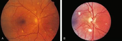 Fig. 2. A, B. Cotton-wool spots in patients with AIDS. |
The underlying abnormality of HIV retinopathy is disease of the retinal vasculature. Trypsin digest preparations and ultrastructural studies reveal selective loss of pericytes, microaneurysm formation, swollen endothelial cells, occluded vascular lumina, and thickened basal lamina,96,100 similar to findings in diabetic retinopathy. Fluorescent microsphere infusion studies have identified breaches in microaneurysms of 200 nm or more, even in the absence of visible hemorrhage; these defects might play a role in the passage of CMV into the retina with development of CMV retinitis.103
The cause of noninfectious microvasculopathy is unknown and likely multifactorial. HIV has been identified within retinal endothelial cells but not in amounts sufficient to account for the widespread changes seen.104 Other proposed mechanisms include deposition of circulating immune complexes,100 release of cytotoxic substances such as monokines or proteolytic enzymes104 and abnormal blood flow.101 Slowing of blood flow probably further contributes to the focal ischemia that results in periodic development of cotton-wool spots.101 The presence of both cotton-wool spots and conjunctival microvasculopathy, consisting of dilated capillaries, vessels of irregular caliber, and sludging of blood flow, has been related to elevated fibrinogen levels and red blood cell aggregation but not with levels of circulating immune complexes or immunoglobulin.101 People infected with HIV have increased neutrophil rigidity, which may also slow blood flow and contribute to microvascular damage.105 Souza Ramalho and associates106 reported a patient with HIV-2 infection and noninfectious retinal vasculitis who had elevated plasma viscosity and red blood cell filterability, both indices of abnormal blood flow. The patient’s clinical and hemorheologic status improved following treatment with prednisone and chlorambucil.
Less commonly, occlusion of larger retinal blood vessels (macrovasculopathy) can occur. Both retinal artery and vein occlusions have been reported, with a prevalence greater than would be expected for age-matched controls.99,107 Hypertension and thrombotic disease are found in a high proportion of affected patients.
Other causes of noninfectious microvasculopathy, such as diabetes mellitus or hypertension, should always be considered. Patients with both diabetes mellitus and HIV disease may have particularly severe noninfectious retinopathy. Reports of hyperlipidemia, glucose intolerance, and accelerated vascular disease among patients treated with protease inhibitors108 have raised concerns that retinal vascular disease may become more frequent in HIV-infected patients. For example, lipemia retinalis has been found in a patient treated with protease inhibitors who had hyperlipidemia.109
Ocular Opportunistic Infections
CMV RETINITIS
CMV retinitis is by far the most common intraocular infection in people with AIDS.99,110 CMV retinitis is rarely the first manifestation of AIDS111,112; most affected patients have CD4+ T-lymphocyte counts of less than 50 cells/μl at diagnosis.111,113 One study found that 85% of people with extraocular CMV infection developed CMV retinitis after a mean follow-up period of 6.4 months.114 Other risk factors for the development of CMV retinitis include high HIV levels in the blood, a previous AIDS-defining illness, homosexual activity as the route of HIV infection, lack of protease inhibitor therapy in antiretroviral regimens, high CMV levels in the blood, and CMV seropositivity.115 Up to 40% of patients with advanced HIV disease developed CMV retinitis in the era before potent antiretroviral therapy.110,111,115,116 Since the introduction of these drugs, the rate is much lower,117 probably on the order of 5% or less. The decline in frequency may reflect not only the widespread use of potent antiretroviral therapy but also the changing demographics of AIDS; heterosexual men are less likely than homosexual men to develop CMV retinitis, perhaps because homosexuals have a higher rate of pre-existing CMV infection.115 Nevertheless, CMV retinitis remains a major cause of morbidity in HIV-infected people. It is especially important to diagnose the infection early, because currently available therapy is usually effective for controlling retinal lesions and because treatment of systemic CMV infection may favorably affect concurrent HIV replication.
CMV retinitis may begin with infection of retinal vascular endothelial cells, with subsequent spread to adjacent glial and neuronal cells as well as to RPE,118 although early autopsy studies failed to identify infected endothelium in all cases.100 It is also possible that infection results from diapedesis of CMV-infected leukocytes in some cases.119
The diagnosis of CMV retinitis is based on clinical signs and symptoms.87,88,99,119,120 Vision is usually good at the time of diagnosis,113 and there is no pain or redness. People with early CMV retinitis usually have floaters and may also complain of photopsias or visual field defects, but between 10% and 75% of patients may be unaware of any symptoms,121,122 especially if the disease is unilateral.98 One study found that over half of new cases of CMV retinitis were diagnosed during routine screening eye clinic visits and that over 75% of affected patients were asymptomatic at the time of diagnosis.122 Routine dilated indirect ophthalmologic examination of patients at highest risk of CMV retinitis, based on a CD4+ T-lymphocyte count of less than between 50 and 100/μl, has been recommended.98,123
Only about 10% of people with AIDS-related CMV retinitis have clinically apparent extraocular CMV disease, although about half have a positive blood or urine cultures for CMV.113 The characteristic retinal lesion has a dry-appearing, granular border surrounding an area of retinal edema and full-thickness retinal necrosis. Findings are similar to those of CMV retinitis described in patients with iatrogenic immunosuppression before the AIDS epidemic began.124 There is a spectrum of lesion appearances; the fulminant or edematous type has prominent necrosis and hemorrhage, often along blood vessels in the posterior pole (Fig. 3). The indolent or granular type is more often found in the retinal periphery and has less dense retinal opacification and little or no hemorrhage. Many cases of CMV retinitis have features of both types. Patients who have an incomplete response to potent antiretroviral therapy may develop CMV retinitis that is less hemorrhagic and less densely opaque. Perivascular sheathing, which may be widespread, occurring in areas of the retina away from the site of necrosis, is found in between 35% and 50% of cases. Bilateral involvement occurs in 30% to 40% of cases at diagnosis.113,122,125 There are usually mild anterior chamber and vitreous inflammatory reactions, and fine keratic precipitates are often seen. Untreated lesions enlarge slowly; spread toward the fovea occurs at a median rate of only 24 μm/day and is somewhat faster toward the ora serrata.126
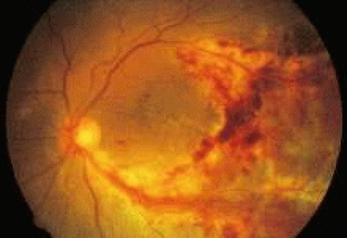 Fig. 3. Cytomegalovirus retinitis in a patient with AIDS; the disorder is characterized by perivascular, white, granular-appearing necrosis and hemorrhage. |
Vision loss may result from primary or secondary optic nerve involvement,127,128 retinal necrosis involving the central macula, rhegmatogenous retinal detachment, retinal vein or artery occlusion, or serous macular exudation.129 Electrophysiologic and psychologic testing suggests that there is a diffuse dysfunction of the retina even when only localized, peripheral lesions are seen clinically.130
Peripapillary CMV retinitis is associated with an increased risk of CMV encephalitis.131 Early detection and treatment may reverse some of these complications and result in improved vision,132 but in most patients the goal of therapy is to prevent further vision loss in affected eyes and to reduce the risk of involvement in uninfected eyes of unilateral cases.133
CMV retinitis occurs less frequently in children with AIDS than in adults,134,135,136 but it is more likely to be associated with symptomatic extraocular CMV disease.137 Perhaps because children are less likely to complain of visual symptoms and are, therefore, less likely to undergo ocular examination than adults, CMV retinitis in children also more commonly presents with bilateral and posterior pole involvement.134,137 Very young children with CMV retinitis may also have high absolute CD4+ T-lymphocyte counts, because the normal CD4+ T-lymphocyte count in the first few years of life is substantially higher than in adults; however, affected children still have counts that are much lower than normal, when adjusted for age.137 Older children with CMV retinitis usually have CD4+ T-lymphocyte counts that are less than 20/μl.134
The differential diagnosis of CMV retinitis includes other necrotizing herpetic retinopathies, as described later, toxoplasmic retinochoroidopathy, candidal chorioretinitis, aspergillic retinitis, ocular cryptococcosis, endogenous bacterial endophthalmitis, syphilitic retinitis, intraocular lymphoma, and Bartonella henselae retinitis (cat-scratch disease).138 Although the distinction between these diseases is usually made clinically and additional workup is unnecessary, there are patients in whom vitrectomy or endoretinal biopsy may be useful diagnostically.139,140,141,142,143,144 In addition, concurrent ocular opportunistic infections with more than one pathogen have been reported in many people with AIDS.
There were four drugs approved by the U.S. Food and Drug Administration (FDA) as of the year 2000 for treatment of CMV retinitis: ganciclovir, foscarnet, cidofovir, and fomivirsen. The first three are available in IV forms; ganciclovir is also available in an oral preparation and as a sustained-release intraocular implant. The first three drugs act by inhibiting CMV DNA polymerase. Fomivirsen is an antisense oligonucleotide that acts by preventing the elaboration of a protein needed for infectivity of the virus; it is delivered by intravitreous injection. All these drugs are virostatic, not virucidal; discontinuation of therapy in persistently immunosuppressed patients, therefore, invariably leads to recurrence of CMV retinitis.
Ganciclovir is a nucleoside analogue. The recommended IV dose is 5 mg/kg every 12 hours for 2 to 3 weeks (induction), followed by 5 to 10 mg/kg once daily (maintenance therapy). Ganciclovir is a bone marrow suppressant; anemia, leukopenia, and thrombocytopenia are common findings associated with its therapy. Foscarnet is a pyrophosphate analogue. The recommended IV dosage is 90 mg/kg every 12 hours for 2 weeks (induction therapy), followed by 90 to 120 mg/kg once daily (maintenance therapy). Foscarnet is nephrotoxic and must be carefully dose adjusted for patients with renal insufficiency. Randomized, controlled studies have shown no therapeutic difference between ganciclovir and foscarnet; both control retinitis initially in about 90% of cases, but reactivation of retinal infection despite prolonged therapy is common, with a median time to progression of retinitis of less than 3 months. Because both IV foscarnet and ganciclovir must be given daily, a permanent indwelling catheter is placed for long-term therapy. Catheter-related infections are a major reason why other options have been developed.
Oral ganciclovir has limited bioavailability but may be effective as maintenance therapy following a course of IV induction therapy for limited periods of time.145 Commonly used doses range from 1 to 1.5 g three times daily. Side effects include GI disturbances and bone marrow suppression. Although use of oral ganciclovir as primary prophylaxis may reduce the risk of CMV infection, including retinitis,146 its use is not routinely recommended because of the cost and associated toxicity.147 An oral prodrug of ganciclovir (valganciclovir) holds the promise of providing serum drug levels comparable with those achieved with IV ganciclovir, while avoiding catheter-related complications; approval by the FDA is anticipated in 2001.
Stay updated, free articles. Join our Telegram channel

Full access? Get Clinical Tree


