CHAPTER 183 Obstructive Sleep Apnea Syndrome
Primary Snoring
Approximately 10% of children snore during sleep on most or all nights, and the majority of these children have primary snoring.2–5 Primary snoring has been defined as snoring during sleep without associated apnea, gas exchange abnormalities, or excessive arousals.6 The factors that predispose children to primary snoring are similar to those described for OSAS and are discussed in detail later in this chapter. Major risk factors for snoring in otherwise healthy children are obesity, decreased nasal patency (rhinitis, septal deviation, nasal obstruction), and adenotonsillar hypertrophy.7,8 Snoring in 6- to 13-year-old children also has been strongly associated with parental cigarette smoking.4,7
Primary snoring does not appear to progress to OSAS in young children and may resolve over time. Investigators have performed polysomnography in children 1 to 3 years after their diagnosis of primary snoring and found no significant difference in the apnea index or nighttime gas exchange.9,10 Ali and coworkers found that 50% of children who were described by their parents as habitual snorers at 4 years of age had stopped snoring without treatment at a 2-year follow-up evaluation.2
In the past, primary snoring was considered a benign condition; however, more recent studies have challenged this assumption. O’Brien and colleagues found that primary snoring was associated with an increased probability of neurobehavioral deficits in children.11 Daytime symptoms such as sleepiness, inattention, aggressiveness, and hyperactivity are reported commonly in habitual snorers and children with OSAS.3,12–15 Disrupted microsleep architecture has now been demonstrated in children with primary snoring, providing a possible link between neurocognitive dysfunction and habitual snoring in children.16,17
Upper Airway Resistance Syndrome
Children with UARS snore and have partial upper airway obstruction that leads to repetitive episodes of increased respiratory effort ending in arousals and sleep fragmentation. Daytime symptoms similar to those seen with OSAS may exist.18–20 Children with UARS have no evidence of apnea, hypopnea, or gas exchange abnormalities on polysomnography. The diagnosis of UARS currently is made with the use of esophageal pressure monitoring to identify increased respiratory effort with related arousals. When polysomnography is performed without esophageal pressure monitoring, snoring with marked paradoxical breathing movements or repetitive arousals may be suggestive of UARS. Treatment options for UARS are identical to those for OSAS and are discussed later in the chapter. The incidence of UARS in children is unknown, but some centers believe it may be more common than OSAS.19
Obstructive Sleep Apnea Syndrome
Approximately 1% to 3% of all children will have OSAS, and as many as 40% of snoring children referred to a sleep clinic or otolaryngologist may have OSAS.21–25 Childhood OSAS has been defined as episodes of upper airway obstruction during sleep that usually are associated with a reduction in oxyhemoglobin saturation or hypercarbia, or both. Sleep-related upper airway obstruction in children may manifest as obstructive apnea or obstructive hypoventilation.26 Obstructive hypoventilation results from continuous partial airway obstruction, which leads to paradoxical respiratory efforts, hypercarbia, and often hypoxemia. This pattern may be more common in children, who are less prone than are adults to collapse of the upper airway.27 Diagnosis of obstructive hypoventilation in children requires end-tidal CO2 monitoring during polysomnography. Despite the absence of complete airway obstruction during sleep, children with obstructive hypoventilation are at risk for all of the reported complications of OSAS.
Obstructive hypoventilation generally is not seen in adults with OSAS. A number of other important differences between adults and children with OSAS are detailed in Table 183-1.
Table 183-1 Childhood versus Adult Obstructive Sleep Apnea Syndrome
| Feature | Adults | Children |
|---|---|---|
| Presentation | ||
| Excessive daytime sleepiness | Main presenting complaint | Infrequent complaint |
| Associated obesity | Majority of patients | Minority of patients |
| Underweight/failure to thrive | Not seen | Frequent finding |
| Daytime mouth-breathing | Not seen | Frequent finding |
| Gender | Male/female = 2 : 1 | Male/female = 1 : 1 |
| Enlarged tonsils and adenoids | Rarely seen | Frequent finding |
| Sleep Pattern | ||
| Obstructive | Obstructive apnea | Obstructive apnea or obstructive hypoventilation |
| Arousal with obstruction | Common | May be less frequent |
| Disrupted | Common | Not often seen |
| Management | ||
| Surgical | Minority of patients with inconsistent results | Definitive in many |
| Medical (positive pressure) | Most common management | Only in selected patients |
Adapted from Carroll JL, Loughlin GM. Diagnostic criteria for obstructive sleep apnea syndrome in children. Pediatr Pulmonol. 1992;14:71.
Symptoms and Signs of Childhood Obstructive Sleep Apnea Syndrome
Children with OSAS almost always present with a history of snoring and difficulty breathing during sleep.23,28,29 Parents often report nighttime sweating, restlessness, and unusual sleeping positions in their affected children. Chest retraction, use of accessory muscles, and paradoxical rib cage motion during inspiration are seen during episodes of upper airway obstruction. Paradoxical breathing (inward movement of the thorax during inspiration) can be normal during sleep in infants and toddlers. During rapid eye movement (REM) sleep, the tonic and phasic activity of the intercostal muscles disappears, and the infant chest wall becomes more compliant. The negative intrathoracic pressures generated during inspiration result in the paradoxical inward movement of the thorax. Paradoxical breathing decreases with age and is rare in healthy children older than 3 years of age.30 Paradoxical respirations in older children are almost always the result of upper airway obstruction.
Enuresis has been associated with OSAS in children but is not a consistent finding.29,31,32 Enuresis associated with OSAS may be related to disrupted, restless sleep patterns that affect arousal. Several studies have reported resolution of nocturnal enuresis after adenotonsillectomy in children with symptoms of nocturnal upper airway obstruction or polysomnography that is diagnostic for OSAS.33–35
Daytime symptoms associated with OSAS include mouth breathing, nasal obstruction, and hyponasal speech. Morning headaches have been associated with nighttime breathing obstruction, but controlled studies comparing children with OSAS with children with normal polysomnography results have found no significant difference in the frequency of this symptom.29,31,36 Adults with OSAS often present with excessive daytime sleepiness, but this complaint is less frequent among children with OSAS.23,31,32,37 OSAS can lead to significant cardiopulmonary complications, poor growth, and problems with learning and behavior. These important sequelae are discussed next.
Complications of Childhood Obstructive Sleep Apnea Syndrome
Growth Impairment
Failure to thrive was commonly described in early reports of children with OSAS.28,29,36 Although overt failure to thrive is now seen less frequently, as a result of earlier diagnosis and management of OSAS, children with OSAS often grow poorly. Improved growth has been reported in childhood OSAS after treatment with adenotonsillectomy.38–40 Marcus and colleagues reported decreased growth in children with even mild OSAS that appeared to be related to the increased work of breathing during sleep. After adenotonsillectomy, the patients had improved growth and decreased energy expenditure during sleep.38 Nocturnal growth hormone secretion also appears to be decreased in children with sleep-related upper airway obstruction.41 OSAS should be considered in the differential diagnosis for failure to thrive or impaired growth that is unexplained by other conditions.
Behavior and Learning Problems
Sleep-related breathing obstruction in children can lead to significant neurocognitive and behavioral complications. A comprehensive review of available data shows that pediatric OSAS is associated with shortfalls in academic performance, deficits in behavior and emotional regulation leading to problems such as hyperactivity and aggressiveness, difficulty with sustained and selective attention, and decreased alertness.42 It also has been suggested that childhood OSAS may lead to problems with memory, intelligence, and executive functioning, but findings are inconclusive to date. Although the exact cause-and-effect relationships are unknown, both sleep fragmentation and intermittent hypoxia are emerging as likely components in the pathophysiology of neurobehavioral morbidity associated with sleep-disordered breathing.43–45 Gozal and associates found that a group of children with OSAS and neurocognitive abnormalities had significantly increased levels of morning C-reactive protein when compared with children with OSAS and no cognitive dysfunction and with control subjects (i.e., children without OSAS).46 This finding suggests that the greater the inflammatory response elicited by OSAS, the greater the risk of neurocognitive dysfunction, possibly secondary to neuronal injury.
In an encouraging trend, several studies have documented post-treatment improvement in children with OSAS who exhibited preoperative disturbances in behavior and cognition.39,47–50 In one study of 12 children with moderate to severe OSAS, a significant reduction in inattention, aggression, and hyperactivity was seen after treatment, as assessed by the Conners Parent Rating Scale. The study authors also noted an improvement in vigilance on a continuous performance task.47 Owens and colleagues found modest improvement after treatment in a small group of children with mild to moderate OSAS who had behavioral problems and mild deficits in executive functioning, attention, and motor skills.50 Gozal reported evidence of learning difficulties in children with OSAS by documenting an increased incidence of snoring and nocturnal gas exchange abnormalities in a group of 297 first-grade children who were in the lowest 10th percentile of their class.48 Of perhaps greatest interest, the children with sleep-associated gas exchange abnormalities who received treatment showed an improvement in academic performance in the second grade, whereas no academic improvements were seen in the affected children whose parents did not seek therapy.
Mitchell and Kelly assessed behavioral abnormalities in children with OSAS using the Behavioral Assessment System for Children before adenotonsillectomy, and again within 6 months after surgery and 9 to 18 months after surgery.51 These investigators found improvements in behavioral measures after adenotonsillectomy that seemed to persist during long-term follow-up, although to a lesser degree than seen shortly after surgery. Similarly, Chervin and coworkers found that children undergoing adenotonsillectomy for any clinical indication (most often, suspected sleep-disordered breathing in this study) had increased difficulty with hyperactivity, inattention, and daytime sleepiness and were more likely to be diagnosed with attention deficit–hyperactivity disorder than control children undergoing other surgical procedures.49 At 1 year after adenotonsillectomy, improvements were seen in all measures.
Clearly, sleep-disordered breathing, even in the mild to moderate range, carries a risk for significant and at least partially reversible neurobehavioral morbidity. It is not clear, however, if the cognitive and behavioral complications of OSAS are completely reversible. Gozal and Pope reported that children with lower academic performance in middle school were more likely to have snored loudly and frequently and to have required an adenotonsillectomy for snoring during early childhood.52 These investigators suggested that the neurocognitive effects of sleep-related upper airway obstruction in young children may be only partially reversible and lead to a “learning debt” that adversely influences subsequent school performance. The diagnosis of OSAS should be considered in children with learning and behavior disorders. Although the ultimate prognosis remains uncertain, early recognition and therapy may help reduce possible life-long neurobehavioral limitations.
Cardiopulmonary Complications
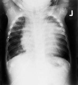
Severe untreated OSAS can lead to pulmonary hypertension and cor pulmonale (Fig. 183-1).28,29 Pulmonary hypertension results from the recurrent, severe, nocturnal hypoxemia, hypercarbia, and acidosis that occur during obstructive hypoventilation and apnea. Cor pulmonale generally is reversible with treatment for OSAS, but perioperative precautions are required in these high-risk patients. Amin and colleagues found that OSAS in children led to structural changes and hypertrophy of both right and left ventricles.53 Most notably, the left ventricular hypertrophy seen in their OSAS patients was related to the degree of severity of the OSAS. These workers noted that left ventricular hypertrophy is a known risk factor for future cardiovascular disease. A follow-up study in children demonstrated a “dose-dependent” decrease in left ventricular diastolic function with increasingly severe OSAS.54 Systemic hypertension, a frequent complication of adult OSAS, also has been reported in children with OSAS.29,55–57 Pediatric OSAS has been shown to be associated with an increased systemic inflammatory response thought to increase the risk for atherosclerotic disease, as demonstrated by elevated levels of C-reactive protein and changes in levels of proinflammatory and anti-inflammatory cytokines.58–60 Improvement in these inflammation indicators was noted after adenotonsillectomy, supporting the importance of early diagnosis and treatment of OSAS to avoid long-term cardiovascular morbidity.58,59
Pathophysiology of Obstructive Sleep Apnea
In otherwise healthy children, adenotonsillar hypertrophy is a significant risk factor for OSAS.36 However, no strict correlation has been found between size of the lymphoid tissues and the presence or severity of sleep-related breathing obstruction.61–63 One child with significantly enlarged tonsils and adenoids may be asymptomatic (i.e., have no difficulty with sleep-related upper airway obstruction), whereas another with only moderate adenotonsillar hypertrophy will have severe OSAS. Furthermore, OSAS develops in only a small percentage of children with adenotonsillar hypertrophy. It is likely that a combination of structural factors and neuromotor abnormalities must be present for OSAS to occur.
Marcus and associates demonstrated that the pediatric upper airway behaves as a Starling resistor.64 In the Starling resistor model, maximum inspiratory flow through the collapsible upper airway is determined by upstream (nasal) pressure and the pressure surrounding the collapsible segment. When airway collapse occurs, flow is independent of the downstream (tracheal) pressure. Airway collapse occurs when the pressure surrounding the airway becomes greater than the pressure within the collapsible segment. The pressure outside the airway is dictated by structural factors, as well as activity of the airway dilator muscles. These investigators found that children with OSAS had greater airway collapsibility than those with primary snoring. Although airway collapsibility was reduced after adenotonsillectomy in children with OSAS, it remained greater than in the patients with primary snoring.
Structural factors other than adenotonsillar hypertrophy may play a role in OSAS in otherwise healthy children. Arens and colleagues used magnetic resonance imaging to study the airway of children with OSAS and found a smaller upper airway volume than in control subjects.65 In a second study, Arens’ group found that the upper airway of children with OSAS was restricted along the upper two thirds, with greatest narrowing in the region of overlap of the tonsils and adenoids.66
Isono and associates studied children with OSAS who were given neuromuscular blockade under general anesthesia. These researchers found that the closing pressure of children with OSAS was positive and greater than that of normal children.67 Collapsibility of the retropalatal and retroglossal segments was significantly increased in their OSAS subjects. They also determined that the cross-sectional area of the narrowest segment of the airway was significantly smaller in children with OSAS.
High-Risk Groups
Adenotonsillar hypertrophy is the leading cause of childhood OSAS. A number of other medical conditions also are strongly associated with OSAS in children, as listed in Table 183-2. Close follow-up is indicated in children in these high-risk groups to look for the development of signs and symptoms that may suggest sleep-related airway obstruction. Such signs and symptoms warrant polysomnographic testing to assess for the presence and severity of OSAS.
Table 183-2 Medical Conditions Associated with Obstructive Sleep Apnea Syndrome*
* Other conditions not listed also may be associated with this syndrome.
Adapted from Marcus CL, Carroll JL. Obstructive sleep apnea syndrome. In: Loughlin GM, Eigen H, eds. Respiratory Disease in Children: Diagnosis and Management. Baltimore: Williams & Wilkins; 1994:475-499. Used with permission.
Obesity
Obese children are at risk for sleep-related breathing obstruction.68,69 Deposition of adipose tissue within the muscles and soft tissues surrounding the upper airway in combination with external compression from the neck and jowls leads to upper airway narrowing in these patients.70 Upward displacement of the diaphragm by the obese abdomen when the affected child is supine and decreased chest wall compliance lead to lower lung volumes during sleep, decreased oxygen stores, and increased risk of desaturation with obstructive events. Obese children are at increased risk for persistent OSAS after adenotonsillectomy. Mitchell and Kelly studied 30 obese children with OSAS and found a decrease in respiratory disturbance index (RDI) (see later under “Polysomnography”) from a mean of 30 preoperatively to a mean of about 12 after adenotonsillectomy.71 Although all children had an improved quality of life as defined by a validated questionnaire, OSAS was decreased in severity but not cured in these obese children.
Craniofacial Abnormalities
Children with skeletal dysplasias such as achondroplasia may have mixed apnea syndromes. Compression of the brainstem at the craniocervical junction may cause abnormalities of ventilatory drive and central apnea. Cervicomedullary compression also can affect upper airway control, leading to airway obstruction during sleep. Adenotonsillar enlargement and the craniofacial features seen in these patients may also contribute to the development of OSAS.72 Sisk and associates diagnosed OSAS in 38% of children with achondroplasia.73 Adenotonsillectomy usually is effective, but additional therapy with continuous positive airway pressure (CPAP) or tracheotomy may be needed in such complicated cases.74 Craniocervical decompression has been used in some patients with achondroplasia, but the effects on OSAS remain controversial.75
Neuromuscular Disease
Cerebral palsy in children is associated with decreased pharyngeal tone, resulting in OSAS. Adenotonsillectomy is a useful initial procedure for OSAS associated with pharyngeal hypotonia.76 These children are at high risk for respiratory complications or persistent OSAS after adenotonsillectomy.77–79 When pharyngeal hypotonia is marked, tracheotomy may be necessary to relieve OSAS. Aggressive craniofacial and pharyngeal surgical approaches have been used to avoid tracheotomy in neurologically impaired patients with OSAS.80 Primary muscular diseases such as Duchenne muscular dystrophy also have been associated with the development of OSAS secondary to loss of upper airway muscle tone.72
Patients with Down syndrome have a high incidence of OSAS. A study of OSAS in these patients found that adenotonsillectomy alone was not adequate therapy for more than 60% of the patients.81 It was concluded that the generalized hypotonia and anatomic abnormalities such as obesity, macroglossia, and midfacial and mandibular hypoplasia all contribute to nocturnal airway obstruction in these patients. Lefaivre and coworkers described a multilevel surgical approach to the management of OSAS in children with Down syndrome, using a combination of facial and mandibular bony advancement and pharyngeal soft tissue surgery to achieve improvement in sleep-related upper airway obstruction.82 Merrell and Shott described the use of lateral pharyngoplasty (plication of the tonsillar pillars), along with adenotonsillectomy, in children with Down syndrome but found polysomnographic abnormalities in 75% of these children after adenotonsillectomy with or without this modification.83
Mucopolysaccharidoses
Mucopolysaccharidoses, such as the Hunter and Hurler syndromes, are associated with severe progressive OSAS in affected children. The head and neck manifestations in these syndromes include short neck, depressed nasal dorsum, macroglossia, adenotonsillar enlargement, and craniofacial abnormalities.84 Bredenkamp and colleagues reported upper airway obstruction in 17 of 45 patients (38%) with mucopolysaccharidoses, 7 (16%) of whom required tracheotomy.85 Leighton and associates found OSAS in more than 90% of patients with mucopolysaccharidoses, with the most severe OSAS found in the Hurler and Hurler-Scheie types.86 Laryngeal tissues can be infiltrated and tracheal size decreased from mucopolysaccharide deposition, leading to airway obstruction at multiple levels.87 Adenotonsillectomy and tracheotomy may be part of the surgical treatment of OSAS in such complicated cases.88 Although its clinical applications are far from clear, enzyme replacement therapy may stabilize mild OSAS in children with Hurler syndrome and may improve upper airway size.89
Acute-Onset or Rapidly Progressive Obstructive Sleep Apnea Syndrome
Abrupt onset or rapid progression of OSAS in children suggests rapid enlargement of lymphoid tissue in the pharynx or other lesions rapidly growing near the airway. Viral syndromes can be associated with marked lymphoid hyperplasia and severe OSAS in children, the common example being infectious mononucleosis secondary to Epstein-Barr virus infection. Acute OSAS associated with Epstein-Barr virus infection is treated with a nasopharyngeal airway and steroids until the lymphoid hyperplasia resolves.90
Diagnosis
Early diagnosis and treatment of OSAS in children will result in decreased morbidity. A study by Richards and Ferdman found that the mean period between onset of symptoms and treatment of OSAS in a group of 45 children was 3.3 years.91 Delays in treatment were related to physician, parent, and third-party factors. Early diagnosis also may be cost-effective: Children with OSAS have been shown to have a 226% increase in health care utilization when compared with control subjects during the 1 year before they receive evaluation and treatment.92 A clinical practice guideline on the diagnosis and management of OSAS in children released by the American Academy of Pediatrics has recommended that all children be screened for snoring as part of routine health care maintenance.93 If snoring is reported, a more detailed evaluation is recommended.
History
Numerous studies have demonstrated that OSAS cannot be distinguished from primary snoring in children, based on clinical history and physical examination alone.23,25,36,94–96 In one report, 89% of parents of children with OSAS observed the child struggling to breathe during sleep, as did 58% of parents of children with primary snoring.23 A majority of parents of children with both disorders were frightened by their child’s nighttime breathing, often staying up nights to watch the child breathe. Goldstein and associates evaluated 30 snoring children referred to a pediatric otolaryngology clinic using a focused history and physical examination in addition to a review of audiotaped breathing of the children during sleep.25 In only one half of the 18 children believed to have definite OSAS clinically was polysomnography diagnostic for OSAS. In a systematic literature review, Brietzke and coworkers found that 11 of 12 articles in the literature concluded that clinical assessment is inaccurate in the diagnosis of childhood OSAS.97 Although the clinical history may not be diagnostic, a thorough evaluation of daytime and nighttime symptoms is helpful in planning subsequent studies and interpreting the findings.
Physical Examination
Routine oral and nasal examination can be supplemented with fiberoptic nasal endoscopy to assess nasal patency, adenoid size, and effect of tonsil size and anatomy on awake respiration.98 Croft and associates used fiberoptic endoscopy to evaluate the site of obstruction in children with polysomnogram-proven OSAS.62 These workers identified distinct adenoidal, adenotonsillar, tongue base, and laryngeal sites of obstruction based on fiberoptic endoscopy and planned successful surgical therapy based on these findings.
Polysomnography
Overnight polysomnography in a sleep laboratory is the “gold standard” method for diagnosing childhood OSAS. An accurate diagnosis of OSAS will ensure that appropriate treatment is provided when needed and will avoid unnecessary surgery in patients with primary snoring. The quantitative data obtained from polysomnography also can help predict which children are at risk for perioperative complications. Severe OSAS on polysomnography has been shown in several studies to be an important risk factor for postoperative respiratory compromise after adenotonsillectomy.78,79
Pediatric sleep laboratories should involve clinicians with expertise in pediatric respiratory disorders or sleep medicine to ensure that the studies are performed and the findings interpreted appropriately for this age group. Overnight polysomnography is recommended. Although a nap study that is positive for obstructive sleep apnea correlates well with the presence of obstructive apnea on an overnight study, a negative nap study result does not exclude the diagnosis of OSAS.99 Nap studies are limited by the short recording time and the possible absence of REM sleep periods. The use of sedatives and sleep deprivation are not recommended because both may increase upper airway obstruction artifactually.100,101
In children, end-tidal CO2 should be monitored to detect obstructive hypoventilation. The consensus statement adopted by the American Thoracic Society, Standards and Indications for Cardiopulmonary Sleep Studies in Children, also recommends measurement of (1) respiratory effort, as assessed by movement of the chest wall and abdomen; (2) airflow at the nose or mouth; (3) measurement of arterial oxygen saturation (SaO2) by pulse oximetry; (4) electrocardiographic recording to monitor cardiac rate and rhythm; (5) electromyography performed in the anterior tibial region to monitor arousals; and (6) appropriate electroencephalography, electro-oculography, and electromyography measurements for sleep staging102 (Table 183-3).
Table 183-3 Polysomnography for Obstructive Sleep Apnea Syndrome in Children
| Sleep State |
| Respiratory Variables |
| Nonrespiratory Variables |
| Recommended but Not Required |
| Audiovideo recording |
Stay updated, free articles. Join our Telegram channel

Full access? Get Clinical Tree


