Nonneoplastic Lesions of the Nasal Cavity, Paranasal Sinuses, and Nasopharynx


Nonneoplastic Lesions of the Nasal Cavity, Paranasal Sinuses, and Nasopharynx


Most specimens from the nasal cavity, paranasal sinuses, and nasopharynx are for nonneoplastic disease. Perhaps because they are so often routine (e.g., chronic sinusitis and inflammatory polyps), they may sometimes receive only cursory histologic examination. That said, a variety of challenging nonneoplastic lesions occur within this region. Furthermore, the accessibility of the nasal cavity allows for biopsy and evaluation of some systemic diseases.

As with many nonneoplastic diseases, the pathologic findings are often nonspecific. The pathologist thus needs to be especially aware of clinical information in such cases. Indeed, both the clinician and the pathologist may need to work together in such cases, as both may be presented with nonspecific findings. With midline destructive lesions, the differential diagnosis of the clinician may be vast, and treatment options may vary greatly depending on the interpretation of the pathologist (Table 13.1).1,2 At the same time, when confronted with granulomatous inflammation, the differential diagnosis of the pathologist may include infectious, autoimmune, iatrogenic, and even neoplastic conditions (Table 13.2).3,4 If the pathologist is to interpret such cases correctly, he will need to be fully armed with clinical and laboratory data. Even in such cases, he may, nonetheless, be sometimes forced to give more descriptive and qualified diagnoses.


SINUSITIS

Acute sinusitis is common following upper respiratory tract infection and is often due to infection by Streptococcus pneumoniae, Haemophilus influenzae, or Moraxella catarrhalis. It is generally treated with antibiotics and is rarely sampled. If sampled, sinus tissue will show abundant edema and infiltration by a mixture of inflammatory cells, with the predominant cell type being neutrophils.


TABLE 13.1 Midline Destructive Lesions


Infectious


Bacterial


Acid-fast bacilli (tuberculosis and leprosy)


Rhinoscleroma (Klebsiella rhinoscleromatis)


Fungal


Fulminant acute invasive fungal infection


Chronic invasive fungal infection


Other


Other infections (spirochetes, parasites, etc.)


Vasculitides


Wegener’s granulomatosis


Allergic granulomatosis and angiitis (Churg-Strauss syndrome)


Other (systemic lupus erythematosus, polyarteritis nodosa, etc.)


Other


Cocaine abuse


Sarcoidosis


Neoplasia (especially Extranodal NK/T-cell lymphoma, nasal type)


Idiopathic midline destructive disease


TABLE 13.2 Granulomatous Inflammatory Lesions


Infectious disease


Mycobacterium tuberculosis or leprosy (rarely other mycobacteria)


Fungal infection


Rhinoscleroma


Leishmaniasis


Vasculitides


Wegener’s granulomatosis


Allergic granulomatosis and angiitis (Churg-Strauss syndrome)


Other


Sarcoidosis


Steroid injection


Neoplasia


Chronic sinusitis, on the other hand, is often sampled, as debridement is sometimes the surgical treatment of choice. The pathogenesis of the disease is thought to be related to multiple factors, including infection, allergy, and the anatomy and physiology of the individual patient.

Microscopically, the respiratory tissue will show variable inflammation composed mostly of lymphocytes, plasma cells, and eosinophils, with the proportions of these cells relative to one another varying from case to case (Fig. 13.1, e-Fig. 13.1).5 Subepithelial and stromal fibrosis can be present, depending on the overall chronicity of the process, with epithelial hyperplasia and even polyp formation often seen. Goblet cells may be more numerous, as may seromucinous glands. Fragments of underlying bone can have sclerotic, reactive changes.


PRIMARY CILIARY DYSKINESIA

A specific cause of chronic sinusitis is primary ciliary dyskinesia, also known as Kartagener syndrome. This disease is associated with male sterility, chronic bronchitis, bronchiectasis, and situs inversus.6,7 It is a genetically heterogeneous disease and typically inherited in an autosomal recessive pattern.

From the pathologic standpoint, assessment of this disease is usually twofold. Functional analysis of the cilia typically involves a wet preparation of scraped nasal epithelium with assessment of the ciliary motility. Normal function should be coordinated and should demonstrate both
intracellular and intercellular synchrony, with cilia typically beating at a frequency of 8 to 20 Hz. Normal function excludes a diagnosis of primary ciliary dyskinesia for all practical purposes. Anatomic analysis of the cilia is performed using electron microscopy. Normal motile cilia are composed of microtubules arranged in a 9 + 2 configuration (Fig. 13.2). Each of the nine outer doublets is attached to the adjacent doublets via nexin links, contains both inner and outer dynein arms, and has radial spokes. A description of all the variants of dysmotile cilia is beyond this book; however, the majority of dysfunction is associated with the absence of inner and/or outer dynein arms.


FIGURE 13.1 Chronic sinusitis with stromal edema and chronic inflammation.


FIGURE 13.2 Electron microscopy of normal cilia showing the classic 9 + 2 microtubule configuration. Outer (white arrow) and inner (black arrow) dynein arms are seen.


FUNGAL DISEASE

Fungi are the causative agents of a number of pathologic conditions involving the nasal cavities.8,9 Commonly, they are related to allergic rhinitis and thus may play some role in the development of chronic sinusitis and secondary complications from such. They also play a role in a number of other pathologic conditions of the sinuses, in which they can be either noninvasive or invasive.


Allergic Fungal Sinusitis

Allergic fungal sinusitis is the most common fungal disease of the sinuses. Clinically, patients with this disease are immunocompetent and may have concomitant asthma, allergic rhinitis, and nasal polyps.10,11 and 12 The disease is
suspected after a protracted history of sinusitis that has been refractory to treatments for bacterial infection. It may be secondary either to hyaline molds such as Aspergillus or Fusarium species or to various dematiaceous molds.10,11,12 and 13

Surgically collected specimens will grossly have tan-brown fragments of mucosa with green-brown mucoid material and have evoked for some the culinary impressions of cottage cheese and peanut butter.
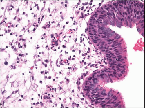
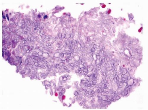
8 Microscopically, samples show abundant mucus with entrapped eosinophils and Charcot-Leyden crystals (allergic mucus) (Fig. 13.3, e-Fig. 13.2).
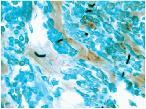
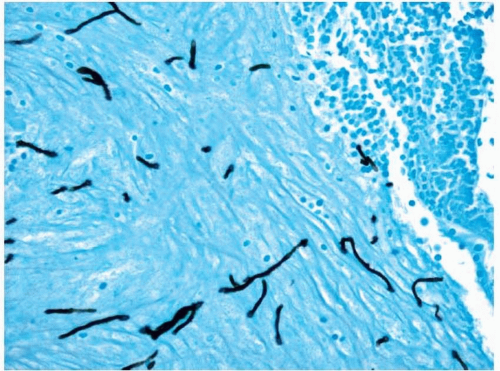
10,12 Although the fungal forms may be difficult to visualize with routine hematoxylin and eosin-stained material, silver staining will show numerous hyphal fragments (Fig. 13.4, e-Fig. 13.3). The hyphal fragments often branch at 45° angles and may have occasional conidia.12 Fragments of edematous and inflamed respiratory mucosa will also be present, showing changes consistent with chronic sinusitis, and should not contain invasive organisms when viewed with special stains.


Sinus Mycetoma (Fungus Ball)

Sinus mycetomas most often occur in the maxillary sinuses of immunocompetent individuals.8,14 Patients typically present with headache, facial pain, nasal obstruction, or a perceived musty odor. They sometimes have concomitant nasal polyps and chronic sinusitis, and they may even develop new-onset seizures. The disease is most often caused by Aspergillus fumigatus.


FIGURE 13.3 Mucus with numerous entrapped eosinophils.


FIGURE 13.4 A silver stain shows occasional hyphal fragments.

Removed material will appear mucopurulent or cheesy.
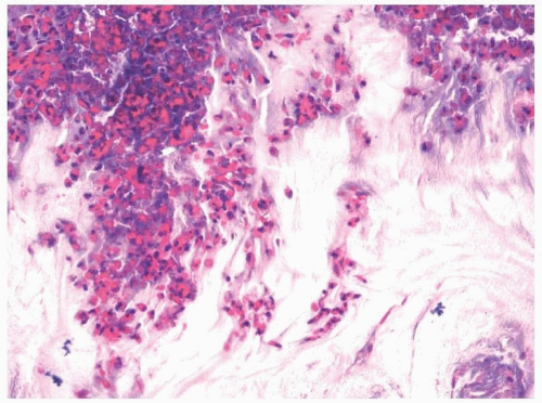
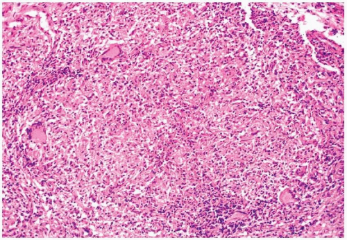
8 On microscopic examination, a dense collection of fungal elements will be seen, devoid of allergic mucus (Fig. 13.5). Silver staining is rarely needed due to the abundance of the organisms (e-Fig. 13.4). The fragments of the sinus tissue will show changes in chronic sinusitis, and fungi should not involve (invade) fragments of respiratory mucosa.


FIGURE 13.5 A fragment of a fungal ball.


Invasive Fungal Sinusitis

Invasive fungal sinusitis is a disease that is generally restricted to immunocompromised individuals, although rare instances are noted in which the disease has affected apparently immunocompetent persons.8,14,15,16 and 17 Acute or fulminant invasive fungal sinusitis may present as a painless, black, nasal septal, or palatal eschar. It is most often caused by fungi of the order Mucorales such as Rhizopus and Mucor, but may also be caused by Aspergillus and Fusarium species, as well as dematiaceous fungi.

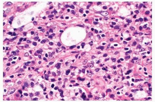
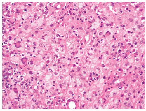
Microscopically, biopsy specimens from patients with acute fungal sinusitis will show invasion of tissue by the hyphal elements, often with involvement of vascular structures (Fig. 13.6, e-Fig. 13.5).8,9,15 A secondary vasculitis and thrombosis with resultant hemorrhage and infarction are common. Surrounding inflammation is often minimal, a reflection of the immunocompromised state of the patient. Some have suggested the use of in situ hybridization for the typing of the etiologic agents.18

Chronic invasive fungal sinusitis follows a more protracted course than acute invasive fungal sinusitis.8,9 Patients are immunocompromised, usually secondary to diabetes mellitus or previous treatment with corticosteroids. The disease may be associated with decreased vision and ocular immobility (orbital apex syndrome). Some believe that the lesion begins as a sinus mycetoma that eventually becomes invasive due to the immunocompromised state of the patient. The disease is most often secondary to infection by A. fumigatus. As with acute invasive sinusitis, histologic sections show tissue and vascular invasion by hyphal elements, often with a limited inflammatory response.


FIGURE 13.6 Invasive fungi seen with silver stain.


Finally, paranasal granuloma or granulomatous invasive fungal sinusitis is a rare chronic fungal disease of immunocompetent patients that is associated with proptosis.8 The disease is secondary to infection by Aspergillus flavus. Most reports of this lesion come from Sudan.

Microscopically, tissue invasion is present, which induces nonnecrotizing granulomatous inflammation with intermixed plasma cells. At the centers of these granulomata, a small amount of fibrinoid necrosis with eosinophils may be present. An associated vasculitis may also be seen.


NASAL TUBERCULOSIS

Involvement of the upper airway in tuberculosis is uncommon, and because of current chemotherapy, it is seldom considered in the differential diagnosis of nasal or pharyngeal disease.19,20 Infection by Mycobacterium tuberculosis can be primary to the site or secondary to a more widespread infection. Infection creates chronic symptoms, with mucorrhea and epistaxis when the nasal cavity is involved and cervical adenopathy is often noted with involvement of the nasopharynx. A discrete mass or ulcer may be present and some ulcers may be deep enough to lead to perforation of the nasal septum. Microscopically, sections will show caseating granulomata with Langhans-type giant cells (Fig. 13.7). Acid-fast stains may reveal the characteristic minute organisms, and immunohistochemical studies with relatively specific antimycobacterial antibodies may also be of value. These techniques, especially the acid-fast stains, are notoriously insensitive, however, and culturing the lesion is essential for the specific diagnosis.


FIGURE 13.7 Necrotizing granulomatous inflammation seen with tuberculosis.


FIGURE 13.8 Mixed inflammation with numerous macrophages seen with leprosy.


NASAL LEPROSY

Leprosy is an infectious, granulomatous disease that involves the slightly cooler regions of the body and therefore typically favors the skin and peripheral nerves and may sometimes involve the mucous membranes and nose.21,22 This is especially true with lepromatous leprosy. It most often involves the inferior turbinates and septum, and crusting, obstruction, and bleeding may be noted. The hallmark microscopic features of leprosy, epithelioid granulomata with foam cells, are often not seen in nasal biopsy specimens. Investigators have noted that sections predominantly show an inflammatory infiltrate rich in macrophages with admixed neutrophils, eosinophils, plasma cells, and mast cells (Fig. 13.8). Bacilli are typically numerous and easily identified with special stains. They tend to be located within macrophages, although nearly all cells may contain the organism (e-Fig. 13.6).


RHINOSCLEROMA

Rhinoscleroma is a chronic, granulomatous inflammatory condition caused by Klebsiella rhinoscleromatis.23,24,25 and 26 It is endemic to Africa, Central and South America, South Central and Eastern Europe, the Middle East, and China. Sporadic cases, however, have been seen elsewhere, including the United States.26 The disease almost always involves the nasal cavity, although it may extend into the palate or pharynx. It is not very contagious and affects primarily immunocompetent individuals.


FIGURE 13.9 Sheets of foamy macrophages seen in a case of rhinoscleroma.

Affected individuals tend to be young, often in their second or third decade of life, and may present with a midfacial necrotizing lesion. They may also initially notice a fetid, purulent rhinorrhea with nasal obstruction. Crusting, epistaxis, and nasal deformity can occur as the disease progresses, which is frequent as early diagnosis is often missed.

The histologic features are characterized by the particular stage: exudative, proliferative, or fibrotic (cicatricial). The early exudative phase is characterized by abundant acute and chronic inflammation with suppurative necrosis or microabscess formation. Edema and granulation tissue may be present. The proliferative or granulomatous phase shows persistent chronic inflammation with groups or even sheets of foamy macrophages (Fig. 13.9, e-Fig. 13.7). Plasma cells may become prominent and Russell bodies are often seen. Special staining will reveal numerous Gramnegative coccobacilli consistent with K. rhinoscleromatis (e-Fig. 13.8). Overlying pseudoepitheliomatous hyperplasia is common. The sclerotic stage is characterized by dense sclerosis with diminished chronic inflammation. Macrophages with definitive organisms may be difficult or impossible to identify at this stage.


RHINOSPORIDIOSIS

Rhinosporidiosis is endemic to South India and Sri Lanka and is caused by the fungus Rhinosporidium seeberi.25,27 The disease usually involves the nasal cavity and is typically unilateral. Clinically, the nasal mucosa often appears polypoid, and the patient may complain of obstruction, epistaxis,

Only gold members can continue reading. Log In or Register to continue

Stay updated, free articles. Join our Telegram channel

Jun 18, 2016 | Posted by in OTOLARYNGOLOGY | Comments Off on Nonneoplastic Lesions of the Nasal Cavity, Paranasal Sinuses, and Nasopharynx

Full access? Get Clinical Tree

Get Clinical Tree app for offline access