Adenocarcinomas, Other Than those that Can Be Classified as Salivary Gland-Type
Most adenocarcinomas of the upper aerodigestive tract can best be classified as salivary gland-type malignancies (Table 7.1). A notable exception occurs in the sinonasal region, where a significant percentage of adenocarcinomas have an intestinal phenotype, and other low-grade and highgrade non-intestinal-type adenocarcinomas also appear distinct from traditional salivary gland-type malignancies. In small biopsy specimens, the distinction between these tumors is not always easy. Nonetheless, the higher grade or worse behaving adenocarcinomas, e.g., the intestinal-type adenocarcinomas and adenoid cystic carcinomas, are usually readily distinguished from the lower grade tumors.
SINONASAL INTESTINAL-TYPE ADENOCARCINOMA
Sinonasal intestinal-type adenocarcinomas are a distinct clinicopathologic entity.1 The tumors most frequently involve the ethmoid sinuses and the nasal cavity; however, they may be found within the other sinuses as well.2,3 and 4 They arise mostly in adult men over a wide age range, although most are diagnosed in the fifth and sixth decades of life. Occasional series that have attempted to report only cases not secondary to environmental exposures have shown no sex predilection, however.1 The tumors are noteworthy for their increased prevalence in persons exposed to hardwood and other dusts, although the risks for such exposure do not appear to be the same throughout the world.2,3,4,5,6 and 7 Sinonasal intestinal-type adenocarcinomas have much higher recurrence rates than lower grade adenocarcinomas and accordingly worse prognoses. Approximately 50% of patients survive for 5 years after their initial diagnosis. Little is known regarding the particular molecular abnormalities associated with these tumors other than the fact that most do not show the abnormalities typical of colorectal adenocarcinomas such as abnormalities of wnt signaling
pathway or mismatch repair protein loss and resultant microsatellite instability.8,9 Comparative genomic hybridization studies have consistently shown gains at 7q, 8q, and 20q with losses at 5q, 17p, and 18q.10,11
pathway or mismatch repair protein loss and resultant microsatellite instability.8,9 Comparative genomic hybridization studies have consistently shown gains at 7q, 8q, and 20q with losses at 5q, 17p, and 18q.10,11
TABLE 7.1 Adenocarcinomas of the Upper Aerodigestive Tract | ||||||||||||||||||||||
|---|---|---|---|---|---|---|---|---|---|---|---|---|---|---|---|---|---|---|---|---|---|---|
| ||||||||||||||||||||||
Grossly, sinonasal intestinal-type adenocarcinomas have been described as papillary and friable and range in color from white-tan to red-purple.1,12 Microscopically, the tumors show intestinal phenotypes that can vary greatly, with some tumors appearing akin to normal small intestinal epithelium (authors have even described an apparent muscularis mucosae) while others are poorly differentiated and have a signet ring phenotype (Table 7.2) (Figs. 7.1, 7.2 and 7.3, e-Figs. 7.1 and 7.2).12,13 and 14 Most tumors (70% to 80%) have a papillary-tubular cylinder cell pattern akin to that seen in most colorectal adenocarcinomas. These usually show papillae or villi that spread across the surface and fold into the stroma. Numerous tubular structures can then be seen on cut surface that infiltrate the underlying stroma, some of which will have intratubular papillary growths. These tumors can produce a variable amount of mucus, and solid and cribriform areas can be seen. The neoplastic cells are tall and columnar with oval, hyperchromatic nuclei that appear stratified. A variable number of goblet cells, Paneth cells, and enterochromaffin cells can also be present. These tumors can be graded as well differentiated, moderately differentiated, and poorly differentiated, much like grading of colorectal adenocarcinomas. Another histologic pattern that may be seen has been termed as the alveolar goblet cell pattern. This pattern is characterized by numerous mucus-filled cavities. These cavities are separated by thin septa
of fibrous tissue and have mucinous epithelial cells either lining the spaces or floating within the cavities. The appearance is virtually identical to that of the so-called colloid carcinomas of the intestinal tract. Finally, less than 5% of these tumors will show a signet ring morphology akin to that seen throughout the enteric tract.
of fibrous tissue and have mucinous epithelial cells either lining the spaces or floating within the cavities. The appearance is virtually identical to that of the so-called colloid carcinomas of the intestinal tract. Finally, less than 5% of these tumors will show a signet ring morphology akin to that seen throughout the enteric tract.
TABLE 7.2 Histologic Types of Sinonasal Intestinal-Type Adenocarcinomas | |||
|---|---|---|---|
|
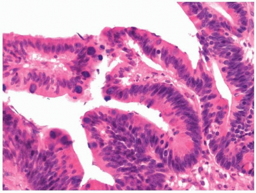 FIGURE 7.1 Sinonasal intestinal-type adenocarcinoma showing a typical papillary pattern. |
The subclassification of sinonasal intestinal-type adenocarcinomas and the grading of the most common pattern, the papillary-tubular type, have
been shown to be predictive of overall survival, with the lower grade papillary-tubular variants showing the longest survival and the highest survival rate at 5 years.13,15,16 Because the tumors are so rare, and because a limited number of therapeutic options are available, subtyping and grading are often not performed. It is unlikely that either is necessary with small biopsies.
been shown to be predictive of overall survival, with the lower grade papillary-tubular variants showing the longest survival and the highest survival rate at 5 years.13,15,16 Because the tumors are so rare, and because a limited number of therapeutic options are available, subtyping and grading are often not performed. It is unlikely that either is necessary with small biopsies.
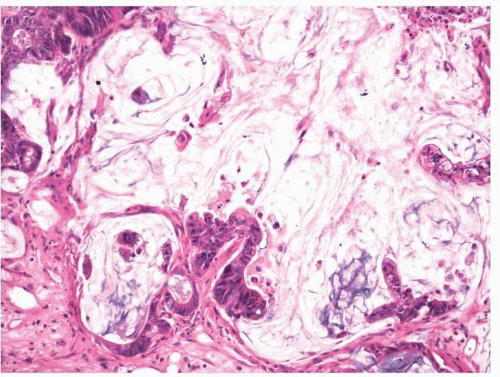 FIGURE 7.2 Sinonasal intestinal-type adenocarcinoma with abundant mucus production. |
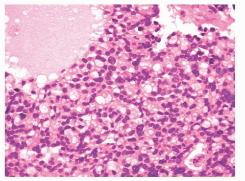 FIGURE 7.3 Sinonasal intestinal-type adenocarcinoma showing a signet ring pattern. |
Immunohistochemically, sinonasal intestinal-type adenocarcinomas show a marked resemblance to true colorectal adenocarcinomas and most tumors will react with antibodies to CK20, CDX2, and MUC2 (Fig. 7.4, e-Figs. 7.3, 7.4 and 7.5).17,18,19 and 20 This allows them to be distinguished from other sinonasal adenocarcinomas. Unlike true colorectal adenocarcinomas, sinonasal intestinal-type adenocarcinomas show greater immunoreactivity with antibodies to neuroendocrine antigens such as neuron-specific enolase and chromogranin, whereas they are less likely to show immunoreactivity with antibodies to carcinoembryonic antigen.21
Stay updated, free articles. Join our Telegram channel

Full access? Get Clinical Tree


