Nodal group
Location
Common areas draining to nodal group
Submental
Inferior to chin, superior to hyoid
Lower lip, floor of mouth, apex of tongue
Submandibular
Inferior to mandible, superior to hyoid
Cheek, lower lip, gums, anterior tongue
Jugulodigastric
Upper neck, near angle of mandible and posterior digastric muscle
Tonsils, posterior oral cavity and oropharynx
Deep cervical
Deep to SCMa and in proximity to the cervical jugular vein
Tongue, trachea, nasopharynx, nasal cavities, palate, esophagus, other nodes in neck
Post auricular
Posterior to auricle and superior to the insertion of the SCM
Auricle
Occipital
Base of scalp at insertion of trapezius
Scalp
Posterior cervical
Posterior to SCM, anterior to trapezius, superior to supraclavicular nodes
Supraclavicular
Above clavicle in lower neck posterior to SCM
Thorax and abdomen
Cervical Fascial Spaces
Another important concept in understanding infectious disease in the neck is the facial envelopes or “spaces” of the neck (Table 14.2) [4, 5]. These fascia divide the neck into a collection of spaces. There is a superficial and a deep cervical fascia. The deep cervical fascia contains three layers, the superficial, the middle and the deep layers. The spaces superior to the hyoid are the parapharyngeal , submandibular, parotid, masticator, and peritonsillar spaces. Inferior to the hyoid is the anterior visceral space. Extending the length of the neck are the retropharyngeal, “danger” space, and prevertebral spaces. The source of infection as well as presenting signs and symptoms are impacted by the contents of the affected space [4]. Infectious processes can spread easily within the fascial space or envelope in which the disease originates, but can also extend to neighboring spaces as disease progresses. Extension of infection into the spaces that extend the length of the neck can spread disease far beyond its origin and thus cause life-threatening complications.
Table 14.2
Cervical anatomy deep neck spaces
Relation to hyoid | Space | Borders of the fascial envelope | Space contents |
|---|---|---|---|
Superior to hyoid | Parapharyngeal (Prestyloid compartment is anterior and Retrostyloid is posterior) | Base of skull superiorly, deep to the pharyngeal constrictor muscle, pretracheal fascia medially, hyoid bone inferiorly, and laterally the mandible, pterygoid muscles and the superficial cervical fascia | Prestyloid: fat, lymph nodes, connective tissue |
Retrostyloid: Carotid sheath; IX, X, XII cranial n; lymph nodes lateral to carotid sheath; and cervical sympathetic chain | |||
Submandibular (Submaxillary portion is above the mylohyoid and Sublingual portion is below) | Mucosa of the floor of mouth and the superficial layer of the deep cervical fascia | Submandibular and sublingual glands, lymph nodes, mylohyoid and digastric m, lingual n, hypoglossal n (cranial n XII) | |
Parotid (incomplete space; connects with parapharyngeal space) | Superficial layer of the deep cervical fascia splits to enclose parotid gland (closure is incomplete) | Parotid gland, cranial n VII, lymph nodes | |
Masticator (anterior and lateral to the parapharyngeal space) | Superficial layer of the deep cervical fascia splits to enclose the mandible and muscles of mastication | Mandible (dental structures), masseter, temporalis, internal pterygoid, inferior alveolar vessels and nerves, mandibular nerve | |
Peritonsillar (bordered by the tonsil, the tonsillar pillars and the superior constrictor muscles) | Not a fascial envelop | Tonsil | |
Inferior to hyoid | Visceral vascular (sometimes referred to as the “highway” of the neck and extends the length of the carotid sheath) | Space within carotid sheath composed of all three layers of deep cervical fascia; connects most neck contents | Carotid a, Jugular v, Vagus n (cranial n X) |
Extend length of neck | Retropharyngeal (extends from the skull base to the first or second thoracic vertebra) | Space within the middle layer of the deep cervical fascia and the alar layer of the deep layer of the deep cervical fascia | Retropharyngeal nodes |
“Danger” space (skull base to posterior mediastinum; deep to the Retropharyngeal space) | Space within the alar and prevertebral layers of the deep cervical fascia and the alar division of the deep layer of the deep cervical fascia | Loose connective tissue | |
Prevertebral (extends from the skull base to the coccyx; deep to the Danger space) | Space deep to the Danger space, within the prevertebral fascia; posterior to the alar division of the deep layer of the deep cervical fascia | Longus colli m |
Clinical Manifestations, Evaluation and Management
Suppurative Cervical Lymphadenitis
Lymphadenitis is seen in association with a variety of pharyngeal, dental, skin or other head and neck infections [6]. Cervical adenitis with abscess formation or suppurative cervical lymphadenitis may develop in some cases of lymphadenopathy. Microorganisms penetrate the skin or mucosal surfaces of the head or neck and are cleared via lymphatic vessels to regional lymph nodes. In the lymph nodes T cell proliferation results node enlargement. In response to invasion of bacterial, viral, mycobacterial or fungal pathogens, additional inflammatory cells, specifically neutrophils, may be recruited to the site of infection. In most instances, disruption of mucosal tissues can result in subsequent invasion by group A streptococcus, other colonizing pathogens or oropharyngeal flora (anaerobes) In other instances pathogens invade the nodal tissue hematogenously, as might be the case with Staphylococcus aureus. Cervical lymphadenitis occurs as a result of host inflammatory responses to this invasion. Lymphadenitis may progress to abscess formation as the host defenses attempt to contain the infection. Typically abscess material consist of cellular debris from necrotic tissue, inflammatory cells and microorganisms. The inflammatory cellular infiltrate may form an abscess wall. Although most cervical lymphadenopathy is viral in etiology, suppuration of an inflamed lymph node is suggestive of primary or secondary bacterial infection with abscess formation. These infections are most often due to group A streptococcus and other streptococci, S. aureus or anaerobes as outlined in Table 14.3.
Table 14.3
Laboratory and diagnostic evaluation
Test | Examples of positive findings | These findings may help diagnose |
|---|---|---|
CBC with differential | Elevated white blood cell count and/or left shift | Infectious lesions |
Atypical lymphocytes | Mononucleosis or malignancy | |
C-reactive protein | Elevated | Non-specific but helpful for following response to therapy; if normal, consider non-inflammatory mass |
Sedimentation rate | Elevated | Non-specific but helpful for following response to therapy |
If normal, consider non-inflammatory mass | ||
Rapid Strep or throat culture | Positive for Streptococcus pyogenes or group A Streptococcus | Strep throat and adenitis |
Rapid influenza test | Positive | Acute influenza adenitis, but does not rule our secondary bacterial adenitis |
Rapid adenovirus test | Positive | Acute adenoviral adenitis but does not rule out secondary bacterial adenitis |
Cat scratch serology | Elevated Bartonella henselae or B. quintana titers | Cat Scratch disease |
EBV serology | Elevated EBV VCA IgM | Acute mononucleosis syndrome |
CMV serology | Elevated CMV IgM | Acute mononucleosis syndrome |
Tuberculin skin test | Induration and erythema | Tuberculosis or non-tuberculous mycobacterial infection (see Chap. 13) |
Interferon gamma release assay (IGRA) | Positive | Tuberculosis (see Chap. 13) |
The diagnostic evaluation of a child with an inflammatory neck mass, suspected cervical neck abscess, or suspected deep neck infection involves a detailed history, physical examination, laboratory studies and if indicated, diagnostic imaging. Helpful historical data includes the age of the child, the duration of illness and mass, the specific associated symptoms involving the upper respiratory tract, and the presence of any symptoms suggestive of a systemic process [7]. Furthermore it is helpful to determine if there has been a change in size or consistency (firm vs. soft) of the mass, presence of fever, recent upper respiratory infection. An exposure history is important to determine if the child has had any animal contact, specifically with kittens or cats, close contact with anyone at risk for tuberculosis, or household members with recurrent boils or soft tissue infection as might be present in cases of methicillin resistant S. aureus (MRSA).
The physical inspection of the neck should include the dimensions and location (midline or lateral) of the mass. If the lesion is lateral then it is further described by nodal group or “level” as outlined in Table 14.1. The presence of an overlying discoloration, sinus or fistula, tenderness, palpable consistency (i.e. soft or firm), and its mobility, including the direction of mobility is also important. Tender lesions are commonly inflammatory and a soft or fluctuant consistency is suggestive of either an infected cyst or an abscess. Careful inspection of nasal passages, pharynx, and oral cavity (including teeth and gums) should be performed to determine a possible source of lymphadenitis. The presence of tonsillar exudate is not diagnostic of bacterial infection and can be present in viral (i.e. EBV or adenovirus) infection (Fig. 14.1). Likewise, the absence of exudate does not exclude infection due to group A streptococcus. Careful examination of the scalp and skin should be performed looking for evidence of infected bites, impetigo, or a papule as may be seen in cat scratch disease.
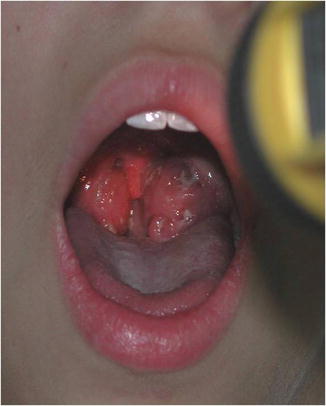
Fig. 14.1
Exudative pharyngitis with EBV infection
The remainder of the physical examination may provide additional clues as to the etiology of an inflammatory neck mass or a systemic illness. For instance, an enlarged spleen is often associated with mononucleosis and a conjunctivitis and rash may be present in children with adenovirus or Kawasaki disease. A scarlatiniform rash or diffuse erythroderma may be consistent with group A streptococcal infection.
Following a thorough history and physical examination additional diagnostic studies and ancillary laboratory testing may be needed prior to surgical intervention. In specific infections (i.e. tuberculosis) this may prevent unnecessary surgery. Table 14.4 summarizes diagnostic and laboratory testing that may be useful in evaluating a child with neck abscess or deep neck infection.
Table 14.4
Diagnostic imaging
Test | Examples of positive findings | Comments |
|---|---|---|
Lateral neck ultrasound | Node enlargement, hypoechoic center suggestive of abscess | No radiation or sedation |
Less specific anatomic information | ||
Computed tomography with contrast | Node enlargement, phlegmon, defined abscess, contiguous inflammation | Radiation, sedation for small children
Stay updated, free articles. Join our Telegram channel
Full access? Get Clinical Tree
 Get Clinical Tree app for offline access

|