Mastoidectomy-Canal Wall Down Procedure

Chapter 17 Mastoidectomy—Canal Wall Down Procedure


image Videos corresponding to this chapter are available online at www.expertconsult.com.


There are two approaches to mastoidectomy in patients with cholesteatoma and chronic otitis media. The canal wall up (also known as intact canal wall) technique is discussed in detail in Chapter 16. This chapter describes the technique for canal wall down surgery. Related topics discussed in this chapter include the role of atticotomy, mastoid obliteration procedures, reconstruction of canal wall down cavities that have not been previously reconstructed, management of dural venous sinus injury during chronic ear surgery, and facial nerve monitoring in chronic ear surgery.


DEFINITIONS


The surgeon may perform a series of mastoidectomy procedures that involve sacrificing a portion or all of the ear canal in continuity with the mastoidectomy.


INDICATIONS FOR CANAL WALL DOWN MASTOIDECTOMY


Mastoidectomy in chronic ear surgery is designed to eliminate mastoid disease in the face of suppurative otitis media and, more commonly, cholesteatoma of the middle ear or mastoid. Generally, canal wall up surgery is preferred to maintain the normal anatomic contours of the mastoid. Certain factors are strong indications for canal wall down surgery, including (1) extensive damage by disease to the posterior canal wall, (2) severely contracted mastoid with low-lying tegmen and far forward sigmoid sinus preventing adequate visualization through a standard canal wall up approach, (3) cholesteatoma in an only hearing ear, and (4) labyrinthine fistula in an ear with extensive cholesteatoma.


Some authors have argued that canal wall down surgery permits excision of cholesteatoma in the sinus tympani region.1 Anatomically, this is not full visualization because the depths of the sinus tympani are medial to the facial nerve. Nonetheless, Hulka and McElveen2 showed that canal wall down procedures do permit additional visualization in the anterior epitympanum and sinus tympani region. The latter is not fully visualized with any technique. Another relative indication for canal wall down mastoidectomy is failure of previous canal wall up procedures with recurrent cholesteatoma from epitympanic retraction pockets. The anatomy of a canal wall down mastoidectomy with full exteriorization of the epitympanum makes retraction pocket recurrences of cholesteatoma unlikely because the whole epitympanum has been exteriorized. Although the use of Silastic in canal wall up facial recess surgery and staging has been significant in reducing the incidence of recurrent cholesteatoma with canal wall up surgery, the presence of a scutal edge and a distinct epitympanum in cases with persistent eustachian tube dysfunction can produce recurrent cholesteatoma.


PREOPERATIVE EVALUATION


A detailed microscopic examination of the ear is necessary preoperatively. Attic retraction pockets should be viewed with suspicion if the surgeon is unable to see the depths of the pocket. Using a right angle pick to feel the depths of such a retraction is often helpful. The use of otoendoscopes has been quite helpful to permit a “fish-eye” view of these pockets. This technique permits wider visualization of the pocket. In these cases, computed tomography (CT) scan can be helpful to define whether what seems to be a small retraction represents the neck of a cholesteatoma or is merely a small retraction. Although the CT scan may underestimate the extent of disease in this area, a large cyst extending into the antrum would be identified with CT scan. An attic pocket in which the depths cannot be palpated that is beginning to retain debris is an indication for surgery. If the CT scan does not show a large cyst in the antrum, an atticotomy may be considered.


Similarly, a distinct attic cholesteatoma with a positive fistula test and the subjective symptom of dizziness is also an indication for imaging. The possibility of a labyrinthine fistula must be considered. Especially if there is a large cholesteatoma, a canal wall down procedure should be considered.


In a pre-existing canal wall down cavity that is draining or is retaining significant debris, the surgeon must evaluate four specific characteristics of the cavity: (1) adequacy of saucerization of the mastoid cortex margins, (2) adequate lowering of the facial ridge, (3) adequate management of the mastoid tip, and (4) adequacy of the meatus. Problems with any of these characteristics can contribute to cavity failures. In chronic drainage situations, areas of persistent mucosalization should be identified so that these are dealt with appropriately in the revision procedure.


Audiometric studies are routinely obtained preoperatively. Generally, we repeat audiometric studies done elsewhere before the patient undergoes surgery. Studies particularly should be repeated if the “outside” audiograms do not coincide with tuning fork tests. Special attention must be given to the adequacy of masking with the audiometric studies performed.


SURGICAL TECHNIQUES


Patient Preparation


The patient is placed supine on the operating table with the head turned away. Hair is shaved approximately 2 to 3 fingerbreadths behind the pinna. Adhesive plastic drapes are applied surrounding the edges of the shaved hair. Approximately 3 to 4 mL of lidocaine (Xylocaine) 1% with epinephrine 1:100,000 is injected in the postauricular region and in the posterosuperior aspect of the ear canal. Sterile preparation is accomplished with povidone-iodine solution, allowing solution to enter the ear canal as well.


In cases in which facial nerve monitoring is used, electromyographic needle electrodes are placed in the orbicularis oculi and orbicularis oris muscles and in the forehead and ipsilateral shoulder to act as ground for monitoring and for stimulating. Sterile towels are placed around the prepared area. The area is dried with a sterile towel, and a large sterile adhesive drape is applied that overlies the sterile prepared area and holds the sterile towels in position.


As mentioned previously, the decision to convert a mastoid operation from canal wall up to canal wall down is usually made intraoperatively. The usual steps for tympanoplasty with mastoidectomy surgery would have been accomplished, including canal incisions, as described in the chapters on tympanoplasty (undersurface graft Chapter 12 and lateral graft Chapter 9). The ear has been reflected forward, the middle ear work has been completed, the tympanic membrane remnant has been prepared for grafting, and the mastoidectomy has begun.


Basic Techniques


The basic principle of canal wall down mastoidectomy surgery is to eliminate all of the disease and exteriorize the mastoid antrum in continuity with the EAC. In addition to removing all diseased air cells, the following four steps are basic to creating a trouble-free mastoid cavity.


Adequate Saucerization


Removal of bone from the edges of the mastoid defect saucerizes or bevels the edges of the defect so that there is no overhanging bone obstructing the wider cavity below (Fig. 17-1). This saucerization ensures that there is no disease tissue laterally. Also, saucerization permits soft tissue surrounding the mastoid to slide into the defect. Paradoxically, removing additional bone around the margins of the mastoid cortex in this saucerization step actually makes a smaller cavity rather than a larger cavity.


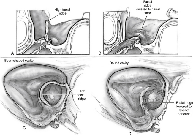
Adequate Lowering of the Facial Ridge


The boundary between the EAC and the mastoid cavity is defined by the height of the facial nerve. Leaving excessive bone overlying the facial nerve (facial ridge) between the EAC and the mastoid cavity creates a situation in which there is a deep trough on the mastoid side (Fig. 17-2). This high ridge is sometimes referred to as a “beginner’s hump.” Occasionally, novice mastoid surgeons leave a large ridge of bone overlying the facial nerve for fear of injuring it. This ridge creates a difficult situation postoperatively with trapped mastoid spaces that are hard to clean in the office, and it prevents natural cleaning. The surgeon should lower the ridge of bone overlying the facial nerve so that the fallopian canal is barely visible with a thin amount of bone overlying the vertical segment of the facial nerve.


Jun 14, 2016 | Posted by in OTOLARYNGOLOGY | Comments Off on Mastoidectomy-Canal Wall Down Procedure

Full access? Get Clinical Tree

Get Clinical Tree app for offline access