Malignancies of the Temporal Bone-Radical Temporal Bone Resection

Chapter 4 Malignancies of the Temporal Bone—Radical Temporal Bone Resection


Primary malignancies of the temporal bone were first recognized in the late 18th century and histologically first confirmed in the 1850s. These lesions are uncommon, with only 250 cases having been reported in the English literature by 1974.1 The overall prevalence in the general population is 6 cases per 1 million.2 Because of their infrequent occurrence, these tumors are often misdiagnosed and treated as chronic external otitis or chronic mastoiditis. They are usually discovered at a later stage, when more radical treatment is required. The infrequent occurrence of the disease poses a challenging obstacle to any attempt at a clinical study regarding treatment.


Secondary malignancies of the temporal bone from regional spread of parotid cancers occur far more commonly. The fissures of Santorini provide a conduit for regional spread through the anterior cartilaginous ear canal.


Basal cell carcinoma and squamous cell carcinoma are the more common malignancies to affect the temporal bone. Basal cell carcinoma is thought to occur secondary to actinic exposure, and commonly involves the external ear or ear canal or both. Squamous cell carcinoma can arise primarily from the external canal or middle ear, or both, or spread into the temporal bone from a primary lesion in the parotid gland. In contrast to squamous cell carcinoma of the upper aerodigestive tract, squamous cell carcinoma of the temporal bone is not related to tobacco or alcohol use. Predisposing factors to these lesions are few. Chronic infection within the temporal bone is the most commonly cited factor.


Adenoid cystic carcinoma can arise either from the ear canal and middle ear or from the parotid gland and spread secondarily into the temporal bone. These lesions have a tendency toward perineural spread. Ceruminous adenoma, adenocarcinoma, and mucoepidermoid carcinoma are other lesions that can affect the temporal bone.


Regional spread to cervical nodes and distant metastases are uncommon. Depending on the extent of the disease, radical resection coupled with radiation treatment offers the best treatment. The efficacy of chemotherapy has not been clearly established, and it may play a role only in recalcitrant disease.


Radical temporal bone resection refers to one of three operations that can be offered to patients with this disease. A lateral temporal bone resection (LTBR) refers to the removal of the external auditory canal (EAC), tympanic membrane, malleus, and incus. Subtotal temporal bone resection (STBR) refers to the additional removal of the otic capsule, and total temporal bone resection (TTBR) refers to the additional removal of the petrous apex with or without the carotid artery.


LTBR is well accepted for lesions that involve the EAC or tympanic membrane or both. Controversy arises in defining the optimal management of neoplasms that invade the mesotympanum. Some authors advocate LTBR with gross removal of middle ear disease followed by radiation therapy, whereas others prefer more radical surgery (STBR or TTBR), followed by radiation therapy. When the tumor has invaded the petrous apex, involvement of dura mater, brain parenchyma, or internal carotid artery (ICA) is usually present.


This chapter focuses on preoperative diagnostic evaluation, the surgical techniques of STBR and TTBR, postoperative management, and potential complications. Rehabilitation and adjuvant treatment for recalcitrant disease are briefly discussed. Finally, we present a literature review of squamous cell carcinoma of the temporal bone in an effort to define the optimal management for middle ear disease and discuss the prognostic significance of dural, brain, and ICA involvement.


DIAGNOSTIC EVALUATION


The diagnostic evaluation begins with a thorough history and physical examination, with special emphasis on the chronology of developing cranial neuropathies. The pathway of tumor spread occasionally can be deduced from a careful history. Facial nerve function, hearing, and balance function should be carefully documented. Examination includes palpation of the parotid gland and cervical lymph glands for the presence of local spread and regional metastases. Patients should be questioned and tested for temporal lobe signs (e.g., memory loss, dysphasia, left-sided neglect, hemiparesis, and olfactory hallucinations) and cerebellar signs (e.g., ocular dysmetria, truncal ataxia, nystagmus, and dysdiadochokinesia).


Imaging allows determination of the extent of tumor involvement. High-resolution axial and coronal computed tomography (CT) imaging at 1.5 mm thickness can identify areas of bony involvement. Enhanced and unenhanced magnetic resonance imaging (MRI) can determine intracranial involvement.


Histologic confirmation of the lesion is essential in further treatment planning. Biopsy specimens of lesions involving the EAC or periauricular skin can be easily obtained. Needle aspiration biopsy of parotid lesions can also be performed.


Angiography is used when involvement of the major vessels is suspected on preoperative imaging, or when surgical exposure of the petrous carotid artery is anticipated. The venous phase of the study can provide important information regarding blood flow through the dural venous sinuses. Embolization of feeding vessels is rarely required because most lesions are relatively avascular.


Cerebral blood flow evaluation is indicated when involvement of the ICA is present. Patency of the anterior and posterior communicating arteries on angiography is an inadequate evaluation for collateral flow. Our current method of preoperative carotid artery testing is described.3 A 30-minute temporary balloon occlusion of the ICA allows identification of patients who would most likely tolerate carotid artery sacrifice. Transfemoral introduction of a nondetachable intravascular balloon, inflated in the ICA, is performed in the patient while sensory, motor, and higher cortical functions are assessed.


Patients who develop a neurologic deficit during temporary occlusion are at high risk for stroke after carotid sacrifice. Preoperative or intraoperative extracranial-to-intracranial arterial bypass should be considered. Repeating the temporary balloon occlusion before surgical extirpation should also be considered. Conservative treatment options should also be discussed with these patients.


Patients who tolerate a 30-minute balloon occlusion of the ICA are at low risk for development of a stroke, provided that a long “distal” stump is avoided. Permanent ICA occlusion can be performed angiographically. Hypotension and hypovolemia in the perioperative period should be avoided if permanent ICA occlusion is performed.


SURGICAL PROCEDURE


Incisions vary according to the extent of the tumor (Fig. 4-1). For lesions contained within the temporal bone, a C-shaped incision extending from the temporal fossa postauricularly into the neck is used. A blind-sac closure of the EAC helps contain the specimen. When tumor invasion of the conchal cartilage or periauricular skin is suspected, an appropriate skin island is incorporated into the overall design. The EAC skin is sutured shut to avoid tumor spillage. The outline of the incisions should preserve the blood supply to the remaining auricle. The anterior and posterior skin flaps are elevated (Fig. 4-2A). The superficial temporal fat pad is elevated with the anterior skin flap in a subperiosteal plane over the zygomatic arch. The superficial temporal and middle temporal arteries are ligated.


The facial nerve can be handled differently, depending on tumor invasion of the parotid gland. When the gland is involved, peripheral branches of the facial nerve are identified with the help of the facial nerve monitor and then divided. The stumps of the anterior segments are secured to the anterior skin flap. The entire parotid gland is dissected off the masseteric fascia, provided that the latter is free of disease, and mobilized posteriorly, while the attachment to the EAC is maintained. When the parotid gland is suspected to be free of tumor, the facial nerve trunk is located in the usual manner at the tympanomastoid suture and divided. The parotid gland, along with the distal stump of the facial nerve, is dissected free of the EAC and mobilized anteriorly off the masseteric fascia.


The jugulodigastric region is explored, and cervical lymph nodes are sent for frozen section pathologic analysis. Regional metastases determine the need for a formal cervical lymphadenectomy. The ninth cranial nerve, the greater auricular nerve, or cervical cutaneous nerves can be used as cable grafts for facial nerve reconstruction. CN IX, X, XI, and XII; the internal jugular vein; and the external carotid artery and ICA are dissected in an inferosuperior direction toward the temporal bone. The sternocleidomastoid and digastric muscles are detached from their attachment to the mastoid.


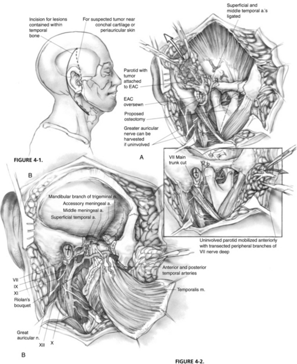
The masseter is detached from the zygomatic arch, allowing exposure of the zygoma and mandible. Zygomatic and mandibular osteotomies (see Fig. 4-2A) can then be performed. The meniscus of the temporomandibular joint is separated from the glenoid fossa, and the chorda tympani nerve emerging from the petrotympanic fissure is divided. The stylomandibular and sphenomandibular ligaments are divided and allow removal of the mandibular segment (Fig. 4-2B). The temporalis muscle is elevated in a subperiosteal fashion and reflected inferiorly. The temporalis muscle must be separated from the lateral pterygoid muscle, and care should be taken not to injure the deep temporal arteries supplying blood to the temporalis muscle. The lateral and medial pterygoid muscles are resected either en bloc with the specimen or separately, depending on tumor invasion.


In a subperiosteal manner, the contents of the infratemporal fossa are elevated off the floor of the middle fossa to expose the middle meningeal artery and vein in the foramen spinosum and the mandibular division of the trigeminal nerve in the foramen ovale. The contents of the foramen spinosum are bipolarly coagulated and divided. Frequently, the venous plexus of the foramen ovale requires bipolar coagulation and packing with oxidized cellulose. The lesser petrosal nerve can be seen emerging from the innominate canal, on its way to the otic ganglion.


The stylohyoid, stylopharyngeus, and styloglossus muscles (Riolan’s bouquet) are detached from the styloid process, which is then rongeured away. The branches of the external carotid artery are dissected in the infratemporal fossa. The anterior tympanic and deep auricular branches of the internal maxillary artery are often divided before identification, and may require bipolar coagulation. The internal maxillary artery is preserved up to the branches of the deep temporal artery. When the internal maxillary artery must be sacrificed, brisk backflow from the anterior stump indicates that the temporalis muscle may derive its blood supply from reversed flow via the pterygoid system. If brisk backflow is not observed, the temporalis muscle cannot be relied on to reconstruct the surgical defect, and microvascular free flap options must be considered. The cartilaginous eustachian tube is divided, and the anterior end is sutured closed to prevent postoperative cerebrospinal fluid rhinorrhea (Fig. 4-3A).


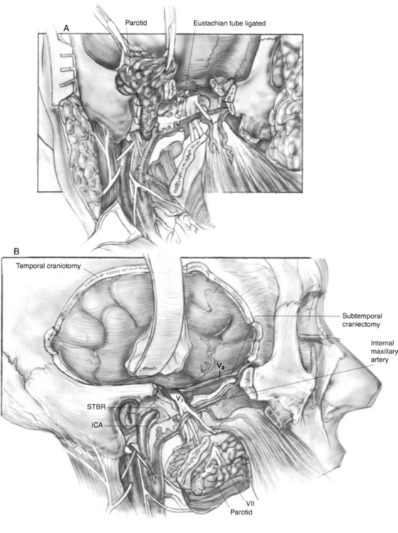
The ICA is dissected toward the carotid canal, and care is taken not to injure CN IX, which crosses its anterior surface. Kerrison rongeurs are used to uncover the vertical and horizontal petrous segments of the carotid artery (Fig. 4-3B). Occasionally, bleeding from the pericarotid venous plexus requires bipolar coagulation. The caroticotympanic artery is also divided when the petrous carotid artery is separated from the specimen. The extent of petrous carotid mobilization depends on whether STBR or TTBR is performed. When STBR is performed, the vertical petrous carotid artery is mobilized from the carotid foramen and canal. When TTBR is performed, the vertical and horizontal petrous carotid artery is mobilized out of the carotid canal to the foramen ovale.


A temporal craniectomy is performed, and the intracranial portion of the middle meningeal vessels is coagulated (see Fig. 4-3B). The patient is hyperventilated to keep the Pco2 at 25 mm Hg for adequate brain relaxation. Mannitol and furosemide can improve brain relaxation. Subtemporal dural elevation proceeds in a posteroanterior direction. The greater superficial petrosal nerve and accompanying petrosal artery are coagulated and divided to lessen traction on the geniculate ganglion. The lesser petrosal nerve and superior tympanic artery are similarly divided. Subtemporal dural elevation proceeds as far medially as possible to expose the superior petrosal sinus. When carcinomatous involvement of the middle fossa dura is suspected, an intradural approach keeps the involved dura attached to the specimen.

Only gold members can continue reading. Log In or Register to continue

Stay updated, free articles. Join our Telegram channel

Jun 14, 2016 | Posted by in OTOLARYNGOLOGY | Comments Off on Malignancies of the Temporal Bone-Radical Temporal Bone Resection

Full access? Get Clinical Tree

Get Clinical Tree app for offline access