Laryngitis
Common
Viruses: adenovirus, influenza
Less common
Viruses: parainfluenza, coronavirus, rhinovirus, RSV, enteroviruses, HSV
Bacteria: Streptococcus pyogenes, Staphylococcus aureus
Uncommon
Viruses: mumps, measlesa, CMV, EBV
Bacteria: Corynebacterium diphtheriae a
Fungi: Candida albicans b
Croup
Common
Viruses: parainfluenza (especially type 1)
Less common
Viruses: RSV, adenovirus, coronavirus, influenza, human metapneumovirus
Uncommon
Viruses: measlesa, rhinovirus, enteroviruses, HSV
Bacteria: Mycoplasma pneumoniae, S. aureus, S. pyogenes, Streptococcus pneumonia, Corynebacterium diphtheriae a
Fungi: Candida speciesb
Epiglottitis
Common
Bacteria: Haemophilus influenzae type b
Less common
Bacteria: H. influenzae non-type b, H. parainfluenzae, S. pyogenes, S. pneumoniae
Uncommon
Bacteria: Pseudomonas aeruginosa b, Klebsiella species
Fungi: Candida speciesb
Clinical Features
The most common symptom in patients with laryngitis is dysphonia with hoarseness and a change in voice at presentation. Other respiratory viral symptoms, such as rhinorrhea, low-grade fever, sore throat, and cough may also be present. Although adenovirus and influenza can be associated with high fevers, if purulent exudate or progressive pain are present, a secondary bacterial infection should be considered.
Diagnostic Evaluation
No specific laboratory testing is usually needed; some clinicians obtain a rapid influenza test or adenoviral polymerase chain reaction (PCR) for highly febrile patients. A rapid strep test and bacterial culture should be obtained if the child has purulent exudate with progressive pain.
Management
Acute laryngitis is typically a self-limited illness that can be treated symptomatically with oral hydration, voice rest, and over-the-counter pain medication. Treatment with antibiotics or steroids is not usually necessary, but antibiotics are indicated in cases of secondary bacterial infection, and steroids if there is considerable concern over airway edema.
Croup (Acute Laryngotracheitis)
Croup is edema and inflammation of the subglottic airway. It is the most common cause of airway obstruction in children aged 6 months to 6 years [1].
Infectious Etiology
Epidemiology
Clinical Features
Viral croup has an incubation period of 2–6 days and typically starts with a prodrome of rhinorrhea, congestion, and low grade fever [3]. Following the prodrome, patients develop the characteristic symptoms of barky cough, hoarseness, and inspiratory stridor. The cough usually resolves after 3 days.
Diagnostic Evaluation
The diagnosis of croup is typically made based on the history and physical exam, however, an anterior-posterior (AP) and lateral radiograph of the neck may help confirm the diagnosis. The typical finding on AP imaging of the neck is the “steeple sign” which occurs secondary to the subglottic edema [4]. The radiographic findings are neither sensitive nor specific, making clinical history the most reliable diagnostic tool [5].
Management
Treatment of viral laryngotracheobronchitis varies depending on the severity of the infection. Patients with a mild episode of croup are typically treated with a single dose of intramuscular (IM) or oral corticosteroids, usually dexamethasone [0.6 mg/kg (maximum dose of 10 mg)], or inhaled budesonide (2 mg) prior to discharge home. However, should a child present with more significant symptoms, they may be admitted for observation and treated with fluids, intravenous corticosteroids, nebulized racemic epinephrine (2.25 %; 0.5 mL in 2.5 mL of saline), and oxygen supplementation if the O2 saturation is <92 %. Nebulized racemic epinephrine can be repeated every 15–20 min if indicated, but the clinical effect only lasts 1–2 h, so children should be carefully observed for symptom return.
The benefits of corticosteroids in the treatment of viral laryngotracheitis has been established in multiple randomized placebo controlled trials demonstrating a variety of benefits including an improvement in croup scores, decreased rates of return to the emergency department (ED) or health care provider, decreased rates of hospitalization, decreased length of hospital stays, and decreased need for respiratory support [6–9]. The majority of patients will respond to treatment within the first 6–12 h [9].
In the most severe cases of croup, patients may require direct laryngoscopy, bronchoscopy and intubation, although this is typically avoided as the endotracheal tube can contribute to the development of subglottic stenosis. Children with a history of multiple episodes of croup or who are under 6 months of age at the time of their first episode should undergo elective laryngoscopy and bronchoscopy when they are healthy to evaluate for a concomitant subglottic disease process (Fig. 11.1).
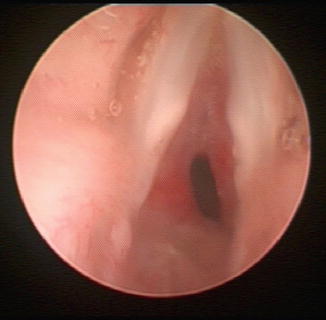
Fig. 11.1
Patient with a history of SGS and acute croup
Epiglottitis
Acute bacterial laryngeal infections represent a relatively rare group of conditions in the pediatric population; however it is imperative that physicians are familiar with and recognize bacterial laryngitis due to its significant potential for morbidity and mortality.
Infectious Etiology
In the post-vaccination era the bacteriology of epiglottitis has changed. Although Haemophilus influenzae type b continues to be the most commonly implicated organism, other organisms are now increasingly found. There have been reports of β-hemolytic streptococci, Staphylococcus aureus, pneumococcus, nontypable Haemophilus influenzae, H. parainfluenzae, and Klebsiella species (Table 11.1) [10].
Epidemiology
Epiglottitis has historically been one of the most devastating pediatric bacterial laryngeal infections. It most commonly affects children between 2 and 7 years of age [5]. Prior to the introduction of the polysaccharide vaccine to Haemophilus influenzae type b in 1985, epiglottitis was routinely encountered in the pediatric setting. Rates in the pre-vaccination era were reportedly between 4.9 and 6.1 cases per 100,000 children per year. These rates have dropped dramatically, and are now reported between 0.02 and 0.3 cases per 100,000 children per year [11]. Indeed, epiglottitis has become almost exclusively a condition of adults, and is often referred to as supraglottitis; however, diligence to identify epiglottitis in children remains necessary due to the potentially fatal outcomes. Contributing factors for continued H. influenzae type b infection are susceptibility of children under the age of 1 who have not completed their vaccination schedule, the increasing frequency of vaccination deferment, and the imperfect rate of immunity conferred by the vaccine.
Clinical Features
The classic teaching is that a child will present toxic appearing, assuming the tripod position (sitting upright, with the chin tilted upwards, and bracing themselves with an outstretched arm). There is typically a history of fever, severe odynophagia, drooling, and muffled speech. Inspiratory stridor is present at times, and signals almost complete airway obstruction. These symptoms tend to develop rapidly over the course of hours.
Diagnostic Evaluation
The diagnosis of epiglottitis is accomplished through history and a non-invasive clinical examination. The patients, having a combination of airway swelling and significant pooling of secretions, can be easily be pushed into airway collapse or laryngospasm. Anxiety provoking maneuvers should be avoided, including intraoral examination to reduce patient anxiety.
Stay updated, free articles. Join our Telegram channel

Full access? Get Clinical Tree


