CHAPTER 70 Laryngeal and Esophageal Trauma
The larynx contains the narrowest region of the ventilatory conduit,1 houses the phonatory apparatus, and functions to protect the airway during swallowing. External laryngeal trauma—the primary focus of this chapter, is an acutely life-threatening injury2–4 that, if not promptly recognized and adequately treated, can also cause significant long-term morbidity in as many as a quarter of patients owing to any combination of dysphonia, aspiration, and airway stenosis.4–6 Laryngeal trauma is often associated with concomitant cervical and intracranial injuries and frequently occurs as part of multisystem polytrauma.4 It is vital therefore that all patients with suspected laryngeal injury be managed by a multidisciplinary trauma team according to Advanced Trauma Life Support guidelines.
Mechanisms of Injury
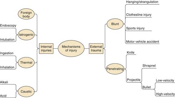
Laryngeal and esophageal trauma can be broadly divided into external trauma, which can be blunt or penetrating, and internal trauma, which can be caused by iatrogenic, thermal, caustic, or foreign body injuries (Fig. 70-1).
Laryngeal Trauma
External Laryngeal Trauma
Incidence
External laryngeal trauma is rare. It has a population incidence of 1 in 137,000 in adults4 and accounts for 0.5% of trauma admissions in children.7 Other studies estimate the incidence of this injury to be between 1 in 50008 and 1 in 30,0009 emergency room visits.
Etiology
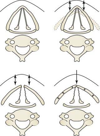
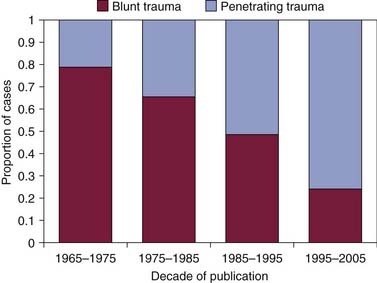
The larynx is well shielded by the mandible superiorly, the sternum inferiorly, the sternomastoid muscles laterally, and the spinal column posteriorly. Further protection is afforded through its mobility. It is however susceptible to crushing anterior cervical trauma, which compresses the organ against the spinal column (Fig. 70-2). Laryngeal injury is also compounded by the process of calcification that begins during the third decade of life and affects men to a greater degree than women.10 Being often asymmetric in respect to the extent of calcification of both the inner and outer surfaces of the thyroid cartilage and each hemi-lamina,11 laryngeal calcification increases the likelihood of fractures after trauma and creates stress risers that can cause framework comminution. Motor vehicle accidents have historically been the main cause of external laryngeal trauma.12 The injury typically occurs when a person not wearing a seat belt or wearing only a lap belt is subjected to rapid deceleration, which thrusts the hyperextended neck onto the dashboard or the steering wheel.13 Other causes of blunt external laryngeal trauma are clothesline injury (in which the rider of a motorcycle encounters a fixed horizontal object at neck level), sports-related injuries,14 and suicidal or homicidal strangulation. Penetrating laryngeal trauma is due predominantly to interpersonal violence in civilian practice and is the main cause of laryngeal injuries sustained in the theater of war.15 Over the past four decades the composition of laryngeal trauma in the United States has changed substantially, due on the one hand to reductions in speed limits, mandatory seat belt laws, and better public education—reducing the incidence of blunt trauma due to motor vehicle accidents—and on the other hand to an increase in penetrating trauma secondary to a greater use of guns and knives (Fig. 70-3).2,3,6,8,9,16–21
Diagnosis
Pertinent Features in the Mechanism of Injury
The mechanism of injury has a significant bearing on the likely pattern and severity of laryngeal trauma. Speed and configuration of the vehicles involved in an accident and the use of lap and shoulder seat belts should be determined. Clothesline injuries are often associated with major trauma to the neck because they impart a large force over a small area.22,23 There are also significant differences in the pattern of injury between suicidal and homicidal strangulation,24 with the latter being more likely to cause laryngotracheal separation and concomitant neurovascular injuries. Strangulation injuries can cause loss of airway due to laryngeal edema some time after the event, despite minimal initial findings. Penetrating trauma is more likely to damage additional cervical structures, with the extent of injury being proportional to both the weight and velocity of the penetrating projectile (E =  mv2, where E is energy, m is mass, and v is velocity). As such, information about the type of weapon used and the distance from which it was fired is useful. With knife injuries, information about the blade and depth of entry is helpful.
Symptoms of Laryngeal Trauma
Laryngeal trauma manifests as a spectrum of symptoms and signs ranging from cardiopulmonary arrest due to airway obstruction to subtle changes in voice quality. The most common presenting feature of laryngeal trauma is hoarseness, followed by dysphagia and pain (Table 70-1).5
Table 70-1 Symptoms and Signs of External Laryngeal Trauma in 33 Cases
| Symptom or Sign | No. of Cases (%) |
|---|---|
| Hoarseness | 28 (85) |
| Dysphagia | 17 (52) |
| Pain | 14 (42) |
| Dyspnea | 7 (21) |
| Hemoptysis | 6 (18) |
From Juutilainen M, Vintturi J, Robinson S, Back L, Lehtonen H, Makitie AA. Laryngeal fractures: clinical findings and considerations on suboptimal outcome. Acta Otolaryngol. 2008;128:213-218.
Examination and Initial Management
Examination and initial management of laryngeal trauma should be part of primary and secondary surveys according to Advanced Trauma Life Support guidelines, to ensure that concomitant injuries are not missed (Table 70-2).4 The over-riding priority is to establish a safe airway with cervical spine protection, which may, and often does, necessitate performing an emergency tracheotomy with the use of local anesthesia. Endotracheal intubation is not a preferred method of airway control in laryngeal trauma, because it can exacerbate a laryngeal injury25 and precipitate total airway obstruction. Endotracheal intubation can be particularly difficult to perform in the presence of concomitant maxillofacial injuries in a patient with an immobile neck. If endotracheal intubation has been performed, it is converted at the earliest opportunity to a tracheotomy to prevent long-term laryngeal injury. In selected cases transtracheal jet ventilation or cricothyroidotomy may be a helpful temporizing measure. No manipulation of the neck is permissible until the cervical spine has been “cleared”—that is, until injuries to the cervical spine have been ruled out in a systematic manner.
Table 70-2 Concomitant Primary Diagnoses Recorded along with Laryngeal Trauma
| Injury | Percentage of Cases |
|---|---|
| Open neck injury | 18 |
| Maxillofacial fractures | 18 |
| Intracranial injuries | 17 |
| Cervical spine fracture | 13 |
| Chest injury | 13 |
| Other facial injury | 10 |
| Skull fracture | 7 |
| Open pharyngeal injury | 4 |
Adapted from Jewett BS, Shockley WW, Rutledge R. External laryngeal trauma: analysis of 392 patients. Arch Otolaryngol Head Neck Surg. 1999;125:877-880.
Use of Computed Tomography
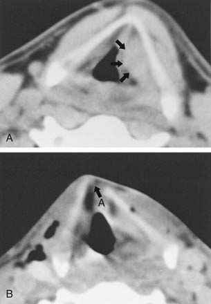
In our practice, multislice computed tomography (CT) (Fig. 70-4) is performed in all patients presenting with “impending airway obstruction” once their airways have been secured, and in those patients with “stable airway” who are found, on flexible nasoendoscopy to have endolaryngeal abnormalities, because imaging may reveal further injuries not readily identifiable with flexible nasoendoscopy. CT scanning can be deferred only in those patients with a history of relatively minor trauma to the neck, no laryngeal tenderness or surgical emphysema, stable airway, and the finding of minimal laryngeal injuries on flexible laryngoscopy. It is important to appreciate that laryngeal trauma is associated with a cervical spine fracture at a rate approaching 10%.4 At the same time, injuries that occur close to the cervical spine are known to be more likely to mask a spinal injury than injuries further away.26 We take the view therefore that all but the most minor of laryngeal injuries constitute a “distracting injury” and mandate cervical spine radiography under National Emergency X-Radiography Utilization Study (NEXUS) criteria (Box 70-1).27 Our practice of using CT to clear the cervical spine irrespective of plain radiography findings is informed by a growing body of evidence demonstrating a much higher sensitivity for CT over plain radiography in diagnosing spinal injuries.28,29 Overall, therefore, detailed radiologic imaging of the larynx is available in the majority of patients with laryngeal trauma both because of primary laryngeal indications and as a by-product of “clearing” of the cervical spine.
Classification
Laryngeal trauma can be classified according the Schaefer-Fuhrman25,30 (Table 70-3) or Lee-Eliashar31 system. We use the Schaefer-Fuhrman system but also formally and separately document the extent of injuries to the laryngeal mucosa and framework, the vibratory mechanism, and the laryngotracheal complex (Table 70-4).
Table 70-3 Schaefer-Fuhrman Classification of Laryngeal Trauma
| Group 1 | Minor endolaryngeal hematomas or lacerations |
| No detectable fracture | |
| Group 2 | Edema, hematoma, minor mucosal disruption without exposed cartilage |
| Nondisplaced fracture | |
| Varying degrees of airway compromise | |
| Group 3 | Massive edema, large mucosal lacerations, exposed cartilage |
| Displaced fracture(s) | |
| Vocal cord immobility | |
| Group 4 | Same as group 3 but more severe with: |
| Severe mucosal disruption | |
| Disruption of the anterior commissure | |
| Unstable fracture, two or more fracture lines | |
| Group 5 | Complete laryngotracheal separation |
Table 70-4 A Systematic Approach to Documenting Laryngotracheal Injuries and Determining the Need for Surgical Intervention
| Laryngeal framework | Stable: |
| Unstable: | |
| Potentially nonviable: | |
| Laryngeal mucosa | Intact/minimally injured: |
| Injured: | |
| Massively injured: | |
| Vibratory apparatus | Intact |
| Injured: | |
| Laryngotracheal junction | Intact |
| Any degree of laryngotracheal separation |
Management
Airway Management
The first priority in managing laryngeal trauma is to assess and secure the airway while protecting the cervical spine. We take a stepwise approach to securing the airway based on information available at the different stages of clinical assessment (Fig. 70-5
Stay updated, free articles. Join our Telegram channel

Full access? Get Clinical Tree


