graduated from Department of Applied Physics, Osaka University, Osaka, Japan in 1982, and received his doctoral degree in applied physics from Osaka University in 1993. In 1989–1994, he was an assistant professor in Department of Applied Physics, Osaka University. He is currently researcher of Miyata Eye Hospital, Miyazaki, and visiting assistant professor of Tokyo Dental Collage Suidobashi Hospital, Tokyo.

17.1 What Is Surface Light Scattering?
Hydrophobic acrylic material has been the majority of intraocular lens (IOL) in recent cataract surgery, since it can be inserted through smaller incisions and the incidence of postoperative capsular opacification (PCO) is lower than other materials. Importance of the postoperative stability such as mechanical fixation within the capsular bag and transparency of the optics has been addressed. Yaguchi et al. first reported increased surface light scattering with a MA60BM (Alcon) that had been implanted over 10 years [1]. Under slit-lamp microscopy observation, strong scatterings are observed on both IOL surfaces. Figure 17.1 shows a typical image of surface light scattering in a pseudophakic eye 10 years postoperatively.
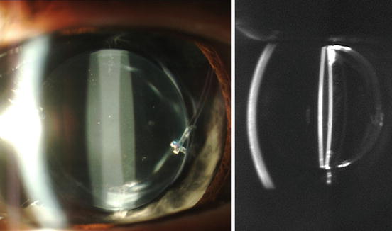
Fig. 17.1
Slit-lamp microscope and Scheimpflug images of an eye with an AcrySof IOL (MA60BM) for 10 years. Intensive light scattering was observed on the IOL surfaces
Besides the surface light scattering, glistenings and optic opacification due to calcification may be observed in a pseudophakic eye. Glistenings are usually observed as bright spots with a size of 1–20 μm under slit-lamp microscope observation and are distributed throughout the entire IOL optic [2]. Glistening and surface light scattering are sometimes confused with one another, but the appearance is different in size and location.
We investigated surface light scattering of four IOLs using a Scheimpflug-camera anterior segment analyzer, EAS-1000 (Nidek Co., Ltd.), up to 3 years postoperatively [3]. Light scattering intensity on the anterior IOL surface was measured with densitometry, since the posterior surface involved the development of PCO. In two IOLs, MA60BM and its 1-piece version SA60AT (Alcon), there was significant increase in surface light scattering after 1–2 years postoperatively. However, other hydrophobic acrylic AR40e and silicone ClariFlex (Abbott Medical Optics, Inc.) did not show such an increase (Fig. 17.2). The results demonstrated that surface light scattering occurred particularly within the AcrySof IOLs.
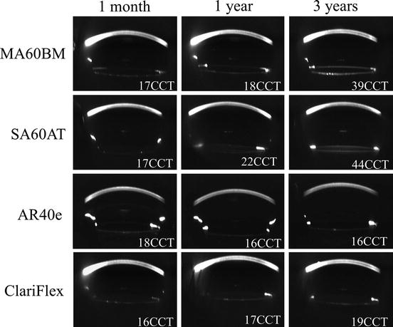
Fig. 17.2
Representative Scheimpflug images with four types of IOLs at 1 month and 1 and 3 years postoperatively. Measurements of light scattering on the anterior IOL surface were denoted. Intensity of surface light scattering increased in AcrySof IOLs. Reprinted from Miyata et al. [3] with permission from B M J Publishing Group
The long-term changes of surface light scattering were assessed by reviewing clinical records of the patient who received AcrySof IOLs [4]. The number of eyes assessed was 406 and the mean follow-up duration was 4.2 years ± 3.4 (SD), ranging 1–15.2 years. Computer compatible tape (CCT) values with AcrySof IOLs showed a continuous increase up to 15 years postoperatively: the increasing rate was 11.5 CCT/year. From the result, influence of increased surface light scattering on the visual function is concerned, that will be discussed in Sect. 17.3.
17.2 Etiology of Surface Light Scatterings
Several years have been spent investigating the etiology of surface light scattering. Explanted AcrySof IOLs had been investigated from many aspects, such as scanning electron microscopy (SEM) with energy-dispersion X-ray analysis and Fourier transform infrared spectroscopy with attenuated total reflectance [5]. However, the etiology could not be identified, since surface light scattering is only observed when the IOL is sufficiently hydrated [6]. It was speculated that the IOL material contains fluid-filled vacuoles in the surface layer when the explanted AcrySof was hydrated. Water aggregates due to this phase separation scattered light on the IOL surface.
Water aggregates located in the surface layer of the hydrated AcrySof IOL were observed using a cryogenic focused ion-beam scanning electron microscopy (FIB-SEM). We observed these water aggregates in an explanted MA60BM that was implanted for 13 years. Half of the IOL was placed in a cryogenic vacuum chamber and sputtered with FIB to create a section below the IOL anterior surface. Immediately after the FIB treatment, sections around the IOL surface layer were observed by SEM. SEM image shown in Fig. 17.3a (magnification of 50,000) revealed the presence of water aggregates within the IOL surface layer. With an unused AcrySof IOL (Fig. 17.3b), SEM images obtained in the same manner did not present such features. Size of the water aggregates observed was in a range of 40–250 nm. The manufacturer confirmed the etiology of the surface light scattering as phase separation of nanometer-ordered water aggregates within the IOL surface layer [5], in which similar FIB-SEM observations of other explanted AcrySof IOLs were presented.
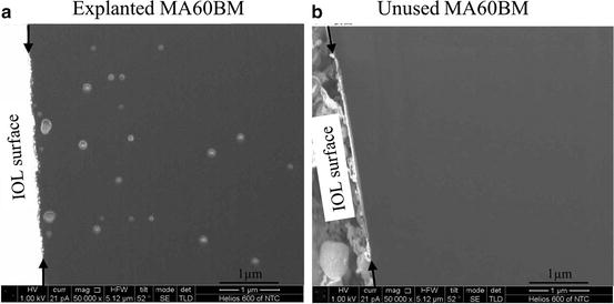
Fig. 17.3
FIB-SEM observations at the surface layer of MA60BM explanted after 13 years (a) and unused (b), with a magnification of 50,000. In the explanted IOL (a), development of nanometer-ordered water aggregates was observed
Compared with glistenings, the location and size are different as illustrated in Fig. 17.4, although both phenomena are caused by development of water aggregates. In surface light scattering, water aggregates are localized below the IOL surface, while glistenings are distributed throughout the entire optic. The sizes range in the nanometer order. Therefore, surface light scattering is also referred to as subsurface nano-glistenings [5]. It is also noted that surface light scattering progressively increased over the years [4], while changes in glistenings are relatively stable after the initial onset [2, 7].