Chapter 9 Imaging the fundus
Von Helmholtz’ invention in 1850 of the first clinical direct ophthalmoscope marked the dawn of modern ophthalmology. Direct ophthalmoscopy and later the development of the binocular indirect ophthalmoscope, the slit-lamp, and a range of high-powered aspheric lenses, enabled the imaging of the human fundus, paving the way for the systematic study of intraocular structures and their diseases by direct observation in vivo.1
Imaging dependent on the state of the media
Confocal scanning laser ophthalmoscopy
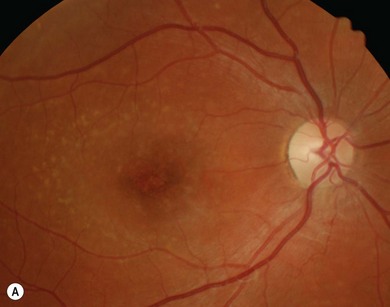
In conventional fundus photography, the entire fundus is flooded with a bright flash to visualize its composite structures. In confocal scanning laser ophthalmoscopy (cSLO), a small focused laser point is rapidly swept across the retina pixel by pixel in a raster pattern. Because imaging is confocal, interfering stray light from adjacent structures is minimized, thereby improving contrast. The use of several laser sources permits the imaging of the retina, RPE, and the optic nerve by different wavelengths according to their respective absorption, reflection, and excitation properties (Fig. 9.1). Confocal imaging also enables in-depth structural analysis of the retina and optic nerve, layer by layer, and three-dimensional digital reconstruction, e.g. in the Heidelberg Retina Tomogram (HRT, Heidelberg Engineering, Heidelberg, Germany). The latest SLOs offer not only facilities for digital fluorescein/indocyanine green angiography, but also autofluorescence, red-free and infrared imaging as well as high-resolution Fourier-domain optical coherence tomography (OCT) all in one machine (Spectralis, Heidelberg Engineering, Heidelberg, Germany).
Autofluorescence
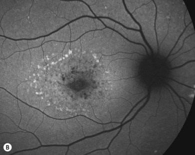
Retinal autofluorescence2 relies primarily on the content of fluorophores in the lipofuscin granules of RPE cells. Therefore, it serves as a non-invasive indicator of the health of the RPE and outer retina: increased autofluorescence indicates abnormal accumulation of lipofuscin in the post-mitotic RPE cell. It therefore serves as an indicator of RPE dysfunction and is seen in a large number of retinal disorders, for instance in Best’s and Stargardt’s disease. Loss of autofluorescence is an indicator of RPE atrophy.
Fluorescein and indocyanine green angiography
Digital SLO angiography provides much greater temporal resolution and detail than is possible with conventional serial photography.3 Unlike in adults, fluorescein (excitation maximum at 490 nm) and indocyanine green (excitation maximum at 805 nm) angiography is uncommonly used in children due to a combination of factors: rarer appropriate pathology and practical concerns of more difficult intravenous access (though oral administration is possible) and administration of intravenous drugs in a child in an eye unit. If angiography in a child is deemed necessary, it must only be carried out with all necessary equipment, drugs, and medical staff trained in pediatric resuscitation available.
Red-free and infrared imaging
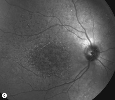
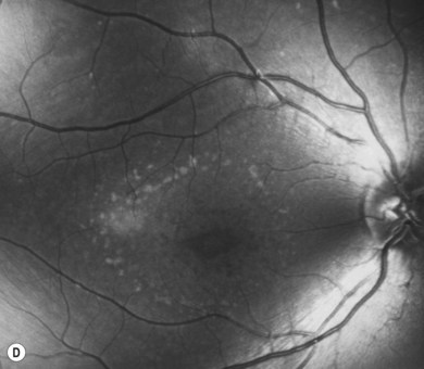
Red-free imaging is particularly useful for highlighting vascular structures and nerve fiber layer defects in the inner retina. Red-free imaging is available with some scanning laser ophthalmoscopes (see Fig. 9.1) and, of course, by using the green filter on the slit-lamp or direct ophthalmoscope. Infrared imaging has been studied in Stargardt’s disease and may play a particular role in visualizing subretinal structures.4,5
Wide-field imaging
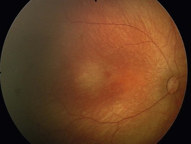
The RetCam system (Clarity Medical, Pleasanton, California, US) provides wide-field imaging of up to 130° (Fig. 9.2). Because it can be used to visualize and document the posterior and much of the peripheral retina, it is common in screening for retinopathy of prematurity and the documentation of non-accidental injuries in babies. In addition to color images, it can also be used for fluorescein angiography. It requires eye contact.
Ultra-wide-field confocal scanning laser
A further technological advance has been the development of ultra-wide-field confocal scanning laser imaging (Optos, Dunfermline, UK). Using an internal parabolic mirror, the scanner can capture up to 200° internal angle (Fig. 9.3
Stay updated, free articles. Join our Telegram channel

Full access? Get Clinical Tree


