Hypoglossal Facial Anastomosis

Chapter 62 Hypoglossal Facial Anastomosis


image Videos corresponding to this chapter are available online at www.expertconsult.com.


Facial nerve paralysis is a debilitating problem cosmetically and functionally. During the course of otologic and neurotologic surgery, sacrifice of the facial nerve is sometimes necessary, and may occur inadvertently, despite meticulous surgical technique. In these cases, one must be prepared to rehabilitate and restore function as much as possible. Direct repair of the injured nerve is currently the best option available to re-establish facial function. If a direct approximation of the nerve ends is impossible, a graft connecting the two ends is the next best choice.


There are situations in which neither of these options is feasible. Perhaps the most common of these involves the extirpation of cerebellopontine angle tumors, in which the facial nerve is severed at the brainstem, and there is no proximal stump present for connection of a graft. There are also cases in which a very attenuated but intact nerve regains no function. In these situations, an alternative to direct repair and nerve grafting is required.


Ideally, facial nerve restoration procedures should provide normal facial tone and symmetry, strong volitional and emotional facial movement, protection of the eye, facilitation of mastication, avoidance of dyskinesias, and no additional motor deficits. Even immediate direct anastomosis cannot fulfill these criteria, however. Several methods to restore some facial function have been developed that require neither direct repair nor grafting. These include cross-facial nerve grafting; nerve/muscle pedicle grafts; and nerve substitutions, such as phrenic, accessory, hypoglossal, and ansa cervicalis.


Connection of a graft with the normal facial nerve followed by redirection of some of these fibers to the paralyzed side is termed cross-facial grafting. This method can provide some symmetry of movement, while avoiding other motor deficits. This procedure partially compromises the normal nerve, however, and provides a scant supply of neural elements to the recipient muscles, leading to inconsistent results.1,2 As a consequence, this procedure has not met with widespread acceptance.3,4 Combinations of cross-facial grafting and traditional nerve substitutions may also be employed.58


Nerve/muscle pedicle grafts such as temporalis muscle transfer have been used with some success, but have also yielded inconsistent results.9 Because the results of nerve/muscle pedicle grafts and cross-facial grafts have been disappointing, modifications of these procedures have been developed that use combination cross-facial grafting and microvascular free muscle flaps for facial reanimation.9,10 More recently, a modification of the traditional temporalis transfer has been used that incorporates the temporalis tendon alone instead of a portion of the muscle.11


Nerve substitution procedures provide a large supply of axons to the recipient muscles, and are generally technically facile. The results are consistent and predictable. The major disadvantages include the lack of emotional facial function and the donor nerve deficit. Because of the close relationship of tongue movement to facial movement, the hypoglossal/facial (CN XII/VII) anastomosis has proved to be a useful procedure in cases of facial paralysis in which a direct repair or graft is impossible. This procedure produces varying degrees of tongue dysfunction, based on the amount of hypoglossal nerve donated for transposition to the facial nerve. Newer techniques have been designed with the goal of preserving tongue function. This chapter focuses on the hypoglossal/facial anastomosis.


PATIENT SELECTION


Patients with facial paralysis must receive a detailed evaluation to determine the etiology of the paralysis. In some cases, the cause is obvious, as in resection of the nerve in the course of removal of a neoplasm. The evaluation of facial palsy of unknown cause is beyond the scope of this chapter. Careful evaluation should precede any reanimation procedure, however, to avoid missing treatable disease, and to avoid destruction of a nerve that has potential for return of function.


In cases of known facial nerve discontinuity in which direct repair or grafting is impossible, the CN XII/VII anastomosis should be performed as soon as reasonably possible. Muscle atrophy and degeneration proceed rapidly after denervation.12 Early repair provides axonal growth to the muscles and limits the amount of muscle degeneration.


The severed nerve also begins to experience fibrosis.13,14 In early anastomoses, new axons fill the nerve sheath before fibrosis and potentially allow a greater supply of axons to the muscles. Although earlier anastomosis gives a better functional result, the CN XII/VII anastomosis is also effective after a prolonged denervation and should be considered up to 2.5 years after injury.4,15 Return of function can occur 4.5 years after injury.16


In other patients in whom the continuity of the nerve is in question, including patients who sustain trauma, have idiopathic palsy, and have nerves damaged in surgery, it is prudent to wait at least 1 year to ensure that no return of function occurs. Electrophysiologic testing is helpful in determining the innervation and viability of facial muscles. A positive response to electroneuronography or evoked electromyography indicates that at least some motor end plates are functional. These patients should be given the longest possible time to show improvement in function. Some of these patients have so few remaining neural elements, however, that they never regain any useful function. In this situation, a CN XII/VII graft helps provide a sufficient amount of neuronal input to the muscles.


Electromyography helps detect polyphasic action potentials indicative of reinnervation, and fibrillation potentials indicative of denervation. In cases of long-standing paralysis (>2.5 years), a muscle biopsy in addition to electromyography may be useful to determine viability, atrophy, and fibrosis. In cases of severe muscle atrophy and neural fibrosis, the results of any reinnervation procedure would be poor, and muscle transfers and other augmenting procedures should be considered.


The clinician should also consider the status of the contralateral twelfth cranial nerve when deciding on the CN XII/VII transposition. Contralateral hypoglossal paralysis is a contraindication to the standard CN XII/VII transposition, as are multiple lower cranial nerve deficits that already compromise swallowing and speech.


SURGICAL TECHNIQUE


In addition to standard head and neck surgical instrumentation, hypoglossal facial anastomosis requires jeweler’s forceps to handle the nerve ends, a Castroviejo needle holder, and microforceps for knot tying. A sterile tongue blade is useful to improve visibility when the ends of the nerves are anastomosed under microscopic vision.


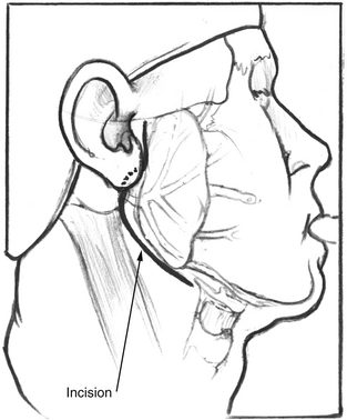
After satisfactory general endotracheal anesthesia has been obtained with the patient in the supine position, the neck is extended, and the face is turned toward the side opposite the paralysis. The ear, face, and neck are prepared and draped using sterile technique. A standard lazy S parotidectomy incision is made in the preauricular crease and extended behind the lobule and then anteriorly about 2 cm below the angle of the mandible (Fig. 62-1). In cases where the patient has had a prior postauricular incision for craniotomy, the postauricular incision may also be used for the superior aspect of the incision. This is particularly true if a mobilization of the facial nerve out of the fallopian canal is planned.


Skin flaps are raised anteriorly and posteriorly. The parotid is mobilized from the anterior border of the sternocleidomastoid muscle and from the external auditory canal. The angle formed by the cartilage of the anterior external canal, known as the tragal pointer, is followed medially to the stylomastoid foramen, where the facial nerve exits the temporal bone. The nerve is dissected from the parotid gland to expose the pes anserinus and free the main trunk from the gland. The nerve is transected at the stylomastoid foramen.


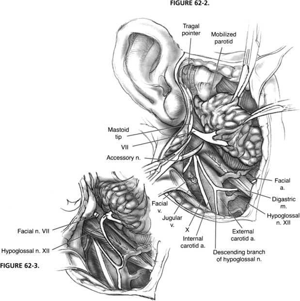
The hypoglossal nerve is identified by retracting the sternocleidomastoid muscle posteriorly and exposing the great vessels of the neck. The posterior belly of the digastric muscle is retracted superiorly, and the hypoglossal nerve is found coursing inferiorly with the great vessels and then turning anteriorly as it supplies the ansa cervicalis, which descends in the carotid sheath (Fig. 62-2). The hypoglossal nerve is followed anteriorly and medially as it enters the tongue muscle. The nerve is freed from its fascial attachments in the neck. The network of veins and arteries entering the internal jugular vein and external carotid artery should be controlled during this maneuver. After the nerve is freed from its attachments, it is divided as far anteriorly as is possible to gain sufficient length. The free hypoglossal nerve is rotated superiorly. Directing the nerve medial to the digastric muscle in this rotation gives the most length, but is unnecessary for a satisfactory anastomosis.


There are many ways to anastomose the ends of the nerves, including collagen trays and fibrin glue, vein sheaths, laser welding, and various suture techniques. It is beyond the scope of this chapter to describe the various methods; however, several principles are almost universally agreed on. The two ends of the nerves should be free of all tension. This requirement is usually not a problem if the technique described earlier is followed. The ends should be cut sharply to provide a flush connection. The anastomosis should be as atraumatic as possible, yet provide strength to prevent disruption. It is also important to ensure, using frozen section histologic evaluation, that the distal facial nerve has not totally fibrosed in cases of long-standing paralysis. A conventional suture technique that yields reliable results is as follows:

Stay updated, free articles. Join our Telegram channel

Jun 14, 2016 | Posted by in OTOLARYNGOLOGY | Comments Off on Hypoglossal Facial Anastomosis

Full access? Get Clinical Tree

Get Clinical Tree app for offline access