Hematopoietic and Lymphoid Disorders
Abundant mucosa-associated lymphoid tissue is present within the upper aerodigestive tract, which can be involved in both nonneoplastic proliferations and malignancies.1,2,3,4,5,6,7,8,9,10,11,12,13,14,15,16,17,18,19,20,21,22,23,24 and 25 The remaining tract may be involved in hematopoietic and lymphoid lesions that are more specific to certain sites (e.g., the sinonasal tract and extranodal NK/T-cell lymphomas), while the entire tract can be involved in other processes (e.g., extramedullary plasmacytomas or diffuse large B-cell lymphoma). This chapter methodically discusses the hematopoietic and lymphoid disorders of this area, first discussing the benign proliferations and then discussing the malignant entities. Those tumors that occur most frequently throughout these sites will be discussed in more detail.
Ancillary studies, including immunohistochemistry, flow cytometry, cytogenetics, and molecular studies that test for immunoglobulin or T-cell receptor rearrangements or various fusion transcripts, are now extremely important for the correct diagnosis of these lesions. Thus, the better one is prepared at the time of biopsy, the better the tissue will be triaged and the more likely one will be able to make a concise and correct diagnosis. Unfortunately, because these diagnoses are often unsuspected, many must be rendered using formalin-fixed tissue and immunohistochemistry.
NONNEOPLASTIC AND BENIGN PROLIFERATIONS
Most benign lymphoid proliferations of the upper aerodigestive tract occur within Waldeyer’s ring, most often in children. Because of obstructive symptoms or recurrent infections, the lymphoid tissue from this area is sometimes removed via tonsillectomy and adenoidectomy. Most specimens from these procedures will show a combination of follicular and interfollicular hyperplasia (Fig. 9.1, e-Figs. 9.1 and 9.2).26 The follicles will be enlarged and somewhat variable in size, with well-formed germinal
centers and mantle zones that typically are “zonated.” The reactive germinal centers will contain abundant mitotic figures and tingible body macrophages. Some degree of interfollicular expansion is also usually present and can show variable changes. Generally, the paracortex and sinuses will have mixed smaller and larger lymphocytes with macrophages, immunoblasts, plasma cells, and acute inflammatory cells. Sometimes the overall growth pattern and particular cell types present allow for the identification of the cause of hyperplasia. Certain reactive conditions can be clinically and histologically worrisome for malignancy.
centers and mantle zones that typically are “zonated.” The reactive germinal centers will contain abundant mitotic figures and tingible body macrophages. Some degree of interfollicular expansion is also usually present and can show variable changes. Generally, the paracortex and sinuses will have mixed smaller and larger lymphocytes with macrophages, immunoblasts, plasma cells, and acute inflammatory cells. Sometimes the overall growth pattern and particular cell types present allow for the identification of the cause of hyperplasia. Certain reactive conditions can be clinically and histologically worrisome for malignancy.
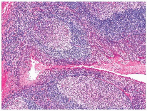 FIGURE 9.1 Follicular and interfollicular hyperplasia. |
Infectious mononucleosis secondary to Epstein-Barr virus (EBV) infection can be associated with massive tonsillar enlargement.5,18,27 Indeed, the portal of entry for this virus is generally believed to be Waldeyer’s ring. Histologically, some degree of follicular hyperplasia is usually seen, together with marked interfollicular expansion by numerous immunoblasts, lymphocytes, and plasma cells (Fig. 9.2). The immunoblasts can be particularly prominent and may surround focal areas of necrosis (Fig. 9.3, e-Figs. 9.3 and 9.4). Some immunoblasts may have multiple nuclei and mimic Reed-Sternberg cells, forcing one to consider a diagnosis of interfollicular involvement by Hodgkin lymphoma.5,18,27 Immunohistochemistry can be helpful here, as immunoblasts should typically react with antibodies to CD20 and not with antibodies to CD15. Confusing the matter, however, is the fact that immunoblasts frequently react with antibodies to CD30 and background granulocytes may react with antibodies to CD15.28 In situ hybridization or immunohistochemistry demonstrating infection due to EBV is not necessarily helpful, as lymphomas also frequently show infection.18,29
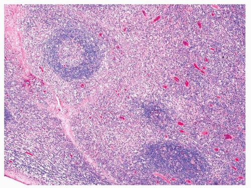 FIGURE 9.2 Marked interfollicular expansion seen in a case of infectious mononucleosis. |
Although infection due to human immunodeficiency virus (HIV) puts patients at a much increased risk for lymphoma, benign HIVinduced hyperplasias are also common. While these lesions may occur in lymph nodes at any site, they can also develop in Waldeyer’s ring.23 Here, they may even represent the initial manifestation of HIV infection of the
patient. These lesions are histologically characterized by florid follicular hyperplasia with markedly enlarged germinal centers that are irregular in shape or “serpentine” (Fig. 9.4). Follicular fragmentation or lysis may be noted, as smaller lymphocytes extend into the germinal centers and give them a somewhat “moth-eaten” appearance. Interfollicular expansion can be present due to numerous monocytoid lymphocytes, immunoblasts, and plasma cells. Interfollicular hemorrhage is also often noted, as are multinucleated giant cells (Fig. 9.5). The giant cells can be located anywhere throughout the tissue but are frequently noted just adjacent to the surface epithelium. These cells have multiple medium-sized nuclei that are usually located at the periphery of the cell. It has been noted that these giant cells are less commonly seen in advanced disease.
patient. These lesions are histologically characterized by florid follicular hyperplasia with markedly enlarged germinal centers that are irregular in shape or “serpentine” (Fig. 9.4). Follicular fragmentation or lysis may be noted, as smaller lymphocytes extend into the germinal centers and give them a somewhat “moth-eaten” appearance. Interfollicular expansion can be present due to numerous monocytoid lymphocytes, immunoblasts, and plasma cells. Interfollicular hemorrhage is also often noted, as are multinucleated giant cells (Fig. 9.5). The giant cells can be located anywhere throughout the tissue but are frequently noted just adjacent to the surface epithelium. These cells have multiple medium-sized nuclei that are usually located at the periphery of the cell. It has been noted that these giant cells are less commonly seen in advanced disease.
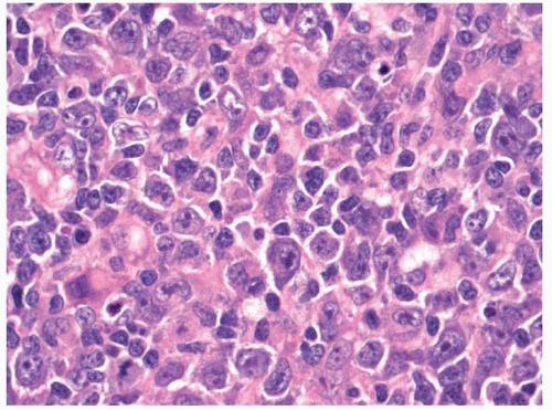 FIGURE 9.3 A proliferation of immunoblasts seen in a case of infectious mononucleosis. |
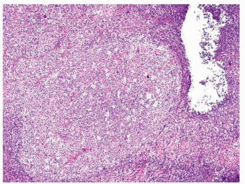 FIGURE 9.4 Marked follicular hyperplasia seen in a patient infected with human immunodeficiency virus. |
Sinus histiocytosis with massive lymphadenopathy is believed to be a reactive condition that may involve lymph nodes or extranodal sites.30,31 The disease may affect individuals at any age; however, it frequently develops in young patients and the mean reported age is around 20 years. Although it affects all races, it is more common in blacks. Sinus histiocytosis with massive lymphadenopathy most commonly presents with cervical lymphadenopathy; however, extranodal sites in the head and neck can be involved either with or without concomitant adenopathy.7,22 In the upper aerodigestive tract, the disease often involves Waldeyer’s ring, but it can also involve other sites, especially the sinonasal area. Symptoms are nonspecific and include fever, obstruction, epistaxis, and changes secondary to mass effect. Although the disease
is benign and nonneoplastic, it frequently recurs after excision either locally or at other sites. Patients rarely die of the disease or because of its treatment.
is benign and nonneoplastic, it frequently recurs after excision either locally or at other sites. Patients rarely die of the disease or because of its treatment.
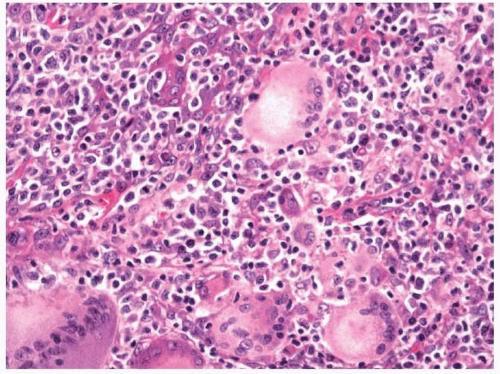 FIGURE 9.5 Multinucleated giant cells can be found in the reactive lymph nodes of patients infected with human immunodeficiency virus. |
Histologically, sinus histiocytosis with massive lymphadenopathy has a biphasic appearance, with darker areas composed mostly of lymphocytes and paler areas composed of histiocytes intermixed with lymphocytes and plasma cells (Fig. 9.6, e-Fig. 9.5).7,22,30 The process diffusely involves subepithelial tissues, while the overlying epithelia and underlying seromucinous glands remain intact. Fibrotic bands can be prominent. The lymphoid areas are composed of small, mature lymphocytes with occasional histiocytes. Germinal center formation is not observed within these areas. The histiocytes, generally better viewed in the less cellular areas, have round to oval nuclei with vesicular chromatin and abundant amphiphilic to eosinophilic, granular to foamy cytoplasm. Prominent emperipolesis characterizes this process, and the phagocytized cells can be lymphocytes, plasma cells, erythrocytes, or neutrophils (Fig. 9.7, e-Fig. 9.6). The process can extend into surrounding soft tissue and bone. If special stains are performed, organisms should not be identified.
A process termed atypical marginal zone hyperplasia of mucosaassociated lymphoid tissue has been described, which primarily affects Waldeyer’s ring in children.1 The affected children presented with unilateral or bilateral tonsillar enlargement. Histologically, these cases were characterized by follicular hyperplasia with expansion of the marginal
zones that can extend into the germinal centers. The expanded marginal zones showed light-chain restriction by immunohistochemistry; however, clonality was not found by molecular methods. The few cases reported received no additional therapy after the initial resection, and all patients remained free of lymphoma during the follow-up period.
zones that can extend into the germinal centers. The expanded marginal zones showed light-chain restriction by immunohistochemistry; however, clonality was not found by molecular methods. The few cases reported received no additional therapy after the initial resection, and all patients remained free of lymphoma during the follow-up period.
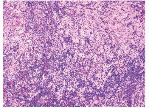 FIGURE 9.6 Sinus histiocytosis with massive lymphadenopathy. |
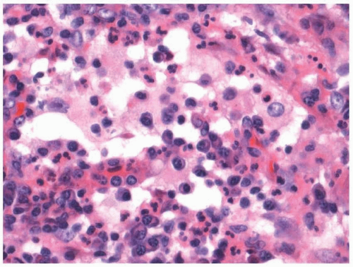 FIGURE 9.7 Marked emperipolesis seen in a case of sinus histiocytosis with massive lymphadenopathy. |
MYELOID MALIGNANCIES
Myeloid sarcomas (extramedullary myeloid tumors, chloromas, etc.) can present anywhere throughout the body and have been noted to involve the upper aerodigestive tract.32,33,34,35,36,37,38 and 39 When they do involve this location, they most commonly affect the oral tissue or mucosal lymphoid tissue of Waldeyer’s ring and are associated with cervical lymphadenopathy. These lesions may be associated with acute myeloid leukemias, myeloproliferative disorders, or myelodysplastic syndromes. Rarely, myeloid sarcomas can occur as isolated lesions. Patients with myeloid sarcomas fare poorly and in some cases the lesions may signify a blast transformation of a preexistent myeloproliferative disorder and myelodysplastic syndrome. When the lesions are isolated, however, they can be associated with good outcomes.32
Myeloid sarcomas are infiltrative lesions that histologically display a sheetlike growth pattern (Figs. 9.8 and 9.9).32,34,36 Neoplastic cells show varying degrees of differentiation and phenotype. Some tumors will be immature and show predominantly myeloblasts, large, atypical cells with scanty cytoplasm, round to slightly convoluted nuclei, delicate chromatin, and prominent nuclei (e-Fig. 9.7). Other cases show more granulocytic or monocytic differentiation. Auer rods are seen in some cases.
Outside of a setting of known hematopoietic neoplasia, the diagnosis of a myeloid sarcoma can be difficult, mostly because it is not considered. Immunohistochemistry is generally needed to confirm a diagnosis and neoplastic cells will react with antibodies to myeloperoxidase (e-Fig. 9.8).14,35,36 Other antigens expressed by the immature myelocytes and blasts can be identified by flow cytometry or immunohistochemistry and include CD13, CD33, and CD117.38 Immunoreactivity with antibodies
to CD34 can be seen in the blasts. Frequently, neoplasms will show immunoreactivity with antibodies to lysozyme and CD68. Cytogenetic abnormalities typical of myeloid neoplasia can be identified in many cases.37
to CD34 can be seen in the blasts. Frequently, neoplasms will show immunoreactivity with antibodies to lysozyme and CD68. Cytogenetic abnormalities typical of myeloid neoplasia can be identified in many cases.37
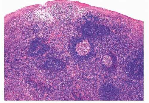 FIGURE 9.8 Partial tonsillar effacement by a myeloid tumor. |
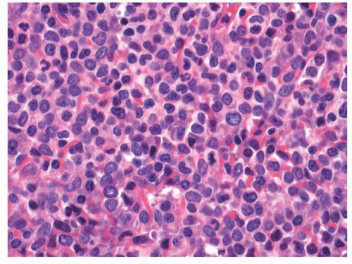 FIGURE 9.9 Sheets of myeloblasts found with a myeloid tumor. |
Precursor B-Cell and T-Cell Malignancies
The most common hematolymphoid malignancies that involve Waldeyer’s ring in children are precursor B-lymphoblastic and T-lymphoblastic lymphomas and Burkitt lymphomas.40 As most precursor B-cell neoplasms also involve the bone marrow and blood, they are usually diagnosed as acute leukemias. Also, although less common, adults can be affected by both precursor B-cell and T-cell malignancies.
Patients with leukemia generally present with systemic symptoms secondary to bone marrow replacement. These include symptoms secondary to anemia, leukopenia, and thrombocytopenia. Patients with involvement of Waldeyer’s ring often have involvement of other lymph nodes and more widespread adenopathy. Symptoms secondary to a pharyngeal mass can also be noted, and patients can have speech, eating, or breathing difficulties. Although patients without bone marrow and blood involvement usually present secondary to more widespread adenopathy, occasional patients will present with symptoms secondary to unilateral tonsillar enlargement. Indeed, although reactive hyperplasia is the major cause for the removal of tonsils in children, most of us have encountered the rare tonsillectomy specimen that is found “incidentally” to contain lymphoblastic lymphoma. We have seen cases submitted for “gross examination only” that were later found to have lymphoblastic lymphoma when the patient presented with more widespread adenopathy.
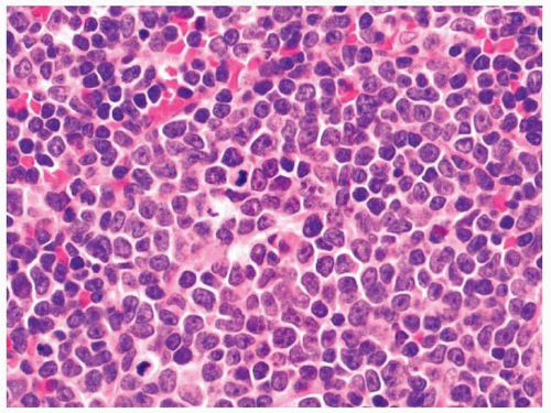 FIGURE 9.10 Sheets of lymphoblasts seen in a case of tonsillar lymphoblastic lymphoma. |
The tonsillar tissue is effaced by sheets of lymphoblasts, although some cases may show only partial nodal involvement (Fig. 9.10).41 The blasts are characteristically small sized to medium sized and have only a small amount of amphiphilic cytoplasm (e-Fig. 9.9). The nuclear contours can vary from smooth to convoluted and the chromatin usually appears fine. Prominent nucleoli may be noted with many mitotic figures. Some degree of maturation is sometimes seen, and some of the lymphocytes can appear smaller, with more condensed chromatin and less prominent nucleoli. Numerous tingible body macrophages can be present, reminiscent of a Burkitt lymphoma.
Antigen expression in precursor B-cell and T-cell leukemias/lymphomas depends on the lineage and maturity of the neoplastic cells. Lesions not involving the bone marrow and blood are more likely to have a precursor T-cell phenotype and express CD3 (cytoplasmic) with variable expression of other T-cell antigens such as CD2, CD4, CD5, CD7, and CD8.41,42 The cells should also express terminal deoxynucleotidyl transferase (TdT) and, often, CD10 (e-Figs. 9.10 and 9.11). Markers of myeloid or B-cell differentiation can also be present and do not necessarily exclude a diagnosis of precursor T-cell lymphoma/leukemia. Lesions that also have bone marrow and blood involvement are more likely to have a precursor B-cell phenotype and will express B-cell antigens such as CD19 and CD79a and others such as CD20 and CD22. The neoplastic blasts should also express TdT and will frequently express CD10. Although these tumors can be diagnosed with immunohistochemistry, flow cytometry is generally the method of choice for assessing antigen expression.
Cytogenetic analysis is very important with these tumors and provides prognostic information that may influence therapy.41 This is
especially true with precursor B-cell malignancies and certain translocations, such as t(9;22)(q34;q11.2), t(4;11)(q21;q23), and t(1;19)(q23;p13.3), are considered “unfavorable,” whereas others, such as t(12;21)(p12;q22), are considered “favorable.” Cases with a hyperdiploid karyotype are also considered “favorable,” whereas those that are hypodiploid are considered “unfavorable.”
especially true with precursor B-cell malignancies and certain translocations, such as t(9;22)(q34;q11.2), t(4;11)(q21;q23), and t(1;19)(q23;p13.3), are considered “unfavorable,” whereas others, such as t(12;21)(p12;q22), are considered “favorable.” Cases with a hyperdiploid karyotype are also considered “favorable,” whereas those that are hypodiploid are considered “unfavorable.”
Mature B-Cell Malignancies
Most types of mature B-cell neoplasia have been noted to involve the upper aerodigestive tract (Table 9.1).3,4,14,15,16 and 17,19,21,24,40,43,44,45,46,47,48,49,50,51,52,53,54 and 55 These tumors, especially the lower grade lesions, primarily affect Waldeyer’s ring. Some, such as diffuse large B-cell lymphomas, can involve both mucosa-associated lymphoid tissue and other sites, while others, such as extramedullary plasmacytomas, often occur outside of Waldeyer’s ring. Of the lesions involving Waldeyer’s ring, diffuse large B-cell lymphomas are the most common, followed by mantle cell lymphomas and follicular lymphomas. Nearly all mature B-cell lymphomas have been noted to involve Waldeyer’s ring, however. Of the mature B-cell malignancies involving the other sites, diffuse large B-cell lymphomas are again the most common malignancies. When involving Waldeyer’s ring, many of these lymphomas efface the normal lymphoid tissue; however, each may only partially involve the tissue. As with other hematolymphoid malignancies, ancillary studies are often necessary for diagnosis. Many of these malignancies can be properly diagnosed with immunohistochemistry; however, flow cytometry is very helpful. Molecular diagnostic studies and cytogenetic analysis can be useful, as many of these tumors show recurrent cytogenetic abnormalities. Finally, immunoglobulin and T-cell receptor gene rearrangement studies can be helpful with very small specimens when a definitive diagnosis of lymphoma cannot be made for one reason or another.
Diffuse large B-cell lymphoma is the most common type of non-Hodgkin lymphoma of adults in Western countries.56,57 Although the lymphoma most frequently occurs in older individuals, it can develop in patients of any age. This lymphoma generally presents as a rapidly growing mass and is usually considered to be primary; that is, there is no clinical knowledge of previous lower grade lymphomas. Sometimes, though, the tumor evolves from lower grade malignancies, such as chronic lymphocytic leukemia/small lymphocytic lymphoma (CLL/SLL), follicular lymphoma, or extranodal marginal zone lymphoma.
Histologically, diffuse large B-cell lymphomas have “large” neoplastic cells intermixed with smaller, more mature, normal-appearing lymphocytes (Fig. 9.11, e-Fig. 9.12).57 In general, the larger, neoplastic cells predominate; however, in some cases they may be few in number and can even be hard to identify, as they are obscured by numerous, small T cells (T-cell rich variant of diffuse large B-cell lymphoma). The tumor nuclei are at least two times the size of a resting mature lymphocyte. They can have scant to moderate amounts of basophilic cytoplasm and
have pleomorphic nuclei with irregular contours and vesicular chromatin (Fig. 9.12, e-Fig. 9.13). Some cases have cells that appear more centroblastic, with more scanty cytoplasm and two to four nucleoli that are often associated with the nuclear membrane. Other cases have cells that appear more immunoblastic, with moderate amounts of cytoplasm and single,
centrally located nucleoli. Regardless of the appearance, mitotic figures are usually easy to identify. Prominent background sclerosis can sometimes be present. Rarely, background necrosis may be noted.
| ||||||||||||||||||||||||||||||||||


