General Considerations of Anesthesia and Management of the Difficult Airway

CHAPTER 9 General Considerations of Anesthesia and Management of the Difficult Airway


Airway management is the essence of clinical anesthesiology. Complex airway management of the patient with an anticipated difficult airway/intubation or the patient with an unanticipated difficult airway/intubation is a multispecialty process that involves anesthesiologists, surgeons, pulmonologists, critical care physicians, emergency physicians, and nursing/technician staff support.


The OLHN surgeon with expertise in rigid laryngoscopy and bronchoscopy, flexible fiberoptic bronchoscopy, and surgical approaches to the airway is uniquely qualified to take the lead surgical role in a team approach with the anesthesiologist to safely manage difficult airway/intubation patients. The goal of airway management is simple: to provide the most expeditious form of management that has the lowest potential for injury and the greatest potential for control of the airway.1


The approach to the patient with a difficult airway varies, depending on whether management is elective or urgent and whether the health care setting is an operating room or a non–operating room environment.


This chapter presents the anesthetic component of complex airway management in a manner that OLHN surgeons can incorporate into their practice. The focus of this chapter is on clinical airway algorithms and the decisions that must be made by the anesthesiologist together with the OLHN surgeon to safely provide anesthesia and airway management for their patient or for a non-OLHN patient seen in joint consultation for airway management. In-depth discussions about the pharmacology and physiology of anesthesiology are beyond the scope of this chapter but can be found in any number of authoritative textbooks of anesthesiology.2,3


Difficult Airway/Intubation: A Multispecialty Problem


Complex airway management is a multifaceted problem involving health care providers in a variety of clinical settings. The consequences of failed airway maintenance, endotracheal intubation, or both, can be devastating to the patient, the practitioner, and the health care system. Critical issues include identification of difficult airway/intubation patients, mobilization of physician and support staff, mobilization of airway management equipment, preparation of the patient, implementation of appropriate airway algorithms, documentation of airway management techniques, dissemination of critical airway information to future health care providers, and quality improvement/medicolegal considerations.


Patient Identification


Controversy regarding predictors and definitions of “difficult” exists in both intraspecialty and interspecialty, dependent and independent of practitioner skill, related to specific techniques, and complicated by changing patient pathophysiology.4 Some patients may be anticipated to be difficult to intubate on the basis of a history of difficulty intubation or clinical predictors of difficult intubation. The ASA Practice Guidelines for Management of the Difficult Airway5 reviews some of the historical or physical examination findings possibly suggestive of a difficult intubation. Some of these predictors of anticipated difficulty with conventional direct laryngoscopy (MAC/Miller) include a large overbite, large tongue, narrow mouth opening, or short chin. Various prediction models, such as correlation with Mallampati oral view I to IV to the Cormack and Lehand laryngoscopic view grades I to IV have been proposed, but none offer 100% sensitivity for prediction of a difficult airway (Figs. 9-1 to 9-4).6 Historically, anesthesiology literature cites an incidence of 1% to 3% for unanticipated difficult airway/intubation in patients undergoing general endotracheal anesthesia.57 The airway management technique used to define “difficult” in this literature was conventional rigid laryngoscopy (Macintosh or Miller blades).


image

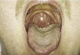
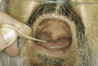
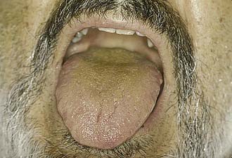
Figure 9-4. Postoperative evaluation of patient in Figure 9-3. Note that without the tongue blade, the patient has a Mallampati class IV airway and should have been considered to be anticipated difficult intubation with conventional laryngoscopy.


Despite advances in airway management techniques and refinement of difficulty predictors, the cited 1% to 3% incidence of unanticipated difficulty has not changed and is still defined by conventional laryngoscopy.69 In an institution where approximately 25,000 general endotracheal anesthetic procedures are done annually, there are potentially 250 to 750 unanticipated difficult airway/intubations per year. Assuming that a full-time practicing anesthesiologist would encounter one unanticipated difficult airway/intubation per year, then, based on ASA membership (which represents 90% of practicing anesthesiologists), there are potentially 30,000 to 90,000 unanticipated difficult airway/intubations annually in the United States. However, these numbers may underestimate the true incidence, because anesthesiologists may not recall the more common “near misses” as vividly as they recall the smaller number of actual difficult airway/intubations in which the outcome was suboptimal. On a national and international level, the scope of this problem and its impact on patients, practitioners, and the health care system is sufficient to warrant vigorous efforts to identify and implement solutions.


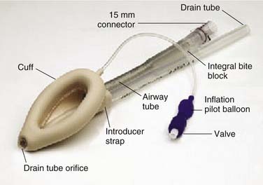
In addition to those patients who have unanticipated difficult airway/intubations on initial presentation, there are cohorts of patients who have anticipated complex airway management; these patients can be successfully managed by a variety of innovative and specialty-specific techniques. Some of these techniques (laryngeal mask airway [LMA]10 and Combitube)11 are readily available (Figs. 9-5 and 9-6), require minimal practitioner education or training, and are inexpensive; other techniques (fiberoptic bronchoscope, surgical airway, specialized rigid laryngoscopes, and fluoroscopic-assisted intubation) are available primarily in specialty centers, may require extensive practitioner skill, and may be relatively expensive. For patients who have undergone head and neck surgery and have visible or hidden implants (e.g., laryngeal stents, thyroplasties), specific considerations for airway management may be unknown to future providers (e.g., thyroplasty patients might require smaller endotracheal tubes than anticipated), thus compromising patient safety and increasing practitioner risk for adverse events.


Successful future management of previously unanticipated difficult airway/intubation patients depends on identification of those patients, and documentation and dissemination of information detailing successful and unsuccessful airway management techniques and primary difficulties encountered. These patients then become anticipated difficult airway/intubations, and the availability of this information promotes quality and safety of care.


Consequences of Difficult Airway Management


The consequences of difficult airway/intubation (with or without an adverse outcome) may be as unsettling as the event itself. The patient may perceive this as a threat to future anesthetic safety or may lack understanding as to the significance of the difficulty. There may be practitioner-perceived threat to professional security. The impact of complex airway management-related events in direct and indirect costs to the health care system is far reaching.


Three studies specifically demonstrated the consequences of difficult airway/intubation management on liability exposure.1214 An analysis of approximately 5000 claims filed in the Maryland legal system over a 15-year period in which one or more anesthesiologists were named as defendants revealed that insertion of an endotracheal tube was the sixth most common medical procedure leading to a liability claim. Most of these claims also included other members of the operating room team (e.g., otolaryngologists, general surgeons, nurse anesthetists, orthopedic surgeons, plastic surgeons, cardiac surgeons, dentists, or nurses) as defendants. One claim (in 1994) resulted in a jury award of $5 million.15 In a 1992 loss analysis study conducted by the Physicians Insurers Association of America (PIAA), files from 43 physician-owned malpractice insurance companies (representing approximately 2000 anesthesiologists nationally) ranked “intubation problems” as the third most prevalent misadventure (behind “tooth injury” and “no medical misadventures”). The average paid indemnity for 175 of 339 files was $196,958.15 The ASA Committee on Professional Liability closed claims study found that respiratory events were the most common cause of brain damage and death during anesthesia, with difficult intubation being the likeliest category for risk reduction. The median payment for respiratory claims was $200,000.12


To put these statistics in perspective, the following must be considered.


Advances in Monitoring


As a previous section made clear, there are serious legal and financial consequences of failed airway management. Fortunately, adverse outcomes related to poor airway management have decreased in frequency with the standardization of anesthetic monitoring. The American Society of Anesthesiology Standards for Basic Anesthetic Monitoring statement was first released in 1986 and further amended in 1998 and 2005.17 These standards call for continual evaluation of the patient’s oxygenation, ventilation, circulation, and temperature during all administered anesthetics. Practically speaking, the standard mandates continuous oxygen analysis of the anesthetic circuit, pulse oximetry, end-tidal capnometry, tidal volume measurement, electrocardiography, and temperature, as well as intermittent (no less frequent than every 5 minutes) measurement of arterial blood pressure and heart rate. In addition, the routine use of the neuromuscular blockade (NMB) monitor (also called the “twitch” monitor) to assess degree of muscle paralysis and return of muscle strength after pharmacological reversal of paralyzing agents significantly contributed to improved patient safety. Compared with earlier eras, this document made the important leap of elevating pulse oximetry and capnometry to standards of care, thus allowing more rapid, accurate recognition of oxygen desaturation and rapid recognition of previously unrecognized esophageal intubation.


The consequences of this change were staggering. Respiratory system adverse events (including airway mishaps) accounted for 36% of claims in the ASA Closed Claims Project data set for the 1970s, but this percentage decreased to 14% of claims in the 1990s after implementation of the routine use of capnometry and pulse oximetry.18 Claims related to death or brain injury likewise had a similar drop in the percentage that was attributable to respiratory adverse outcomes. Caused in part by continuous pulse oximetry and capnometry, anesthesiology has been a leader in the patient safety movement in the past decade.


Standard Anesthetic Pharmacology


This section provides a brief introduction to the array of drugs used by anesthesiologists to facilitate anesthesia and maintain control of the airway. An in-depth discussion of these agents is not within the scope of this chapter; however, certain features of these drugs that are particularly applicable to airway management are highlighted.


Induction Agents and Volatile Anesthetics


In most adult patients, a peripheral intravenous line is started preoperatively to administer fluid and drugs. Therefore, most adult patients receive an intravenous induction of anesthesia. The most commonly used intravenous induction agents are thiopental, propofol, etomidate, and ketamine. Each of the intravenous induction agents has the advantage of quick onset, producing unconsciousness within 1 to 2 minutes when given in standard induction doses. Of note, thiopental and propofol are both associated with negative inotropic effects and a related drop in blood pressure when used for induction of general anesthesia. Etomidate’s effects are considered to be more hemodynamically stable, but this drug has the drawbacks of potential adrenal suppression and myoclonic activity. Thiopental, propofol, and etomidate all produce apnea along with unconsciousness. In contrast to the other three agents, ketamine is notable for not producing apnea with administration. The maintenance of spontaneous ventilation with ketamine is an important consideration in the management of patients with potentially difficult airways and other subsets of patients presenting for surgery. In addition, ketamine has the advantage of being able to be given intramuscularly in patients without intravenous access. The major disadvantages of ketamine include emergence delirium and the sympathomimetic effects of the drug, and thus can cause tachycardia and hypertension, which limit its role in the treatment of patients with cardiovascular disease. Additionally, ketamine produces exaggerated secretions, and an antisialogue such as glycopyrrolate should be coadministered if the airway is not secured immediately after induction.


For most adults, the volatile anesthetic agents (e.g., halothane, isoflurane, sevoflurane, and desflurane) are used for maintenance of anesthesia rather than induction. Their role in induction of anesthesia is limited by slower onset of activity and patient inability to tolerate the scent of the anesthetic gas. However, in pediatric anesthesia, wherein most patients are intolerant of intravenous placement before coming to the operating room, “mask inductions” with volatile agents is common. Sevoflurane is typically the agent used for mask induction because it is considered less noxious than the other agents. All of the volatile agents have the significant advantage of maintaining spontaneous respiration while producing unconsciousness.


Adjuncts to Induction: Sedatives and Opioids


Benzodiazepines are also commonly given in the immediate preoperative period for their anxiolytic and amnestic effects. Midazolam is the most commonly used benzodiazepine, because it has onset of activity in 2 to 4 minutes. In larger doses, benzodiazepines can also be used as induction agents themselves. In sedative doses, the benzodiazepines do not typically produce significant respiratory depression. However, in combination with opioids, the respiratory depression can be synergistic. Furthermore, the response to benzodiazepines can be idiosyncratic, and a sedative dose can produce unconsciousness and apnea in sensitive patients.


Opioids are used intraoperatively to provide analgesia and a balanced anesthetic. When given as part of anesthetic induction, they are useful in blunting the sympathetic response to laryngoscopy and intubation. They also have a role during sedation of patients, because they can produce a sense of well-being, with decreased responsiveness to noxious stimuli. Commonly used opioids are fentanyl, sufentanil, remifentanil, morphine, and hydromorphone. Opioids produce a dose-dependent central respiratory depression with increased PaCO2 and diminished respiratory drive. This respiratory depression can often be offset in the awake patient by asking the patient to consciously breathe deeply. However, the combination of opioids with benzodiazepines can result in a patient with central apnea who is unresponsive to instructions to breathe. Both opioids and benzodiazepines can be antagonized at the receptor level by naloxone and flumazenil, respectively.


Lidocaine is sometimes used as adjunct during anesthetic induction, although not in doses large enough to be an induction agent itself. Propofol can cause venous irritation during administration, and pretreatment with lidocaine into the same vein may decrease patient discomfort. In addition, lidocaine does have its own anesthetic effects and may decrease sympathetic response to laryngoscopy and intubation. Doses are kept to 1 to 1.5 mg/kg to avoid potential toxicity of local anesthetics, which are described later.


Paralytic Agents


Paralysis of the patient eases endotracheal intubation by relaxing the jaw and stopping vocal cord motion. Furthermore, paralysis is often necessary for the surgical procedure itself. There are two classes of paralytics, depolarizing agents and nondepolarizing neuromuscular blockers.


The depolarizing agent used in the United States is succinylcholine. Succinylcholine acts at the acetylcholine receptor in the neuromuscular junction, activating the receptor but then occupying it and therefore prolonging the refractory period before the muscle can contract again. The drug eventually diffuses away from the receptor and is metabolized and deactivated by pseudocholinesterase. Succinylcholine does produce fasciculation of the muscle, which can cause postoperative myalgia. In addition, the original opening of the receptor causes potassium efflux from the muscle, which raises the serum potassium transiently by approximately 0.5 mEq/L. This increase in potassium is exaggerated in patients with up-regulated amounts of acetylcholine receptors, such as after differentiation caused by stroke or other central nervous system injury. In patients with already elevated serum potassium levels, succinylcholine can precipitate ventricular dysrhythmias.


The major advantage of succinylcholine is its very fast onset of action. Paralysis sufficient for endotracheal intubation can be reliably produced within 45 to 60 seconds. Another advantage is its short duration of action, because clinical paralysis usually dissipates within 5 minutes of an intubating dose. It has been thought that this quick return of strength would allow resumption of spontaneous respirations if positive-pressure ventilation were not successful. However, a past study shows that deleterious oxygen desaturation may occur before resumption of spontaneous respirations.19 In addition, the small percentage of patients who are pseudocholinesterase deficient will have prolonged paralysis after administration of succinylcholine. Vigilant use of the NMB monitor has led to increased diagnosis of patients with atypical cholinesterase activity, which varies with the population but is cited to be 1 : 2800 in the general population in the United States, with a 1 : 1 male/female ratio.3 Confirmatory blood laboratory diagnosis is made by determining the patient’s dibucaine number. Succinylcholine has also been identified as the most common muscle relaxant trigger for malignant hyperthermia (MH).20 Primary contraindications for the use of succinylcholine include known or suspected MH, increased intracranial pressure, increased intraocular pressure, and elevated potassium. Although not contraindicated in patients with pseudocholinesterase deficiency, administration should be monitored with the NMB monitor to verify full return of strength before extubation.


The other group of paralytics is the nondepolarizing neuromuscular blockers. These drugs work in the neuromuscular junction by preventing the binding of acetylcholine to its receptor and subsequent muscle contraction. There are many different nondepolarizing agents, clinically distinct because of their different times of onset, durations of action, and different routes of metabolism. None of these agents work as quickly as succinylcholine. In the patient in whom there is a contradiction to the use of succinylcholine, the nondepolarizer of choice for rapid sequence intubation is rocuronium, which has an onset of action between 60 and 75 seconds. However, when given in doses sufficient for intubating conditions, the effects of rocuronium persist for 30 to 40 minutes (and cannot be pharmacologically reversed for 20 to 30 minutes), which can be a major problem if the initial attempts to intubate the trachea are unsuccessful. Establishment of mask ventilation is then essential, and although the risk of aspiration is now greater, there are no other options available to the practitioner.


Local Anesthetics


Local anesthetics are discussed here because of their use surgically as an adjunct for analgesia, and because of their use for topical and regional anesthesia of the airway in awake patients. Lidocaine and bupivacaine are the most commonly used local anesthetics for local infiltration or nerve blocks at our institution. The surgeon must be aware of the maximum dose allowable, given the risk of local anesthetic toxicity, manifested first by central nervous depression and seizures, followed by cardiovascular dysrhythmias, and potentially ventricular fibrillation. The maximum dose of lidocaine is 5 mg/kg and up to 7 mg/kg can be given safely if epinephrine is used in the solution to slow uptake through subcutaneous tissues into the central circulation. A 2% lidocaine solution contains 20 mg/mL of lidocaine, so a 70-kg patient should receive no more than 17.5 mL of this solution. The effects of intravenous lidocaine administered during induction are additive to the amount of lidocaine absorbed from local infiltration, topical application, or regional block, so communication between the anesthesiology and surgical teams is crucial to avoid potentially toxic overdoses. The maximum dose of bupivacaine is 2 to 3 mg/kg, with the upper end of the range reflecting the addition of epinephrine to the solution during local infiltration. As a word of caution, toxic effects are seen with much lower doses of local anesthetics administered directly into the circulation, so careful aspiration must be done before injection of these drugs during infiltration or regional blocks. Injection into the carotid artery during extraoral glossopharyngeal block can produce immediate seizures and loss of consciousness.


Cocaine is also used for topicalization of the airway during head and neck surgery. The advantage of cocaine applied to the nasal mucosa is its vasoconstrictive properties in addition to its anesthetic properties. However, the side effects of cocaine include tachycardia and hypertension, which can be particularly deleterious in patients with coronary artery or other cardiovascular disease. The addition of phenylephrine to lidocaine jelly offers similar vasoconstrictive properties with fewer risks than cocaine. In addition, cocaine has significant addictive properties, so its use during surgery must be intensively monitored.


Antihypertensives


Airway management in the last decade has been radically advanced by the increased understanding of the pathophysiology of ischemia and the judicious perioperative use of antihypertensives for patients at increased risk of ischemic events. Intraoperative hypertension and tachycardia can be a direct response to agents used in topicalization of the airway for awake airway management techniques, specifically cocaine, epinephrine in lidocaine mixtures, and phenylephrine in lidocaine mixtures. In the asleep patient, translaryngeal intubation of the trachea stimulates laryngeal and tracheal receptors, resulting in marked increase in the elaboration of sympathomimetic amines. This sympathetic stimulation results in tachycardia and an increase in blood pressure. In normotensive patients, this increase is approximately 20 to 25 mm Hg; it is much greater in hypertensive patients. This increase in blood pressure results from vasoconstriction, owing to unopposed alpha stimulation in hypertensive patients taking β-blocking agents.


The most commonly used antihypertensives for intraoperative control of hypertension and tachycardia related to airway management include the β-blockers esmolol and metoprolol, and the α- and β-blocker labetalol. Most blood pressure and heart rate changes occur about 15 seconds after the start of direct laryngoscopy and become maximal after 30 to 45 seconds. Esmolol is especially effective in blunting these responses because of its almost immediate onset of action, ease in titration, and short action of duration with half-life of 9 minutes. Labetalol is comparable in attenuating hemodynamic effects, but is less immediate in onset of action and has a half-life of 5 hours.


Standard Induction versus Rapid Sequence Induction


After the patient with an expected routine airway is monitored and anesthesia is induced, the next step is appropriate management of the airway.


An important question to ask before this point is whether the patient is at risk for aspiration of gastric contents into the airway, an event that can be potentially catastrophic. The patient’s risk of aspiration of gastric contents helps determine whether the patient should be managed with rapid-sequence induction (RSI) and intubation or with a nonrapid sequence of events. The increased risk of aspiration is due to the presence of gastric contents and is the reason anesthesiologists are concerned about the length of time patients have been without food or drink before surgery. The ASA has published guidelines for preoperative fasting that are based on the time required for gastric emptying in healthy patients.21 As surgeons, your familiarity with these guidelines can prevent the delay or cancellation of elective surgery.


The summary of fasting recommendations is 2 hours for clear liquids, 4 hours for breast milk, and 6 hours for other food or beverage, including infant formula and milk. In patients with delayed gastric emptying, such as diabetic gastroparesis, further fasting may be necessary for reduced risk of aspiration. In addition to adherence to fasting guidelines, pharmacologic agents given preoperatively may reduce risk of aspiration and include clear antacids (30 mL of 0.3 M sodium citrate), anticholinergic agents (atropine or glycopyrrolate), metoclopramide (to stimulate gastric emptying and to increase lower esophageal sphincter tone), and H2-receptor antagonists (cimetidine or ranitidine) to decrease further secretion of additional acid.


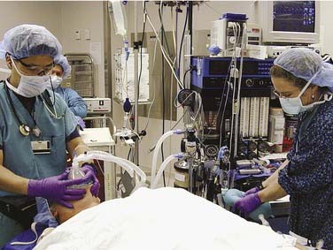
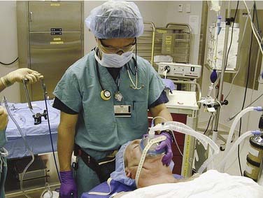
In patients without increased risk of aspiration, a controlled and stepwise approach is taken with induction and intubation. After monitoring and preoxygenation, general anesthesia is induced. Once the patient is unconscious, positive-pressure mask ventilation is performed (Fig. 9-7). Only after successful mask ventilation is established is a paralyzing agent given. This stepwise approach to the airway increases patient safety because, even if intubation cannot be performed successfully, it is known that the patient can be mask ventilated and oxygenated while the paralytic wears off or alternative intubation techniques are readied. The ability to ventilate a patient is more crucial than the ability to intubate a patient, and bag-valve-mask ventilation is a lifesaving skill that every anesthesiologist must master. After successful mask ventilation, the paralytic is given, and intubation is performed after the paralytic takes effect.


RSI and intubation are done for patients with an increased risk of aspiration, such as a patient with a full stomach or a patient with a significant history of gastroesophageal reflux. During an RSI technique, mask ventilation is not done, because it can fill the stomach with air and increase the risk of aspiration even further. Instead, the paralytic agent is given immediately after the induction agent. Cricoid pressure is held throughout, and the patient is not ventilated for the time it takes the paralysis to take effect.


Proper preoxygenation allows most apneic patients to maintain oxygen saturation during this minute. The patient is intubated once paralysis is achieved, usually by means of direct laryngoscopy. After confirmation of proper endotracheal tube placement by end-tidal CO2 and auscultation of bilateral breath sounds, the endotracheal tube cuff is inflated, and cricoid pressure can be released.


The risk of an RSI is that intubation may not be successful and the ability to mask ventilate the patient has not been previously established. The most dangerous result of failed RSI could be a paralyzed patient who cannot be ventilated or intubated (see Case 8).


Therefore, the stepwise approach to the induction of anesthesia and establishment of mask ventilation before paralysis is the safer and preferred technique for a patient without increased risk of aspiration and without an expected difficult airway. The stepwise approach for a patient with risk for aspiration and with an expected difficult airway requiring general anesthesia is awake versus RSI, with immediate backup in the event of failed intubation.


Difficult Airway/Intubation: Advances in Airway Devices and Techniques


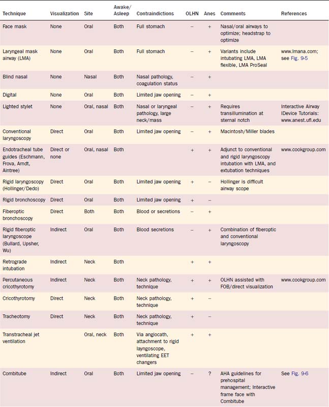
In parallel with organizational responses to difficult airway management, individual practitioners responded with inventions of new airway devices and with innovative combinations of existing techniques. In Table 9-1, a brief review of many of these devices is presented, with identification of primary use by anesthesiology and of OLHN. For a more in-depth discussion of the devices and appropriate techniques, refer to Airway Management: Principles and Practice22 and references identified in Table 9-1.


To appreciate the scope and magnitude of these efforts, consider that in the early 1990s, most anesthesiologists in the United States had the following airway management techniques: awake blind nasal, awake nasal fiberoptic intubation, and awake and asleep conventional laryngoscopy. Select difficult patients would undergo spontaneous ventilation “breathe-down” inductions without paralysis to facilitate optimal conditions for the OLHN surgeon to attempt to secure the patient’s airway with either rigid laryngoscopy or bronchoscopy. Complications of this technique included laryngospasm, aspiration, lost airway because of inability to maintain spontaneous ventilation, or difficult positive pressure mask ventilation, and inability to intubate by the OLHN surgeon.


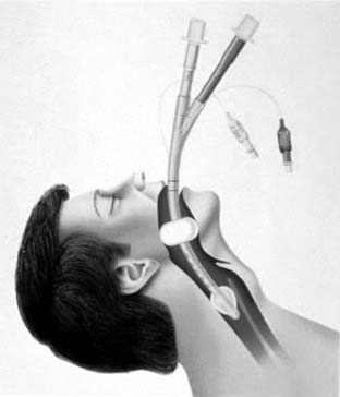
In the early 1990s, OLHN surgeons had the following airway management techniques: awake and asleep surgical airway, awake fiberoptic bronchoscopy, and asleep rigid laryngoscopy and bronchoscopy (Fig. 9-8).


Indisputably, the most significant invention in the recent history of airway management was the LMA (see Fig. 9-5).10 Although introduced into the United States in the early 1990s as an alternative to elective face mask ventilation (Fig. 9-9) in general anesthetics not requiring intubation, the value of the LMA as a rescue device for the most devastating situation “cannot intubate/cannot ventilate” quickly became realized. In the 1993 ASA Practice Guidelines for Management of the Difficult Airway, the LMA was the airway device that routed the Difficult Airway Algorithm into the significant branch points. In the 2003 amended Guidelines, the LMA was promoted as a first choice device for cannot-ventilate rescue options.23


In the past decade, for elective difficult airway management, the “family” of LMAs (Classic, ProSeal, Flexible, and Fastrach) has had a major impact on the decision branches with the Difficult Airway Algorithm. Specifically, many practitioners decide that if they can mask the patient with an LMA, definitive placement of an endotracheal tube (ETT) can be achieved with either fiberoptic bronchoscopy or ETT exchanger assistance. Backup plans, however, must be in place, because these techniques can be difficult and unsuccessful, as described in Case 7.


Table 9-1 briefly presents many of the advances in airway management devices discussed previously and throughout this chapter. References, including links to more comprehensive websites, are provided for additional information. Cases 1 to 10 span 10 years of experiences with difficult airway patients and advancements of airway management techniques.


The Johns Hopkins Departments of Anesthesiology and Critical Care Medicine and Otolaryngology—Head and Neck Surgery Airway Management Initiative and Formation of a Hospital-Wide Airway Service


The following section briefly outlines ongoing efforts of the Departments of ACCM and OLHN Surgery Airway Management Initiative and formation of a hospital-wide Airway Service.


Conceptual Framework


The Johns Hopkins Medical Institutions Departments of ACCM and OLHN Surgery responded to the release of the ASA guidelines by developing an Airway Management Initiative that has evolved over the past 15 years into the present-day Airway Management Service, education program, and hospital-wide difficult airway management response system. For the first 6 years, both departments provided financial support. However, in 1999, the Legal Department issued the following statement regarding the Airway Management Initiative and multidisciplinary Airway Service that resulted in full financial support by The Johns Hopkins Medical Institutions24:


Preoperative Evaluation and Identification of “Airway Alert”


Approximately 50% of patients seen for surgery are evaluated in our Preoperative Evaluation Center (PEC) several days or weeks before their scheduled procedure by the PEC staff, which consists of an attending anesthesiologist, anesthesiology resident, and nurse practitioner. The PEC anesthesiologist identifies the difficult airway/intubation patient on the basis of prior documented history, physical examination, or specific surgical needs. If consultation with an otolaryngologist or other specialist is needed to facilitate airway management, this is arranged. On the basis of the PEC staff evaluation, the patient is identified as an “Airway Alert” on the Preanesthesia Assessment Form and entered into the hospital’s operating room scheduling system (ORSCIS) with “Airway Alert” identified in the free text field associated with the surgical procedure. Whenever possible, an anesthesia attending is identified who will be available for assistance with airway management on the day of surgery and who has experience with the specific condition or airway difficulty the patient has and the recommended technique. However, the designated anesthesiologist and surgeon caring for the patient during the surgical procedure determine the final airway management plan. When appropriate, postoperative intensive care unit beds are requested to facilitate extubation management. The PEC anesthesiologist, and Airway Service members if consulted, discuss with the patient overall concerns and probable airway management plans A, B, and C. Discussions include any potential for the procedure to be aborted if issues arise during airway management that are not appropriately planned for. Written informed consent is obtained, including for elective tracheotomy if considered part of the airway management plans. For select patients, recommendations are made for enrollment into Medic Alert.


Before surgery, the anesthesiologist reviews the Preanesthesia Assessment Form to specifically identify the following information: surgical procedure, airway requirements (e.g., nasal/oral ETT, jaw wired at end of procedure, tracheotomy as part of procedure), patient’s yes/no response to the presence of gastroesophageal reflux/hiatal hernia (to assess aspiration risk and potential for awake vs. RSI technique), previous anesthesia experiences/airway difficulties, height and weight, and attempted-standardized airway examination (oral excursion in fingerbreadths [scale of 1 to 4], temporomandibular distance in fingerbreadths [scale of 1 to 4], upper and lower dentition [normal, dentures, caps, decayed], Mallampati I to IV, neck extension/flexion [scale of normal to decreased by 1 to 2]). The final patient interview and examination are conducted, and the anesthesiologist and surgeon (with assistance from Airway Service, as requested) formulate their actual plans A, B, and C.


Formulation of Intraoperative and Postoperative Plans A, B, and C


Members of the Airway Service are familiar with the ASA difficult airway/intubation algorithm and our institutional experience with rigid laryngoscopy as a primary otolaryngology technique within the ASA algorithm.


The first question the Airway Service asks is, “What is the surgical expectation at the end of the procedure?” Specific answers need to be provided for questions about if and at what stage of the procedure the patient might have a tracheotomy done, if the jaw will be wired closed, whether direct manipulation of the airway will result in edema or significant bleeding, whether positioning could cause airway edema, what will total fluid management entail, and what other considerations might suggest continued intubation at the end of the procedure. For these patients, precise extubation strategies are planned to include the location of extubation (intensive care unit vs. returning to the operating room for extubation) and physicians, staff, and equipment resources that must be available.


Next, the Airway Service explores the concept of “difficult.” Although the ASA attempts to distinguish between difficult mask and difficult intubation, when the patient does not have a documented prior airway history and is assessed to be difficult for either mask or intubation, considerations for awake intubation techniques are discussed. Obvious limitations to awake techniques are severe mental status issues (e.g., intoxication, noncooperative, mental retardation), severe respiratory/hemodynamic compromise, and unstable trauma. In our experience, patient refusal of an awake intubation has rarely been an issue, primarily because of the positive approach to awake intubation taken by the anesthesiologist during the discussion with the patient.


The next question that the Airway Service asks is, “Should the intubation be awake, oral, nasal, or surgical?” followed by, “What is the surgical preference for the ETT (cuffed or uncuffed, smaller-than-expected size, right-angle endotracheal (RAE) or nasal RAE tube, reinforced, laser, double lumen, microlaryngeal, or other specialty ETT)?”


Finally, the Airway Service addresses the issue of the patient’s surgical attending airway management expertise and comfort with specific airway management techniques. Specifically, if the situation deteriorates and an emergency surgical airway becomes necessary, who will do it: the patient’s surgical attending or OLHN member of the Airway Service?


Based on this knowledge, plans A, B, and C are developed by the Airway Service and communicated to the surgeon, patient, and nursing and support staff. Modifications to the plans are made jointly, before the initiation of any airway management.


Carts: Anesthesia Fiberoptic Cart and Adult Emergency Airway Cart


Two airway carts were created to address specific airway management situations: the Anesthesia Fiberoptic Cart and the Adult Emergency Airway Cart. Both are available with and without a monitor. Both carts include airway alert wristbands and information on how to enroll patients in the in-hospital registry, enter an airway note into the Electronic Patient Record (EPR), and enroll into Medic Alert. The complete contents for each cart are listed on the Adult Airway Management Card.


The Anesthesia Fiberoptic Cart is specifically set up for elective fiberoptic intubations or for fiberoptic verification of double-lumen ETT placement. It has both an LF 1/2 Olympus (3.5/4.0 mm) bronchoscope and adult bronchoscope, Olympus (5.0/6.0 mm) assorted airway management equipment, and topical anesthesia supplies. This cart is specifically designed to be used by anesthesiologists in the operating room.


The Adult Emergency Airway Cart is specifically set up for elective procedures in the operating room in which the following techniques are part of the airway management plan and for which the otolaryngologist has primary involvement: fiberoptic bronchoscopy, rigid laryngoscopy, rigid bronchoscopy, jet ventilation, and surgical airway. Adult Emergency Airway Carts are also located in all intensive care units and the Emergency Department. To note, monitors are included with these carts.


For an emergency in the operating room, our otolaryngologists prefer to use the Adult Emergency Airway Cart without the monitor and organized on a single flat-surface cart without numerous drawers. It has the same equipment as the preceding Adult Emergency Airway Cart, but the critical equipment, rigid laryngoscopes and adult fiberoptic bronchoscope, is already assembled for immediate use.


Staff: Anesthesiologist, Specialty Nursing Staff, Surgical Technologists, and Airway Equipment Specialist


For years, anesthesiologists were taught to believe that all aspects of airway management were the responsibility of the anesthesiologist, without specified assistance from nursing or support staff. At The Johns Hopkins Medical Institutions, it was the practice of otolaryngologists and pulmonologists to schedule patients with complex airway pathology to have a diagnostic fiberoptic bronchoscopy in the operating room, with monitored sedation by an anesthesiologist. It became increasingly apparent that successful management of these patients required a team of four professionals: an anesthesiologist to monitor and provide sedation, a surgeon or pulmonologist to perform the bronchoscopy, a nurse to assist with the bronchoscopy, and an equipment specialist to set up the equipment. We found that if any one of the team members failed to perform these duties, the patient’s airway management was compromised. We realized that it was not reasonable to expect that an anesthesiologist (or other physician with airway expertise) working alone could provide all the components of complex airway management in a timely and successful manner. When the OLHN surgeon was not immediately required, the Airway Service provided an additional anesthesiologist to provide patient monitoring and sedation while the airway management was being accomplished by the Airway Service and/or operating room anesthesiologist facile with complex airway management. The goals were successful airway management, patient safety, patient satisfaction, and efficient use of operating room resources.


The Airway Service currently has support staff from the Departments of ACCM, Surgery, and Nursing, including specialty operating room nurses, airway equipment specialists, certified surgical technologists (CST), and critical care technicians. Their primary responsibilities are to assist physicians in difficult airway management, maintain and upgrade airway equipment, and ensure that the organizational process of a functional airway team is maintained. They are also responsible for the education other operating room staff, specifically as to their roles in airway emergencies.

Stay updated, free articles. Join our Telegram channel

Jun 5, 2016 | Posted by in OTOLARYNGOLOGY | Comments Off on General Considerations of Anesthesia and Management of the Difficult Airway

Full access? Get Clinical Tree

Get Clinical Tree app for offline access