G
GABA See neurotransmitter.
galilean telescope See telescope, galilean.
galvanic nystagmus See nystagmus.
ganciclovir See antiviral agents.
ganglion An aggregation of nerve cell bodies found in numerous locations in the peripheral nervous system. Plural: ganglia.
ciliary g. A small reddish-grey body about the size of a pinhead situated at the posterior part of the orbit about 1 cm from the optic foramen between the optic nerve and the lateral rectus muscle. It receives posteriorly three roots: (1) the long, nasociliary or sensory root (or ramus communicans), which contains sensory fibres from the cornea, iris and ciliary body and some sympathetic postganglionic axons going to the dilator muscle; (2) the short (or motor root or oculomotor root) which comes from the Edinger–Westphal nucleus through the third nerve (oculomotor). It carries fibres supplying the sphincter pupillae and ciliary muscles; (3) the sympathetic root, which comes from the cavernous and the internal carotid plexuses. It carries fibres mediating constriction of the blood vessels of the eye and possibly mediating dilatation of the pupil. The ciliary ganglion gives rise to 6–10 short ciliary nerves. Syn. lenticular ganglion; ophthalmic ganglion.
See reflex, pupil light.
gasserian g. Sensory ganglion of the fifth nerve located in a bony fossa on the front of the apex of the petrous temporal bone. It receives the sensory portion of the fifth nerve (trigeminal) in the posterior part of the ganglion. From its anterior part the three divisions of the fifth nerve are given off: the ophthalmic (which contains the sensory fibres from the cornea and the eye in general), the maxillary and the mandibular nerves. Syn. semilunar ganglion; trigeminal ganglion.
See herpes zoster ophthalmicus.
lenticular g.; ophthalmic g. See ganglion, ciliary.
semilunar g. See ganglion, gasserian.
superior cervical g. One of the uppermost and largest ganglion in the two chains of sympathetic ganglia lying alongside the vertebral column. It is located just below the base of the skull between the internal carotid artery and the internal jugular vein. It gives rise to the internal carotid nerve, which forms the internal carotid plexus.
trigeminal g. See ganglion, gasserian.
gangliosidosis See disease, Sandhoff’s; disease, Tay–Sachs.
ganzfeld A visual stimulus that consists of completely homogeneous and colourless luminance conditions throughout. It is used especially when recording the standard electroretinogram.
gap junction See synapse.
Gardner Reversal-Frequency Test See test, developmental and perceptual screening.
gasserian ganglion See ganglion, gasserian.
gaussian approximation See ray, paraxial.
gaussian optics See optics, paraxial.
gaussian points See points, cardinal.
gaussian space See paraxial region.
gaussian theory See theory, gaussian.
gaze To fixate steadily or continuously.
See cardinal positions of gaze.
gene The unit of heredity which determines, or contributes to, one inherited feature of an organism (e.g. eye colour). Physically, a gene is composed of a defined DNA sequence, located at a specific place (locus) along the length of a chromosome and transmitted by a parent to its offspring. The DNA sequence of nucleotide bases (adenine, cytosine, guanine and thymine) encodes a specific sequence of amino acids corresponding to a particular protein. If the DNA sequence at one locus is identical on a pair of homologous chromosomes the organism is referred to as homozygous (homozygote) and if the DNA sequence is not identical it is referred to as heterozygous (heterozygote). The total effect of all genes influences the development and functioning of all organs and systems in the body.
See chromosome; genome; inheritance; mutation; pedigree.
gene-environment interaction A term used to indicate that an effect is due to a mixture of environmental factors (nurture) and genetic factors (nature). Most traits show gene-environment interactions, such as myopia, IQ test results, skin colour etc. Blood type and iris colour are predominantly genetically transmitted, whereas language is predominantly environmental.
See theory, biological-statistical; theory, nativist.
gene therapy A therapeutic method in which a defective gene is replaced by a normal copy of itself, thus restoring its function. There are several ways in which a new gene is carried into a diseased cell. A common method uses a retrovirus, an adenovirus or an adeno-associated virus as vectors to introduce genes into cells and DNA. This therapy has been used in the treatment of several eye diseases, especially retinoblastoma and retinitis pigmentosa, but so far with limited success.
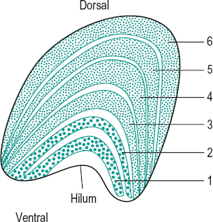
geniculate body, lateral (LGB) Ovoid protuberances lateral to the pulvinar of the thalamus in the diencephalon of the forebrain and into which the fibres of the optic tract synapse on their way to the visual cortex. However, because of the semidecussation of the optic nerve fibres in the optic chiasma, the lateral geniculate body in the right thalamus receives the fibres originating on the temporal retina of the right eye and the nasal fibres of the left. Each body appears, in cross-section, to consist of alternating white and grey areas. The white areas are formed by the medullated nerve fibres of the optic tract, while the grey areas consist largely of the nerve cells in which the fibres of the optic tract terminate (synapses) and from which arises the fibres of the optic radiations. There are six grey areas or layers of cells, with layer 1 being the most ventral and layer 6 the most dorsal (or posterior). Layers 1, 4 and 6 receive the crossed or nasal fibres from the contralateral retina, while layers 2, 3 and 5 receive the uncrossed or temporal fibres of the ipsilateral retina. The neural components of the LGB without the blood vessels and covering layer form the lateral geniculate nucleus (LGN).
There are two main types of cells in the LGN: in layers 1 and 2 (those most ventral) the cells are substantially larger than in the other four layers and are called magno cells and the layers, magnocellular layers. The main input to these cells are the retinal rods and the magno ganglion cells. In the other four layers (those most dorsal) the cells are smaller and are called parvo cells and the layers, parvocellular layers. The main input to these cells are the retinal cones and the parvo ganglion cells. The cells of the parvocellular layers seem to be mainly responsible for transmitting information about visual acuity, form vision, colour perception and low contrast targets. The cells in the magnocellular layers seem to be mainly responsible for transmitting information about motion and flicker perception, stereopsis and high contrast targets. The magnocellular and parvocellular cells project to different cells in the primary visual cortex (V1), where they retain the same segregation as in the lateral geniculate bodies. The receptive fields of the cells in the LGN are circular with either an ‘on’ or ‘off’ centre with the opposite behaviour in the surround, but they are more sensitive to contrast than the retinal ganglion cells (Fig. G1).
See brachium; cell, M; cell, P;fibres, visual; pathway, visual.
general refraction formula See paraxial equation, fundamental.
geniculocalcarine tract See radiations, optic.
geniculostriate pathway See pathway, geniculostriate.
Gennari, line of See line of Gennari.
genome The complete set of genes in an individual. In humans it is estimated at approximately 30 000 genes and over three billion base pairs (two nucleotides joined together across a double helix) of DNA.
genotype The complete genetic constitution of an individual at a particular location (locus) in the genome. At many locations (loci) throughout the genome, the chromosomal DNA sequence differs subtly between individuals. Each of the various DNA sequences at one locus is called an allele: for instance, if there are three sequence variants present, then there are three alleles. Offspring inherit one homologous chromosome from each parent. Thus, a genotype comprises two alleles: the allele inherited from the father (carried on the paternal chromosome) and the allele inherited from the mother (carried on the maternal chromosome).
gentamicin See antibiotic.
geometrical axis; optics See under the nouns.
gerontoxon See corneal arcus.
Gerstmann syndrome See syndrome, Gerstmann.
giant cell arteritis See arteritis, temporal.
giant papillary conjunctivitis See conjunctivitis, giant papillary.
giantophthalmos Megalocornea associated with an enlargement of the anterior segment of the eye.
See keratoglobus.
Giles–Archer lantern See test, lantern.
glabella 1. A prominent area of the frontal bone situated above the root of the nose. 2. The skin between the eyebrows, which is usually hairless. Syn. intercilium.
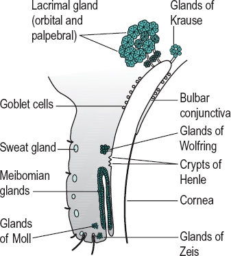
gland An aggregation of cells which secretes or excretes a substance. There are two main groups of glands: (1) The endocrine glands which have no duct and whose secretion (a hormone) is absorbed directly into the blood. Examples: adrenal gland, pineal gland, pituitary gland, thyroid gland. (2) The exocrine glands whose secretion reaches the surface by means of ducts. There are three main types of secretion by exocrine glands: the serous glands which secrete a watery substance rich in proteins (e.g. lacrimal gland, sweat glands), the mucous glands which secrete mucus, a viscous product (e.g. goblet cells), and the sebaceous glands which secrete a lipid substance (e.g. meibomian glands).
accessory lacrimal g’s. They are the glands of Krause and Wolfring. These glands are histologically identical to the main lacrimal gland, but are located within the eyelids. These glands are responsible for basal (not reflex) tear secretion and appear to be under sympathetic neural control.
g’s. of Ciaccio See glands of Wolfring.
ciliary sebaceous g’s. See glands of Zeis.
ciliary sweat g’s. See glands of Moll.
conjunctival g. Any gland that secretes a substance into the conjunctiva, such as the lacrimal, meibomian, Krause and Wolfring glands or a goblet cell.
g’s. of Henle These are not really glands. They are folds in the mucous membrane of the palpebral conjunctiva, situated between the tarsal plates and the fornices, in which there are goblet cells (Fig. G2). Syn. crypts of Henle (strictly speaking this term refers only to the pit-like depressions).
g’s. of Krause Accessory lacrimal glands of the conjunctiva having the same structure as the main lacrimal gland. They are located in the subconjunctival connective tissue of the fornix, especially the superior fornix (Fig. G2).
lacrimal g. A compound gland situated above and to the outer side of the globe of the eye. It consists of two portions: (1) a large orbital or superior portion; and (2) a small palpebral or inferior portion. It secretes the
See dacryoadenitis; dacryops; fossa for the lacrimal gland; nerve, zygomatic; tear duct.
g’s. of Manz Tiny glands located near the limbus. They secrete mucin. The existence of these glands in man is not established.
meibomian g’s. Sebaceous glands located in the tarsal plates of the eyelids whose ducts empty into the eyelid margin. They are arranged parallel with each other, perpendicular to the lid margin, about 25 for the upper lid and 20 for the lower. They secrete sebum. This sebaceous material provides the outermost oily (or lipid) layer of the precorneal tear film. It prevents the lacrimal fluid from overflowing onto the outer surface of the eyelid. It also makes for an airtight closure of the lids and prevents the tears from macerating the skin. The meibomian glands can be seen showing through the conjunctiva of fair-skinned people as yellow streaks (Fig. G2). Meibomian gland dysfunction (MGD) may be induced by blepharitis, chalazion, contact lens wear (particularly soft lenses) and ageing. The most common sign is a cloudy or absent secretion upon expression with symptoms of a mild dry eye. Hot compresses and lid massage will cure more than half of the patients; oral tetracycline will help in many of the others. Syn. palpebral follicles; tarsal glands.
See blepharitis, posterior; chalazion; film, precorneal; hordeolum, internal; keratoconjunctivitis sicca; meibomianitis; tarsus; Tearscope plus.
g’s. of Moll Sweat glands of the eyelids. They are situated in the region of the eyelashes (Fig. G2). Syn. ciliary sweat glands.
tarsal g’s. See glands, meibomian.
g’s. of Wolfring Accessory lacrimal glands of the upper eyelid situated in the region of the upper border of the tarsus (Fig. G2). Syn. glands of Ciaccio.
g’s. of Zeis Sebaceous glands of the eyelids which are attached directly to the follicles of the eyelashes. Their secretion contributes to the oily layer of the precorneal film (Fig. G2). Syn. ciliary sebaceous glands.
See blepharitis, marginal; hordeolum.
glare A visual condition in which the observer feels either discomfort and/or exhibits a lower performance in visual tests (e.g. visual acuity or contrast sensitivity). This is produced by a relatively bright source of light (called the glare source) within the visual field. A given bright light may or may not produce glare depending upon the location and intensity of the light source, the background luminance, the state of adaptation of the eye or the clarity of the media of the eye.
direct g. Glare produced by a source of light situated in the same or nearly the same direction as the object of fixation.
disability g. Glare which reduces visual performance without necessarily causing discomfort.
discomfort g. Glare which produces discomfort without necessarily interfering with visual performance.
eccentric g. See glare, indirect.
indirect g. Glare produced by an intense light source situated in a direction other than that of the object of fixation. Syn. eccentric glare.
g. source See glare.
g. tester An instrument for measuring the effect of glare on visual performance. There exist several (e.g. Brightness Acuity Tester (BAT), Miller–Nadler Glare Tester, Optec 1500 Glare Tester). Glare testing is valuable in patients with corneal and lenticular opacities before and after surgery and in elderly patients in whom adaptation to glare is usually more difficult. The Miller–Nadler Glare Tester consists of a glare source surrounding a Landolt C. The instrument contains 19 black Landolt C, all of the same size, 6/120 (or 20/400). Each Landolt C is presented in one of four orientations and from the highest to the lowest contrast at which the subject can no longer judge in which direction the letter appears. The contrast threshold is expressed in percentage disability glare.
The Brightness Acuity Tester (BAT) is a
Stay updated, free articles. Join our Telegram channel

Full access? Get Clinical Tree


