S
saccade See movements, fixation.
saccadic eye movement See movement, saccadic eye.
saccadic oscillation See flutter, ocular; myoclonus, ocular; opsoclonus.
saddle bridge See bridge, saddle.
safety glass See glass, safety.
sag Abbreviation for sagitta or sagittal depth; the height of a segment of a circle or sphere.
See lens measure; vertex depth.
sagittal depth See sag; vertex depth.
sagittal focus See astigmatism, oblique.
sagittal plane See plane, sagittal.
saline, physiological A 0.9% sterile solution of sodium chloride in water. This concentration of sodium chloride is considered approximately isotonic with the tears. It is used to store and rinse soft contact lenses, to irrigate the eye, etc. Syn. normal saline; NaCl 0.9%.
See eyewash; irrigation; solution, isotonic.
‘salt and pepper’ fundus See fundus, salt and pepper.
Salzmann’s nodular degeneration See degeneration, Salzmann’s nodular.
sample See sampling.
sampling The selection of a group of subjects from a population. This is usually done for the purpose of experimentation. The part of the population selected is called the sample: it is usually considered to be representative of a given population. A good sample must be random, i.e. every possible member of that population has an equal chance of being selected. Otherwise, it is said to be biased. Sampling can extend either across geographical areas (spatial sampling) or over a period of time (temporal sampling).
Sampaolesi’s line See line, Sampaolesi’s.
Sandhoff’s disease See disease, Sandhoff’s.
sarcoidosis An idiopathic, multisystem granulomatous disorder in which the eye is affected in about 20% of patients. Ocular manifestations include anterior uveitis with aqueous flare, keratoconjunctivis sicca, lacrimal gland lesion, ‘mutton-fat’ keratic precipitates, posterior synechia, iris granulomas and nodules, intermediate uveitis, posterior uveitis or optic neuropathy.
See dacryoadenitis; iridocyclitis; keratopathy, band; keratopathy, exposure; syndrome, Mikulicz’s.
sarcoma Malignant tumour formed by proliferation of mesodermal cells.
Sattler’s layer See layer, Sattler’s.
Sattler’s veil Clouding of vision accompanied by seeing coloured haloes around lights caused by corneal oedema resulting from contact lens wear, more frequently the hard lens type (PMMA). The cause has been shown to be due to a circular diffraction pattern formed by the basal epithelial cells and the extracellular spaces. Syn. Fick’s phenomenon.
See clouding, central corneal.
saturation Attribute of a visual sensation, which permits a judgment to be made of the proportion of pure chromatic colour in the total sensation. Note: This attribute is the psychosensorial correlate, or nearly so, of the colorimetric quantity purity (CIE).
scale, Snell–Sterling visual efficiency See visual efficiency scale, Snell–Sterling.
scanning laser polarimetry See polarimetry, scanning laser.
scatter illumination, sclerotic See illumination, sclerotic scatter.
scatter, Tyndall See effect, Tyndall.
scattering, Rayleigh Diffusion of radiation in the course of its passage through a medium containing particles the size of which is small compared with the wavelength of the radiation (CIE).
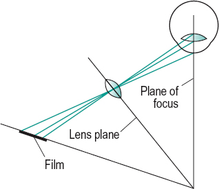
Scheimpflug photography An imaging technique that provides an assessment of the anterior segment of the eye in a sagittal plane. The principle of the technique is that the film plane, lens plane and subject plane (plane of focus) are tilted in such a way that all three planes intersect along a straight line. In this condition a subject will be completely in focus. The technique can be used to detect and monitor opacities in the media of the anterior segment (e.g. in the lens), to document these changes and to carry out biometric evaluation (e.g. depth of the anterior chamber, lens thickness measurement) (Fig. S1).
Scheiner’s disc; experiment; test See under the nouns.
schematic eye See eye, schematic.
Schiötz tonometer See tonometer, impression.
Schirmer’s test See test, Schirmer’s.
Schlemm’s canal See canal, Schlemm’s.
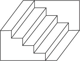
Schroeder’s staircase This is an ambiguous figure that gives rise to two different perceptions. It consists of a drawing of parallel step-like lines extending from the upper corner of a parallelogram to the lower opposite corner. One either sees a staircase from underneath or a staircase from above and, upon continuous viewing, the impression alternates (Fig. S2). Syn. Schroeder’s staircase visual illusion.
See figure, Blivet; Necker cube; Rubin’s vase.
Schwalbe, anterior limiting ring of; line See ring of Schwalbe, anterior limiting.
Schwann cell See cell, Schwann.
science of vision See vision science.
scimitar scotoma See scotoma, arcuate.
scintillans, synchisis See synchisis scintillans.
scintillating scotoma See scotoma, scintillating.
scissors movement See movement, scissors.
sclera The tough, white, opaque, fibrous outer tunic of the eyeball covering most of its surface (the cornea contributes 7% of, and completes, the outer tunic). Its anterior portion is visible and constitutes the ‘white’ of the eye. In childhood (or in pathological conditions) when the sclera is thin, it appears bluish, while in old age it may become yellowish, due to a deposition of fat. The sclera is thickest posteriorly (about 1 mm) and gradually becomes thinner towards the front of the eyeball. It is a sieve-like membrane at the lamina cribrosa. The sclera is pierced by three sets of apertures: (1) the posterior apertures round the optic nerve and through which pass the long and short posterior ciliary vessels and nerves; (2) the middle apertures, 4 mm behind the equator which give exit to the vortex veins; and (3) the anterior apertures through which pass the anterior ciliary vessels. The tendons of insertion of the extraocular muscles run into the sclera as parallel fibres and then spread out in a fan-shaped manner. The sclera is commonly considered to be divided into three layers from without inward: (1) the episclera, (2) the scleral stroma and (3) the suprachoroid (lamina fusca) which is interposed between choroid and sclera. Syn. sclerotic. Note: some authors consider the suprachoroid as belonging to the choroid. However, when choroid and sclera are separated part of the suprachoroid adheres to the choroid and part to the sclera.
See cribriform plate; evisceration.
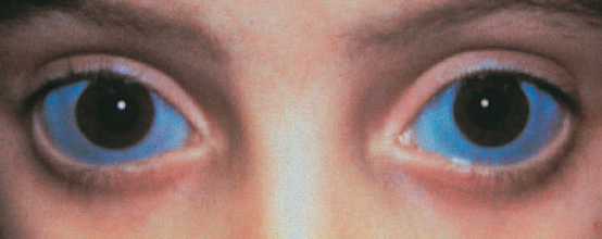
blue s. A hereditary defect in which the sclera has a bluish appearance. The sclera is thinner than normal and is susceptible to rupture if the person engages in contact sports. It is often associated with fragility of the bones and deafness as part of a condition called osteogenesis imperfecta (fragilitas ossium, van der Hoeve’s syndrome), with keratoconus or with acquired scleral thinning (e. g. necrotizing scleritis). Syn. blue sclerotic (Fig. S3).
See syndrome, Ehlers-Danlos; syndrome, Marfan’s.
scleral buckling See retinal detachment, rhegmatogenous.
scleral crescent See crescent, myopic.
scleral contact lens; ectasia; indentation; rigidity; ring See under the nouns.
scleral size, back Maximum internal diameter of the back surface of a scleral contact lens before the outer sharp edge has been rounded. Syn. back haptic size.
scleral spur A ridge of the sclera at the level of the limbus interposed between the posterior portion of Schlemm’s canal and the anterior part of the ciliary body. The scleral spur is the structure to which some of the ciliary muscle fibres are attached.
See gonioscopy; sulcus, internal scleral.
scleral zone The portion of a scleral contact lens designed to lie in front of the sclera. Syn. haptic.
sclerectasia See ectasia, scleral.
sclerectomy Surgical removal of a portion of the sclera performed in glaucoma surgery. A common procedure is to make a conjunctival flap at the limbus, followed by a full thickness scleral opening and iridectomy.
deep s. A type of non-penetrating filtration surgery aimed at lowering intraocular pressure by dissecting a superficial scleral flap and excising a deeper partial thickness scleral flap below leaving a thin membrane consisting of trabeculum and Descemet’s membrane through which the aqueous humour diffuses. It then drains from the anterior chamber to the subconjunctival space or though Schlemm’s canal. A collagen implant is usually inserted under the superficial flap to improve drainage of aqueous humour.
scleritis Inflammation of the sclera, which in its severe necrotizing or in the posterior type may cause sight-threatening complications such as keratitis, uveitis, angle-closure glaucoma or optic neuropathy. It affects females more commonly than males in the fourth to sixth decades of life. Like episcleritis it has a tendency to recur. It is characterized by pain, which can be severe, redness, tearing and some patients may develop nodules (nodular scleritis). It is often associated with a systemic disease (e.g. rheumatoid arthritis, Wegener’s granulomatosis, polyarteritis nodosa, lupus erythematosus, ankylosing spondylitis, syphilis, herpes zoster). It can involve part of the sclera, e.g. anterior scleritis (which is the most common, and it is classified as diffuse non-necrotizing or nodular non-necrotizing) or posterior scleritis. Treatment includes topical and systemic steroids and immunosuppressive drugs for very severe cases.
See keratitis, acute stromal; syndrome, Brown’s superior oblique tendon sheath.
necrotizing s. The most severe form of scleritis, much less common than the other types. About half the patients have one of the following diseases: rheumatoid arthritis, Wegener’s granulomatosis, polyarteritis nodosa, systemic lupus erythematosus, or herpes zoster. It is characterized by pain, and white, avascular areas next to damaged areas through which one can see the brown colour of the underlying uveal tissue, and to congested areas of the sclera. In most cases visual acuity is decreased. The necrosis gradually spreads around the globe. Treatment typically consists of topical steroids, immunosuppressive agents and occasionally surgery to repair scleral or corneal perforation.
See keratolysis; scleromalacia. s.
necroticans See scleromalacia.
posterior s. Inflammation of the sclera involving the posterior segment of the eye. The condition is often associated with a systemic disease (e.g. rheumatoid arthritis). It is characterized by pain and reduced visual acuity. The severity of the visual impairment depends on the involved tissue and its location. Signs include eyelid oedema, proptosis, limitation of ocular movements and, if anterior scleritis is present, redness. The ocular fundus may present disc swelling, choroidal folds, macular oedema and serous retinal detachment. Treatment consists mainly of systemic steroids and immunosuppressive agents.
See choroidal folds.
sclerochoroiditis Inflammation of the sclera and the choroid. It can occur as a result of pathological myopia and it presents with a posterior staphyloma in the region of the optic disc.
scleroconjunctival Pertaining to the sclera and the conjunctiva.
scleroconjunctivitis Inflammation of the sclera and of the conjunctiva.
sclerocornea A rare, congenital condition in which the sclera and cornea are considered as a single layer. The limbus is ill defined and portions of opaque scleral tissue with conjunctival vessels cover the cornea. The condition is usually bilateral and frequently associated with cornea plana. Visual acuity is reduced and often it is merely light perception if the entire cornea is involved. The eye is usually hyperopic. Systemic associations include mental retardation, deafness and craniofacial abnormalities. Treatment includes correction of the refractive error but in cases of central corneal opacification keratoplasty may be indicated.
scleroderma Thickening and hardening of the skin due to new collagen formation. It occurs either in localized or in systemic disease. It may very occasionally produce keratoconjunctivitis sicca. Syn. dermatosclerosis.
scleroiritis Inflammation of the sclera and of the iris.
sclerokeratitis Inflammation of the sclera and of the cornea.
scleromalacia A bilateral and painless degenerative thinning of the sclera occurring in people with rheumatoid arthritis. In this condition rheumatoid nodules may develop in the sclera and cause perforation (scleromalacia perforans). Syn. necrotizing scleritis without inflammation; scleritis necroticans.
sclerosis, central areolar choroidal See dystrophy, central areolar choroidal.
sclerosis, multiple (MS) An autoimmune disease in which there are disseminated patches of demyelination and sclerosis (or hardening) of the brain, spinal cord and peripheral and optic nerves causing paralysis, tremor, disturbance of speech, nystagmus, diplopia due to involvement of the extraocular muscles, and frequently retrobulbar optic neuritis.
See myokymia; oscillopsia; potential, visual evoked cortical; pupil, Marcus Gunn; Uhthoff’s symptom.
sclerotic See sclera.
sclerotic scatter illumination See illumination, sclerotic scatter.
sclerotomy Surgical incision of the sclera. It may be performed to extract an intraocular cyst, or to drain a choroidal haemorrhage.
scopolamine hydrobromide See hyoscine hydrobromide.
scotoma An area of partial or complete blindness surrounded by normal or relatively normal visual field.
See angioscotoma; hemianopia; quadrantanopia.
absolute s . A scotoma in which vision is entirely absent in the affected area.
See retinoschisis; scotoma, relative.
annular s. See scotoma, arcuate; scotoma, ring.
arcuate s . Scotoma running from the blind spot into the nasal visual field and following the course of the retinal nerve fibres. A double arcuate scotoma extending both in the upper and lower part of the field may join to make an annular scotoma or ring scotoma. A common cause is glaucoma. Syn. comet scotoma; scimitar scotoma.
See fibres, arcuate; retinal raphe; scotoma, Bjerrum’s.
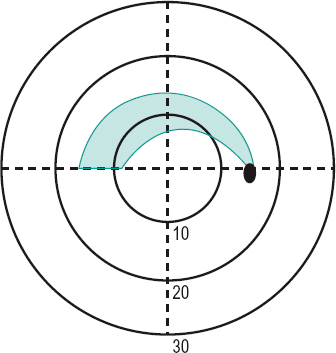
Bjerrum’s s . An arcuate scotoma extending around the fixation point (usually located between the 10° and 20° circles), which occurs in open-angle glaucoma. It often extends from the horizontal midline to the optic disc (Fig. S4). Syn. Bjerrum’s sign.
See Roenne nasal step; scotoma, Seidel’s.
central s. A scotoma involving the fixation area.
comet s. See scotoma, arcuate.
congruous s’. Scotomas in the two visual fields that are identical. They form a single defect in the binocular visual field. Such scotomas are often the result of lesions in the visual cortex.
flittering s. See scotoma, scintillating.
incongruous s’. Scotomas in the two visual fields that differ in one or more ways. Such scotomas are often the result of lesions in the optic tract.
junction s. A visual defect due to a lesion (e.g. a pituitary tumour) at the junction of one optic nerve with the chiasma where it is believed that the inferior nasal fibres of the contralateral optic nerve loop before passing backward to the optic tract. The visual defects typically consist of an upper temporal quadrantanopia in the field of the contralateral eye with, usually, a temporal hemicentral scotoma in the ipsilateral eye. Some authors attribute these visual defects to prechiasmal compression of one optic nerve plus compression of the whole chiasma.
See Wilbrand’s knee.
negative s. A scotoma of which the person is unaware. The physiological blind spot is an example of a negative scotoma but it is usually referred to as a physiological scotoma.
paracentral s. A scotoma involving the area adjacent to the fixation area.
physiological s. See scotoma, negative.
positive s. A scotoma of which the person is aware.
relative s. A scotoma in which there is some vision left or in which there is blindness to some stimuli, but not to others.
See scotoma, absolute.
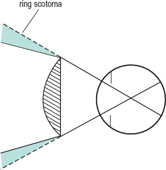
ring s. 1. An annular scotoma surrounding the fixation point. It may be formed by the development of two arcuate scotomas. Syn. annular scotoma. 2. A circular area in the peripheral field of view at the edge of a strong convex spectacle lens which is not seen (Fig. S5). This scotoma is due to the prismatic effect at the edge of the lens and unlike other scotomas, not from a pathological condition. When the head turns the ring scotoma also turns and it is then called a roving ring scotoma.
See phenomenon, jack-in-the-box.
roving ring s. See phenomenon,jack-inthe-box; scotoma, ring.
scimitar s. See scotoma, arcuate.
scintillating s. The sudden appearance of a transient, shimmering scotoma with a zigzag outline of brightly coloured lights (also called a fortification spectrum or fortification figures). It usually occurs as one of the first symptoms of a migraine attack. Syn. flittering scotoma.
See migraine; teichopsia.
Seidel’s s. An arcuate scotoma extending above and below the blind spot found in glaucoma. Syn. Seidel’s sign.
scotometer An instrument such as a campimeter or a perimeter for detecting and plotting the position and magnitude of a scotoma.
See campimeter; perimeter.
scotopia See vision, scotopic.
scotopic eye See eye, dark-adapted.
scotopic sensitivity syndrome See syndrome, Meares-Irlen.
scotopic vision See vision, scotopic.
screen, Bjerrum’s See screen, tangent.
screen, Hess A black tangent screen for measuring and classifying strabismus. It consists of a chart divided by red lines into small sections of 5° separations. As the screen is flat, the lines are curved in a pincushion pattern (i.e. the middle of the lines is closer to the centre of the screen than the ends of the lines). At the respective positions on the lines in the eight major meridians are small red dots indicating positions 15° and 30° from the fixation point. Two green threads extend from the upper corners of the screen and meet a third green thread, which originates on the end of a pointer. The three threads form a figure Y. The patient, wearing a red lens in front of one eye and a green lens in front of the other, moves the pointer until the common origin of the green threads is superimposed on each of the red dots. The discrepancy between the two on the screen indicates the extent of the deviation in various directions of gaze. It is a particularly useful test to indicate an incomitant strabismus as the field plots obtained with each eye are compared and an estimate of which muscle/s is affected can be made. Moreover, if the inner (15°) and outer (30°) plots are affected differently the incomitancy is of mechanical origin. Note: there exists a computerized version of the Hess screen.
screen, Lees An instrument similar to the Hess screen. It consists of two internally illuminated screens placed at right angles. One eye views one screen directly, the other views the second screen via a mirror; hence, the eyes are dissociated. When one eye fixates a point on one illuminated screen, its position as seen by the second eye is indicated with a wand on the second, non-illuminated screen. When this is illuminated, the position of the wand relative to the screen markings indicates whether a muscle or set of muscles is paretic or not.
screen, tangent A large plane surface for detecting and plotting the central visual field (about 50° in diameter) by moving the position of a stimulus (e.g. a white 1 mm pinhead). It consists of dull black cloth or other material perpendicular to the line of sight and placed usually 1 m away from the subject (2 m gives more accuracy). In the centre of the screen is a white spot that provides a fixation point and a series of radial and circumferential lines are sewn or drawn to facilitate the localization of the stimulus. Syn. Bjerrum’s screen.
See campimeter; perimeter.
screener, vision See vision screener.
SEAL See staining, fluorescein.
sebaceous gland carcinoma See carcinoma, sebaceous gland.
sebum See glands, meibomian.
seclusio pupillae A complete blocking of the anterior chamber from the posterior chamber by a posterior annular synechia.
See glaucoma, inflammatory; pupillary block.
secondary deviation; glaucoma; position See under the nouns.
secretion 1 . The substance produced by a cell or organ (e.g. a gland). 2. Production by a cell or organ of a physiologically active substance. This flow out of a cell is driven by an osmotic pressure gradient across the membrane, which is created by active transport of one or more ion species from one side to the other.
See transport, active; ultrafiltration.
see 1 . To perceive by the eye. 2. To discern. 3. To note: to understand.
see-saw nystagmus See nystagmus.
segment height The vertically measured distance from the lowest point on the lens to the top of the segment of a bifocal (or trifocal) ophthalmic lens. Syn. seg height.
segment of a bifocal lens (seg) An area of a bifocal lens of a power different to that of the main portion. Syn. portion. There is the distance portion, which has the correction for distance vision, and the near or reading portion, which has the correction for near vision.
See portion, intermediate.
segment of the eye, anterior Portion of the eye comprising all the structures situated between the front surface of the cornea and the front surface of the vitreous. The eyelids are sometimes included in this definition.
segment of the eye, posterior Posterior portion of the eye comprising the vitreous humour, the retina, the optic disc, the choroid and most of the sclera.
Seidel aberration See aberration, monochromatic.
Seidel’s scotoma See scotoma, Seidel’s.
semidecussation See hemidecussation.
semi-finished lens See lens, semi-finished.
semilunar fold See plica semilunaris.
senile macular degeneration See macular degeneration, age-related.
senilis, arcus See arcus, corneal.
sensation The conscious response to the effect of a stimulus exciting any sense organ.
See perception.
sensation, visual A sensation produced by the sense of sight.
sense Any faculty (or ability) by which some aspect of the environment is perceived. The five main senses are those of sight, hearing, smell, taste and touch. The sense of sight may be further divided into the colour sense, the form sense, the light sense, the space sense, etc.
sense organ A structure especially adapted for the reception of stimuli and the transmission of the relevant information to the brain. The organ of sight is the eye, in which light is transduced into nerve signals in the photoreceptors of the retina.
sensitive period See period, critical.
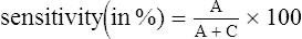
sensitivity 1. The capability of responding to or transmitting a stimulus. 2. The reciprocal of the threshold. 3. The extent to which a test gives results that are free from false negatives (i.e. people found not to have the defect when they actually have it). The fewer the number of false negatives, the greater is the sensitivity of the test. It is usually presented as a percentage of the number of people truly identified as defectives, referred to as true positives, A (or hit), divided by the total number of defective people tested. The total number includes all the true positives, A, plus the false negatives, C (or miss). Hence
See specificity.
sensitivity, contrast
Stay updated, free articles. Join our Telegram channel

Full access? Get Clinical Tree


