F
f number Designation for a photographic lens which gives the ratio of the focal length to the diameter of the effective aperture or entrance pupil. Example: f/8 means that the lens has a focal length eight times the diameter of the entrance pupil. Syn. f/stop; f-value; focal ratio; lens speed.
facet, corneal See corneal facet.
factor, daylight The ratio of daylight illuminance received at a given point inside a room to the simultaneous illuminance on a horizontal plane outside exposed to an unobstructed sky. It is usually expressed as a percentage, 5% or more being considered a well-lit room.
factor, reflection See reflectance.
factor, spectral transmission See spectrophotometer.
facultative hyperopia See hyperopia, facultative.
Faden procedure See procedure, Faden.
fallen eye syndrome See syndrome, fallen eye.
false macula See macula, false.
false negative See sensitivity.
false positive See specificity.
familial Pertaining to a condition or trait, either hereditary or acquired, which is found in more members of a family than would be expected by chance.
See acquired; congenital; hereditary.
familial autonomic dysfunction See syndrome, Riley–Day.
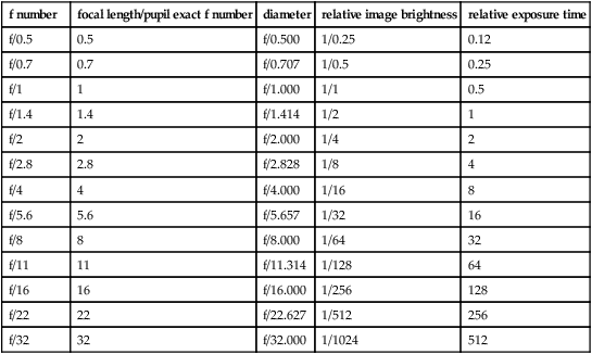
Table F1
Sequence of f numbers used in photography with the corresponding relative image brightness and exposure time to maintain constant film exposure. The relative image brightness is equal to the square of the f number fraction
| f number | focal length/pupil exact f number | diameter | relative image brightness | relative exposure time |
| f/0.5 | 0.5 | f/0.500 | 1/0.25 | 0.12 |
| f/0.7 | 0.7 | f/0.707 | 1/0.5 | 0.25 |
| f/1 | 1 | f/1.000 | 1/1 | 0.5 |
| f/1.4 | 1.4 | f/1.414 | 1/2 | 1 |
| f/2 | 2 | f/2.000 | 1/4 | 2 |
| f/2.8 | 2.8 | f/2.828 | 1/8 | 4 |
| f/4 | 4 | f/4.000 | 1/16 | 8 |
| f/5.6 | 5.6 | f/5.657 | 1/32 | 16 |
| f/8 | 8 | f/8.000 | 1/64 | 32 |
| f/11 | 11 | f/11.314 | 1/128 | 64 |
| f/16 | 16 | f/16.000 | 1/256 | 128 |
| f/22 | 22 | f/22.627 | 1/512 | 256 |
| f/32 | 32 | f/32.000 | 1/1024 | 512 |
fan and block test See test, fan and block.
fan chart See chart, astigmatic fan.
far point of accommodation See accommodation, far point of.
far point of convergence See convergence, far point of.
far point of the eye See accommodation, far point of.
far point sphere See sphere, far point.
far sight See hyperopia.
Farnsworth–Munsell 100 Hue test See test, Farnsworth.
Farnsworth test See test, Farnsworth; test, lantern.
fascia A sheet of connective tissue covering, partitioning or binding together muscles and certain other organs, such as the lacrimal sac, the orbital septum and other organs within the orbit, the sclera (e.g. Tenon’s capsule), etc.
fascia bulbi See Tenon’s capsule.
fascia, palpebral See orbital septum.
fasciculus, medial longitudinal One of a pair of nerve fibres, one on each side of the midline and extending from the upper midbrain to the cervical spinal cord. It is composed largely of ascending fibres from the vestibular nuclei ascending to the motor nuclei (third, fourth and sixth) and innervating the extraocular muscles; and to a lesser extent of descending fibres from the medial vestibular nuclei, the reticular formation, the superior colliculi and nucleus of Cajal innervating the musculature of the neck. Syn. medial longitudinal bundle; posterior longitudinal bundle.
fast eye movements See movements, saccadic eye.
fatigue, visual A feeling of weariness resulting from a visual task. It can be of ocular, muscular or psychic origin. However, there does not seem to be objective proof of a reduction in visual aptitude (e.g. visual acuity) accompanying visual fatigue.
See asthenopia.
feathers A cluster of fine bubbles or particles commonly arising from foreign material or from a fold in the glass in a molten or plastic state (British Standard).
Fechner’s colours See Benham top.
Fechner’s law See law, Fechner’s.
Fechner’s paradox Subjective impression of a decrease in the brightness of a field when viewing it binocularly, after one eye that was closed looks through a dark filter (about 5% transmission). This is paradoxical since more light is received by the eyes when the field is viewed binocularly, as compared to monocularly.
felderstruktur muscle fibres See fibres, felderstruktur muscle.
fenestration See lens, fenestrated; lens, scleral contact.
Fermat’s law See law, Fermat’s.
Ferry–Porter law See law, Ferry–Porter.
fibre A long thread or filament constituting human and animal tissues (e.g. nerve axon, muscle fibre, the filament of connective tissue).
arcuate f’s. Axons of the ganglion cells of the retina which are temporal to the optic disc and pass above and below the papillomacular bundle in an arcuate course. Syn. arcuate nerve fibres bundle.
See retinal raphe; scotoma, arcuate.
cilio-equatorial; cilio-posterior capsular f’s. See Zinn, zonule of.
circular f’s. See muscle, ciliary.
felderstruktur muscle f’s. A type of extraocular muscle fibres whose effect is to produce slow, and tonic contraction. They are mainly responsible for maintaining smooth pursuit movements. The fibres are located in the superficial portions of the extraocular muscles and are unique to this type of muscle.
fibrillenstruktur muscle f’s. A type of extraocular muscle fibres whose effect is to produce fast and twitch type of contractions. They are mainly responsible for saccadic eye movements. The fibres are located deep within the extraocular muscles and are the type usually found in skeletal muscles.
Henle’s f. See layer of Henle, fibre.
lens f’s. Long, six-sided bands containing few organelles and mostly lacking a nucleus, derived from epithelial cells just within the capsule of the crystalline lens and attached to an anterior and to a posterior suture. New lens fibres are continuously produced throughout life thus contributing to lens growth with age.
longitudinal f’s. See muscle, ciliary.
macular f’s. See fibres, papillomacular.
medullated nerve f’s. See fibres, myelinated nerve.
meridional f’s. See muscle, ciliary.
f’s. of Mueller See cell, Mueller’s.
myelinated nerve f’s. Anomalous congenital extension onto the retina of the myelin sheaths covering the optic nerve fibres. This myelination beyond the lamina cribrosa normally disappears soon after birth. Ophthalmoscopically, it appears as whitish, striated feather-shaped patches which may or may not obscure retinal vessels. Vision in these areas may be reduced, although visual acuity is not affected as the patches are most frequently located adjacent to the optic disc and sometimes in the periphery. The most characteristic sign may be an enlargement of the blind spot. Syn. medullated nerve fibres; opaque nerve fibres.
See cribriform plate.
orbiculo-anterior capsular; orbiculo-posterior capsular f’s. See Zinn, zonule of.
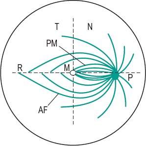
papillomacular f’s. Axons of the ganglion cells of the macular region of the retina which enter the temporal portion of the optic disc and travel in the central region of the optic nerve. In the optic chiasma, the temporal macular fibres remain on the same side, while the nasal ones cross to the other side. These fibres make up the papillomacular bundle (Fig. F1).
pupillary f’s. Axons of the optic nerve which branch off from the visual portion of the optic tract, before the lateral geniculate body, to run in the superior brachium towards the pretectal region anterior to the superior colliculus. They mediate the pupillary reflexes.
See pretectum; reflex, pupil light.
radial f’s. See muscle, ciliary.
visual f’s. Axons from the ganglion cells of the retina, making up the optic nerves and optic tracts. They synapse in the lateral geniculate body and then project to the region of the calcarine fissure of the cortex conveying the nervous impulses associated with vision.
zonular f’s. See Zinn, zonule of.
fibrillenstruktur muscle f’s. See fibres, fibrillenstruktur muscle.
fibrinoplatelet See plaques, Hollenhorst’s.
fibrocyte, corneal See corneal corpuscle.
fibroplasia, retrolental See retinopathy of prematurity.
fibrosis, preretinal macular Proliferation of glial cells over the surface of the internal limiting membrane of the macular region of the retina. Ophthalmoscopically the retina presents a glinting reflex. The condition may occur after trauma, eye surgery, retinal vascular disease (e.g. branch retinal vein occlusion) and inflammation and with any of the causes of retinitis proliferans and most commonly in elderly patients. Initially the patient is asymptomatic or reports some distortion of vision (metamorphopsia). This stage is often called cellophane maculopathy. As the condition develops, visual acuity diminishes, there is retinal wrinkling and the preretinal membrane becomes denser obscuring some retinal vessels in ophthalmoscopy. Some patients may also develop a macular hole and posterior vitreous detachment. If vision is significantly reduced, the main treatment is by vitreous surgery with removal of the layer of preretinal proliferative tissue. Syn. epiretinal membrane; macular epiretinal membrane; macular pucker; premacular fibrosis; preretinal membrane; preretinal vitreous membrane; surface wrinkling retinopathy.
See retinopathy, proliferative.
Fick, axes of See axes of Fick.
Fick’s phenomenon See Sattler’s veil.
field A limited area.
binocular visual f. An approximately circular zone of radius about 60° centred on the point of fixation (slightly larger in the lower part of the field) in which an object stimulates both retinas simultaneously. Beyond that area on each side, the visual field is monocular.
f. of excursion See field of fixation.
f. of fixation The area in space over which an eye can fixate when the head remains stationary. The field of fixation is smaller than the field of vision. It extends to approximately 47° temporally, 45° nasally, 43° upward and 50° downward. Syn. field of excursion; motor field.
See field of view, apparent; field of view, real; field, visual.
f. glasses See binoculars.
keyhole visual f. A term used to describe a visual field defect in which there is a bilateral homonymous hemianopia with macular sparing. An occipital lobe lesion sparing the posterior tips of the occipital lobe usually causes this lesion.
f. lens See eyepiece.
motor f. See field of fixation.
receptive f. The retinal area within which a light stimulus can produce a potential difference in a single cell. Retinal ganglion
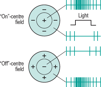
receptive fields are circular, often with a response different in the centre than in the periphery (also referred to as on-centre/off-centre or centre/surround organization). Ganglion cell receptive fields are very small in the macular region and large in the periphery of the retina. Receptive fields also exist in the lateral geniculate bodies where they are similar to those of the retina. In the visual cortex they have various shapes and sizes and may only respond to either a vertical bar or a black dot moving in a given direction and at a given speed, etc. Receptive fields reflect the interaction between excitation and inhibition between neighbouring neurons. The term can also describe the region of space that induces these neural responses (Fig. F2).
See cell, complex; cell, hypercomplex; cell, simple; inhibition, lateral; summation.
f. stop See diaphragm.
surrounding f. That area of the field of view surrounding any object.
f. of view The extent of an object plane seen through an optical instrument.
f. of view, apparent Angle subtended by the exit port of a sighting instrument or an empty frame aperture at the centre of the entrance pupil of the eye. Syn. apparent peripheral field of view. Note: when referring to the apparent field of fixation, the reference point is the centre of rotation of the eye. Syn. apparent macular field of view (Fig. F3).
See field of fixation.
Table F2
Average extent of the normal visual field (in degrees) of one eye of a young adult looking in the straight-ahead position, and measured with a white target subtending 1.0° under normal room illumination
| temporally | 94° |
| down and temporally | 88° |
| down | 70° |
| down and nasally | 54° |
| nasally | 60° |
| up and nasally | 56° |
| up | 54° |
| up and temporally | 64° |
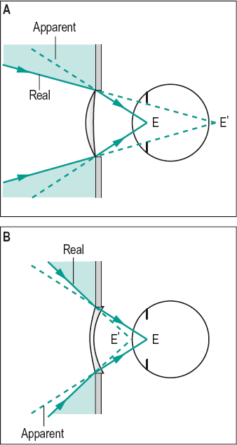
f. of view, real Angle subtended by the effective diameter of a lens at the point conjugate with the centre of the entrance pupil of the eye. Syn. real peripheral field of view; true field of view. Note: when referring to the real field of fixation, the reference point is the centre of rotation of the eye. Syn. real macular field of view (Fig. F3).
See phenomenon, jack-in-the-box.
f. of vision See field, visual.
visual f. (VF) The extent of space in which objects are visible to an eye in a given position. The extent of the visual field tends to diminish with age. The visual field can be measured either monocularly or binocularly. In the horizontal plane meridian the visual field extends to nearly 190° with both eyes open, the area seen binocularly, that is the region where both eyes can see the simulus is about 120°, and the area seen by one eye only is about 154°. Syn. field of vision.
See field, binocular visual; perimetry, kinetic; perimetry, static; test, confrontation; vision, island of.
visual f. expander An optical system designed to enlarge the field of vision. The most common types are reverse telescopes (e.g. looking through the objective of a galilean telescope), which minify objects being viewed but present more information by means of the enlarged visual field. They are usually of low power because of the reduction in visual acuity induced by the minification of the image. Prisms can also be used to expand the visual field. These systems are used mainly to improve mobility in patients with glaucoma and retinitis pigmentosa who have constricted visual fields or tunnel vision.
fifth cranial nerve See nerve, trigeminal.
figure A part or pattern in the visual field which has the perceptual attribute of completeness and is perceived as distinct from the rest of the field which forms the ground. Example: a printed word against a background page.
ambiguous f. An image or drawing arranged in such a way that its perception oscillates or flips involuntarily between, usually, two interpretations even though the retinal image remains constant, thus indicating that higher cortical processing are involved. Syn. reversible figure.
See figure, Blivet; figure, Kanizsa; illusion; Necker cube; Rubin’s vase; Schroeder’s staircase.
Blivet f. An ‘impossible’ figure in which three apparently solid tubes are attached at one end of a rectangular base which projects only two bars (Fig. F4).
See Necker cube; Schroeder’s staircase; Rubin’s vase.
fortification f. See scotoma, scintillating.
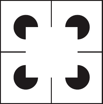
Kanizsa f. An ambiguous figure in which the illusory contour of a square (or triangle) appears in the middle of four (or three) truncated solid squares (or circles). It is an illustration of the perceptual ability to make sense of an incomplete figure by creating a ‘whole’ image from the separate elements (Gestalt organization). Some people cannot perceive the contour. Syn. Kanizsa square (Fig. F5).
reversible f. See figure, ambiguous.
filamentary keratitis See keratitis, filamentary.
film, anti-reflection See anti-reflection coating.
film, precorneal The field covering the anterior surface of the cornea which consists of lacrimal fluid and of the secretion of the meibomian and conjunctival glands. Its total thickness was thought to be about 9 μm but recent investigations have questioned that value and point to a much larger figure. It is composed of three layers: (1) The deepest and densest is the mucin layer (or mucous layer) which derives from the conjunctival goblet cells, as well as some secretion from the lacrimal gland. (2) The watery lacrimal fluid is the middle layer, called the lacrimal (or aqueous layer). It is secreted by the lacrimal gland and the accessory glands of Krause and Wolfring. It forms the bulk of the film and contains most of the bactericidal lysosyme and other proteins, inorganic salts, sugars, amino acids, urea, etc. (3) The oily layer (or lipid layer) is the most superficial and is derived principally from the meibomian glands in the lids as well as some secretion from the glands of Zeis. It greatly slows the evaporation of the watery layer and may provide a lubrication effect between lid and cornea (Fig. F6). Note: Some authors have suggested that the precorneal film is made up of only two layers; an innermost aqueous and mucin gel layer and an outer lipid layer. Syn. lacrimal layer; preocular tear film; tear film; tear layer.
Stay updated, free articles. Join our Telegram channel

Full access? Get Clinical Tree


