E
‘E’ game A technique used to evaluate visual acuity in young children. The letter ‘E’ is shown to the child, who is instructed to either state or point his or her fingers in the direction of the open side of the letter. The ‘E’ is subsequently rotated left, right, up and down randomly as its size is decreased, and acuity is obtained when the smallest letter is just recognizable.
See chart, illiterate E; test, Cardiff acuity.
Eales’ disease See disease, Eales’.
eccentric fixation; viewing See under the nouns.
eccentricity Term referring to the angular distance from the centre of the visual field or from the foveola of the retina. Example: the maximum density of rods in the retina is at a retinal eccentricity of about 20°.
echo See ultrasonography.
echography See ultrasonography.
echothiophate iodide See acetylcholinesterase.
eclipse blindness See blindness, eclipse.
econazole See antifungal agent.
ectasia, corneal A forward bulging and thinning of the cornea. It may result from a disease of the cornea (e.g. keratoconus), trauma, atrophy, raised intraocular pressure or as a complication of photorefractive surgery in which the corneal stroma has been left thinner than about 250 μm. If uveal tissue is included in the protrusion, the condition is called a staphyloma. If the ectasia is limited to a peripheral part of the cornea, it is called Terrien’s disease or Terrien’s marginal degeneration. It is due to degeneration of marginal corneal tissue with superficial vascularization and lipid deposition. It affects adult males more commonly than females and the eye has progressive astigmatism, typically against the rule. Therapy includes rigid contact lenses and occasionally keratoplasty. Syn. keratectasia; keratoectasia; kerectasis.
See degeneration, pellucid marginal; keratoglobus; staphyloma.
ectasia, scleral A bulging and thinning of the sclera, due to disease, trauma, atrophy or raised intraocular pressure. It may be total as in buphthalmos, or partial as in staphyloma. Syn. sclerectasia.
ectoderm The outermost of the three primary germinal layers of an embryo (the other layers being mesoderm and endoderm) from which the eye is derived. It differentiates into outer surface ectoderm and inner neuroectoderm, which gives rise to neural crest cells. The surface ectoderm gives rise to the crystalline lens, the lacrimal gland, the meibomian glands, the corneal and conjunctival epithelium and the epidermis of the eyelids. The neuroectoderm (neural ectoderm) will form the retina, retinal pigment epithelium, the pigmented and non-pigmented layers of the ciliary and iris epithelium, the dilator and sphincter muscles of the iris and the optic nerve fibres. Neural crest cells will form the corneal stroma and endothelium, sclera, iris and choroidal stroma, ciliary muscle and trabecular meshwork.
See cup, optic; mesoderm; vesicles, optic.
ectopia lentis See luxation of the lens.
ectopia of the macula See macula, ectopia of the.
ectopia pupillae See corectopia.
ectropion Outward turning of the eyelid margin. The most common cause is a loss of tonus of the pretarsal orbicularis muscle combined with laxity of the medial and lateral canthal tendons, which occurs in old people and affects only the lower eyelid (involutional ectropion). Tears collect in the lacrimal lake and overflow onto the skin of the face. Other causes of ectropion are scarring, burns, trauma (called cicatricial ectropion), spasm of the orbicularis muscle, which may affect either the upper or lower eyelid, or paralysis of the orbicularis muscle in which only the lower eyelid is affected. Ectropion may lead to exposure keratitis as the lower part of the cornea remains exposed. Management includes instilling an ocular lubricant and patching the eye during sleep as a temporary measure, but, if severe, the treatment is surgical.
cicatricial e.; involutional e. See ectropion.
congenital e . A rare congenital eversion of the eyelid (most often the lower). It may be due to a deficiency of the anterior eyelid lamina. It is usually associated with other disorders, such as blepharophimosis syndrome or Down’s syndrome.
e. uveae A turning of a portion of the posterior pigment epithelium of the iris growing or being drawn around the pupillary margin onto the anterior iris surface. It may be acquired (e.g. following iris neovascularization, neovascular glaucoma, iris melanoma) or congenital (e.g. neurofibromatosis).
eczema An inflammatory disease of the skin characterized by a rash of red spots, rough scaling, dryness and soreness of the skin sometimes leading to the formation of blisters. It often gives rise to itching or to a burning sensation. It may occur on the skin of the face where parts of spectacles rest. Frames should be cleaned regularly to avoid causing skin irritation. Syn. contact dermatitis.
edema See oedema.
edge clearance A small, peripheral gap between the edge of a rigid contact lens and the cornea. It is important as it allows tear exchange and eases lens removal. The absence of edge clearance in rigid contact lenses may lead to superficial corneal damage.
edge detection See contour.
edge lift Deviation of the posterior surface of a contact lens from a sphere at a given diameter. This is produced by either the peripheral curve(s) or the edging process. Edge lift provides peripheral clearance of a rigid contact lens, which is assessed by fluorescein pattern. If the edge lift is specified axially (as an extension of the back central optic zone, measured parallel to the axis of symmetry) it is referred to as axial edge lift. If specified radially as an extension along the back optic zone radius it is referred to as radial edge lift.
See optic zone diameter; optic zone radius, back.
edging Grinding the edge of a lens to the finished shape and size required, at the same time imparting the desired edge form, e.g. flat, bevelled, etc. This is accomplished with a machine called an edger, either by hand with a grinding wheel or automatically operating from a lens pattern or former.
See cribbing; former; glass cutter; glazing.
Edinger–Westphal nucleus See nucleus, Edinger–Westphal.
Edridge–Green lantern An occupational colour vision test that consists of small round and variable sized coloured lights produced by coloured and neutral density filters.
edrophonium chloride See anticholinesterase.
effect The result of an action or condition.
Aubert’s e. See , phenomenon Aubert’s.
Bezold–Brücke e. See phenomenon, Bezold–Brücke.
Broca–Sulzer e. The brightness produced by a flash of a given luminance depends upon its duration. It is maximum for durations around 30–40 ms when the flash luminance is photopic.
Brücke–Bartley e. An increased brightness produced by an intermittent light source (usually around 8–10 Hz) compared to the same light source viewed in steady illumination. Syn. brightness enhancement.
Cheshire cat e. A form of binocular rivalry in which a moving object seen by one eye can cause the entire image, or parts of the image, of a stationary object seen by the other eye to disappear. The effect can be observed by dividing the field of vision with a mirror placed edge-on in front of the nose at a slight angle. One eye looks straight at a stationary object, such as a sleeping cat, while the other eye sees a reflection through the mirror of a white wall or background. If a hand is waved on the mirror side in the region of the field where the cat is seen, the whole cat or part of it may be seen to disappear.
See retinal rivalry.
Craik–O’Brien–Cornsweet e. A phenomenon in which the brightness of an area on one side of a transition strip appears greater than the brightness of the area on the other side of the strip, although both areas outside the transition strip have exactly the same luminance. The transition strip consists of two opposing luminance gradients that meet along a linear edge (called Cornsweet edge); on one side the luminance gradually increases to the edge and on the other side the luminance gradually decreases to the edge. The area adjoining the gradient of increasing luminance appears brighter than the area adjoining the gradient of decreasing luminance. One possible explanation is that the edge information predominates and the visual system and brain ‘fill-in’ the area next to it to construct a higher brightness percept. Note: by covering the transition strip it is easy to confirm that the two areas have the same luminance. Syn. Craik–O’Brien–Cornsweet illusion; Cornsweet illusion.
crowding e. See phenomenon, crowding.
differential prismatic e. The difference in prism power induced by a pair of ophthalmic lenses of different power when the eyes look in various directions of gaze (except through the optical centres). Large amounts of differential prismatic effect can hinder fusion and give rise to diplopia. Example: A patient’s right eye is corrected by +5 D, the left eye by +2 D. When the eye rotates upward so that the visual axes intersect the lenses 1 cm above the optical centres, the induced prism power becomes 5 Δ base down on the right and 2 Δ base down on the left. The differential prismatic effect is 3 Δ base down in front of the right eye, probably too large for fusion to be maintained. Syn. prismatic imbalance; relative prismatic effect.
See anisophoria; law, Prentice’s.
Gelb e. In a faintly illuminated room a piece of black paper (or a rotating black disc) is illuminated by a high intensity projector. The beam of the projector falls exactly on the area of the black surface. The paper or disc will then appear to be white. A reversal of the perception is accomplished by placing a small piece of white paper near the disc in front of the projected light, at which time the paper or disc reappears in its true colour, i.e. black.
kinetic depth e. An impression of a three-dimensional structure of a moving two-dimensional shadow cast by a three-dimensional object. It is most easily demonstrated by casting a shadow onto a translucent screen.
Mandelbaum e. A tendency for the accommodative response to be altered when interposing a conflicting visual stimulus to the one being viewed. If the eyes are viewing a distant object through a dirty window or a wire fence, the actual accommodative response will tend to be raised. If the eyes are viewing a near object in front of a dirty window or wire fence the actual accommodative response will be less than if there were no conflicting stimulus.
McCollough e. A visual after-effect of colour that is seen when viewing, for a minute at least, two differently oriented and differently coloured gratings, such as a vertical grating with blue and black stripes and a horizontal grating with orange and black stripes. After adapting to these the subject looks at a figure containing a grating of vertical black and white stripes and a grating of black and white horizontal stripes of the same size as the original coloured gratings. The white stripes will then appear to be of the complementary colour, that is, the vertical stripes appear pinkish and the horizontal stripes appear bluish.
moiré e. An illusory shimmering movement produced by moving one pattern superimposed on another pattern very similar to it. The phenomenon occurs because parts of the periodic patterns are in phase in some locations, and out of phase in other locations. Examples: passing by a set of railings; if a transilluminated square wave grating is superimposed on an identical grating but cross each other at an angle of less than 45°, moiré fringes will appear at the intersections. Syn. moiré pattern.
See Toposcope.
oblique e. In central vision, contours with oblique orientations are perceived and discriminated less easily than those close to the horizontal or vertical.
Pulfrich e. See stereophenomenon, Pulfrich.
Raman e. In certain substances scattered light may be of a slightly different wavelength from that of the incident light.
Stiles–Crawford e. Variation of the luminosity of a pencil of light stimulating a given receptor with the position of entry of the pencil through the pupil. The maximum luminosity occurs for pencils passing through the centre of the pupil and stimulating the receptor along its axis. This phenomenon is attributed to the particular shape of the cone cells of the retina and occurs only in photopic vision.
Tyndall e. Diffusion of light by the particles present in a liquid or gas. It is because of this effect that heterogeneities (e.g. increased proteins) of the media of the eye can be seen, as occurs in iris and/or ciliary body inflammation. Syn. Tyndall scatter.
effective power See power, effective.
efferent Carrying nervous impulses away from the central nervous system to the periphery.
See afferent.
efficacy, luminous The amount of light emitted by a lamp for each watt of power consumed. It is expressed in lumens/watt.
efficiency scale, Snell–Sterling See visual efficiency scale , Snell–Sterling.
efficiency, spectral luminous (of a monochromatic radiation of wavelength λ) Ratio of the radiant flux at wavelength λm to that at wavelength λ such that both radiations produce equally intense luminous sensations under specified photometric conditions and λm is chosen so that the maximum value of this ratio is equal to one. Symbols: V(λ) for photopic vision. V’(λ) for scotopic vision (Fig. E1). Note: unless otherwise indicated, the values used for the spectral luminous efficiency in photopic vision are the values agreed internationally in 1931 by the CIE and adopted in 1933 by the International Committee on Weights and Measures. For scotopic vision the CIE in 1951 provisionally adopted new values for young observers (CIE). Syn. luminosity curve.
Efron grading scales See grading scales.
Egger’s line See ligament of Wieger.
egocentre A point of reference in the self usually located between the eyes. Absolute judgement of distances and visual directions of objects fixated binocularly are referred to the egocentre.
See localization; oculocentre.
egocentric localization See localization.
Ehlers–Danlos syndrome See syndrome, Ehlers– Danlos.
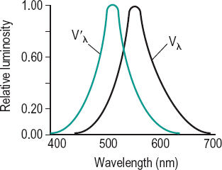
Table E1
Photopic and scotopic relative luminous efficiency factors. The data are based on an average from a large number of individuals agreed by the CIE in 1931 for the photopic factor Vλ and in 1951 for the scotopic factor V’λ
| wavelength (in nm) | Vλ | V’λ |
| 380 | 0.000 0 | 0.000 589 |
| 400 | 0.000 4 | 0.009 292 |
| 420 | 0.004 0 | 0.096 61 |
| 440 | 0.023 0 | 0.328 1 |
| 460 | 0.060 0 | 0.567 2 |
| 480 | 0.139 0 | 0.793 0 |
| 500 | 0.323 0 | 0.981 8 |
| 507 | – | 1.000 0 |
| 520 | 0.710 0 | 0.935 2 |
| 540 | 0.954 0 | 0.649 7 |
| 555 | 1.000 0 | – |
| 580 | 0.870 0 | 0.121 2 |
| 600 | 0.631 0 | 0.033 15 |
| 620 | 0.381 0 | 0.007 374 |
| 640 | 0.175 0 | 0.001 497 |
| 660 | 0.061 0 | 0.000 312 9 |
| 680 | 0.017 0 | 0.000 071 55 |
| 700 | 0.004 1 | 0.000 017 80 |
| 720 | 0.001 05 | 0.000 004 78 |
| 740 | 0.000 25 | 0.000 001 379 |
| 760 | 0.000 06 | 0.000 000 425 |
| 780 | 0.000 00 | 0.000 000 139 |
eidetic image See image, eidetic.
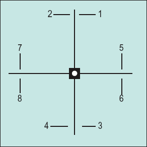
eikonometer Instrument for measuring aniseikonia. The direct comparison eikonometer (or standard eikonometer) uses as a target a cross with a small white disc at the centre of a black square at its intersection (Fig. E2). Four pairs of opposing arrows are placed four degrees away from the centre of the cross with the even numbered arrows polarized in one direction and the odd numbered ones in the other direction. The subject wears polarizing lenses so that each set of four arrows is seen by one eye. If the subject has aniseikonia, one set of arrows will not appear aligned with the other. Aniseikonia either in one or more meridians can be measured by means of an adjustable magnifying device before one eye. There is also a space eikonometer in which the parts of the target are seen three-dimensionally in space. An office model of this type has been manufactured. The space eikonometer is based on a modification of stereopsis. Aniseikonia will make the target appear tilted. The amount of aniseikonia is indicated by the power of the size lens that swings the target back into a frontoparallel plane. Syn. aniseikometer.
See lens, aniseikonic; plane, frontoparallel.
electrodiagnostic procedures Methods such as the electroretinogram, the electrooculogram and the visually evoked cortical potentials which are used to facilitate the diagnosis of some ocular diseases (e.g. retinitis pigmentosa) or the objective measurement of some visual functions (e.g. refractive error, visual acuity).
electroluminescence See luminescence.
electromagnetic spectrum See spectrum, electromagnetic.
electromyogram (EMG) Recording of electrical activity of a muscle associated with contraction and relaxation. This is obtained by placing a microelectrode within a muscle. The recording process is called electromyography.
See law of reciprocal innervation, Sherrington’s.
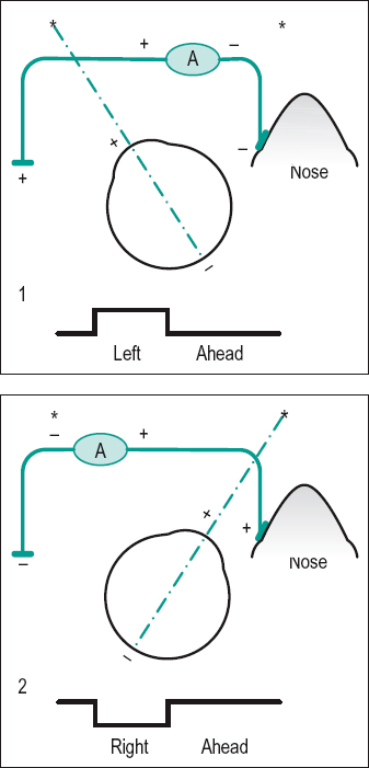
electrooculogram (EOG) Recording of eye movements and eye position provided by the difference in electrical potential between two electrodes placed on the skin on either side of the eye. The EOG consists of two potentials: the standing potential (resting potential, dark phase, dark current) which is evoked by moving the eyes in the dark and originates from the retinal pigment epithelium and the light potential (light rise) which is evoked by moving the eyes in a lighted environment and originates from the photoreceptors. Clinically, the ratio between the light and dark potentials (sometimes also called the Arden index or Arden ratio) is assessed. If that ratio is less than 1.8 it indicates a malfunction of the structures from which the potential originates. The EOG is also used to monitor eye movements (Fig. E3).
See disease, Best’s; fundus flavimaculatus; potential of the eye, resting.
electroretinogram (ERG) Recording of mass electrical response of the retina when it is stimulated by light. It is recorded by placing an electrode in contact with the cornea (often with the aid of a contact lens) or around the eye under the eyelid. A second electrode is placed either on the forehead or the face. The response is complex as many cells of various types contribute to it and varies according to whether the eye is dark or light adapted, the colour of the stimulus, the health of the retina, etc. The curve consists of two major components: a negative a-wave and a positive b-wave. The a-wave originates in the photoreceptors while the b-wave originates in the bipolar and Mueller cells. Both waves also have a photopic and a scotopic component.
pERG indicates that this potential is pattern-elicited. The ERG and pERG are useful indicators of the health of all the layers of the retina and can differentiate between the functioning of the rods and cones.
See alternating checkerboard stimulus; Leber’s congenital amaurosis; potential, early receptor; potentials, oscillatory; retinitis pigmentosa.
electroretinography, multifocal (mfERG) Simultaneous recording of the electroretinogram from small retinal areas (e.g. 103 hexagonal areas in the central 50 degrees of the retina) which are independently light stimulated according to a binary m-sequence (e.g. a random reversal at 75 Hz). This method enables evaluation of specific regions of the retina (e.g. macular degeneration) and mainly of the cone pathway.
elephantiasis oculi 1. Enlargement of the eyelids due to lymphatic obstruction. Syn. elephantiasis palpebral. 2. Extreme exophthalmos.
elephantiasis palpebral See elephantiasis oculi.
elevation of the eye
Stay updated, free articles. Join our Telegram channel

Full access? Get Clinical Tree


