Purpose
To compare the complications and outcomes of Descemet stripping automated endothelial keratoplasty (DSAEK) when the tissue is either folded and inserted with a forceps or inserted using a platform injector device without folding.
Design
Prospective, randomized, masked clinical trial.
Methods
DSAEK was performed in 100 eyes of 79 patients undergoing DSAEK surgery for Fuchs corneal dystrophy. Fifty eyes were randomized to have the donor tissue inserted with Charlie II insertion forceps (Bausch & Lomb Surgical) and 50 eyes were randomized to have the donor tissue inserted with the Neusidl Corneal Inserter (Fischer Surgical Inc). All other steps of the surgical procedure were exactly the same. Surgical problems, postoperative complications, and central endothelial cell density at 6 months were recorded and then measured by a masked observer. The study’s main outcome measures were total central endothelial cell density and percentage of donor endothelial cell loss from before surgery to 6 months after surgery and rate of complications (graft dislocation and primary graft failure).
Results
No primary graft failures occurred in either group and only 1 dislocation occurred in the series (Neusidl group). One late failure occurred at 6 months (Neusidl group). There was no difference in the preoperative endothelial cell density between the Neusidl and forceps groups, but there was a higher percentage of cell loss with the Neusidl group (33%) than with the forceps group (25%) at 6 months ( P = .017).
Conclusions
The Neusidl Corneal Inserter yielded a low immediate complication rate for DSAEK surgery for novice and experienced surgeons. Although still at an acceptable level, short-term endothelial survival was significantly worse after Neusidl tissue insertion than that after forceps tissue insertion.
Descemet stripping automated endothelial keratoplasty (DSAEK) has emerged as the most common method of surgical treatment for Fuchs corneal dystrophy and other conditions in which endothelial dysfunction is the primary cause of visual loss. It is now well established that the endothelial cell loss in the first 6 months after DSAEK surgery usually is higher than that associated with standard full-thickness penetrating keratoplasty. Donor endothelial cell death is related directly to the trauma that occurs at the time of surgery, and varying surgical techniques can have varying effects on the degree of cell loss and even early graft survival.
A critical step of DSAEK surgery is the insertion of the donor tissue into the recipient anterior chamber. One of the most common methods of insertion of the tissue is with the use of a forceps. It is now well established, both in the laboratory and clinically, that the size of the incision through which the tissue is placed has a great influence on the degree of endothelial damage and cell loss, with a 5-mm incision causing less cell loss and less donor failure than a smaller 3-mm incision. Avoiding the compression of the donor tissue by the recipient wound should result in less injury to the endothelium and should improve postoperative endothelial cell counts.
One method of avoiding wound compression would be a method of delivering the tissue to the anterior chamber with a tissue injector device. Although there are several tissue insertion devices on the market today for DSAEK surgery, none of these devices has undergone a prospective, randomized, double-masked clinical trial to compare the device performance directly with that of a standard forceps insertion technique. The purpose of this study was to compare the complications and 6 month outcomes of the Neusidl Corneal Inserter device (Fischer Surgical Inc, St. Louis, Missouri, USA) with those of a forceps insertion technique (Charlie II forceps; Bausch & Lomb Surgical, St. Louis, Missouri, USA) for DSAEK surgery in the treatment of common, uncomplicated Fuchs corneal dystrophy eyes.
Methods
Protocol
This randomized clinical trial was approved prospectively by the Legacy Health System Institutional Review Board, and patient data were collected and maintained in accordance with Health Insurance Portability and Accountability Act guidelines. The study is registered at http://www.ClinicalTrials.gov under clinical trial number NCT01357122 . An institutional review board-approved and Health Insurance Portability and Accountability Act-compliant clinical protocol and surgical consent form for endothelial keratoplasty were developed and enrollment was initiated for patients with endothelial dysfunction. All patients signed this institutional review board-approved clinical research consent form (see Supplemental Figure ).
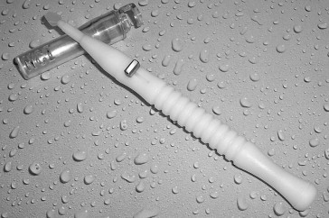
All cases in this report are patients who underwent DSAEK for Fuchs corneal dystrophy with surgery performed in the 18-month period between July 2009 and December 2010. There were a total of 100 cases of DSAEK for Fuchs corneal dystrophy performed during this time for this prospective, randomized, masked clinical trial to compare the results of tissue insertion with a Neusidl Corneal Inserter versus insertion with a standard Charlie II insertion forceps. See Figures 1 and 2 for pictures of both instruments.

Fuchs corneal dystrophy eyes with either a cataract or prior phacoemulsification cataract surgery with posterior chamber intraocular lens were candidates for this prospective study. Fuchs dystrophy eyes that had the accompanying risks of anterior chamber intraocular lenses, filtering blebs, tubes, or other anterior segment abnormalities that may influence postoperative endothelial survival were not entered into the study.
No specific requests were made of the eye banks to provide tissue for endothelial keratoplasty with any different donor characteristics than what we normally request for our full-thickness penetrating keratoplasty tissue. We accept donor tissue with an endothelial cell density (ECD) of more than 2000 cells/mm 2 , any age between 4 and 75 years, and death to transplantation time of up to 12 days.
The diameter of the donor tissue was chosen by the surgeon at the time of surgery and was not randomized. The donor diameter chosen was individualized for each patient and was based on the largest diameter circle that could be fit on the individual cornea without overlap of the edge of the tissue over the 2 limbal paracentesis sites or the corneal portion of the main insertion wound. No tissue was more than 8.5 mm in diameter in this study.
Surgery was performed by 4 corneal surgeons at Devers Eye Institute (2 experienced surgeons (M.A.T., N.S.) and 2 novice surgeons (M.D.S., J.M.G.) who were corneal fellows), with the senior surgeon (M.A.T.) performing 56% of the cases and the other experienced surgeon performing just 3 cases. All surgeons used the exact same surgical technique for every case, with the exception of the method of tissue insertion. The senior surgeon performed more than 20 practice surgeries using the Neusidl Corneal Inserter with cadaver eyes before this study and was first assistant for every case performed by the novice surgeons to assure that the surgical steps were performed exactly the same every time. All cases were randomized prospectively to insertion of the tissue using either Charlie II insertion forceps or the Neusidl Corneal Inserter. The surgeon was not aware of the method of insertion until the randomization envelope was opened at the time of donor preparation. The study was masked in that the patient was unaware of the method used for tissue insertion, and although the surgeon obviously was aware at the time of surgery which device was used, at the time of 6-month data acquisition, there were no identifying notes made in the clinic chart to allow the surgeon to recall which device had been used. Both the technician acquiring the endothelial specular image at 6 months and the observer performing the endothelial cell count analysis were masked to the insertion device used.
Prospective data collection was performed before surgery and at 6 months after surgery by a masked observer for the outcome measure of central ECD. Complications such as iatrogenic graft failure and donor dislocation requiring rebubbling also were recorded in a masked fashion.
Specular Microscopy Data
The vast majority of donor tissue came from the Lions VisionGift eye bank that used an EB-3000 XYZ eye bank specular microscope (HAI Laboratories Inc, Lexington, Massachusetts, USA). These preoperative cell counts were obtained using an apices digitized method and the manufacturer’s calibrations for magnification. The apices of at least 100 cells from the endothelial images of each cornea were counted. Preoperative donor tissue specular microscopy was performed by 3 trained, independent eye bank technicians who all had at least 1 year of experience with this technology. Postoperative specular microscopy measurements of ECD (Konan SP4000 noncontact specular microscope; Konan Medical Corp, Fairlawn, New Jersey, USA) were performed in our clinic at Devers Eye Institute at 6 months after surgery. A certified ophthalmic technician performed all postoperative testing of patients using the same specular microscope each time. Postoperative ECD was obtained using the center method of manually marking the center of each cell and using the manufacturer’s calibrations for magnification to calculate ECD. The automated method of determining cell borders and ECD specifically was not used. Analysis of endothelial pleomorphism and polymegathism was not performed in this study.
Surgical Procedure
DSAEK was performed with our standard technique as previously published. In all cases, the tissue was precut by an eye bank technician and then provided to the surgeon, usually within 26 hours of precutting. A video of our DSAEK technique using precut donor tissue is shown in Ophthalmology ‘s online video section (available at http://aaojournal.org ) from our previous report on the use of precut tissue in DSAEK.
In brief, our standard DSAEK procedure involves the use of a 5-mm temporal scleral access wound through which the donor tissue is placed into the eye. This wound is located approximately 0.5-mm peripheral to the temporal limbus with a sclerocorneal tunnel of approximately 2 mm in length until entry into the anterior chamber. To accommodate the Neusidl Corneal Inserter, all incisions in this study were determined by calipers to be 5.25 mm. This incision size of 5.25 mm was used for both the Neusidl and the forceps insertion cases.
The recipient’s Descemet membrane was stripped, the peripheral recipient bed was scraped with a Terry scraper (Bausch & Lomb Surgical), and the pupil was constricted. The donor tissue was prepared under the microscope at a separate donor table.
For the purposes of this study, the randomization envelope was not opened and the surgeon was not informed of the designated use of either forceps or Neusidl Corneal Inserter for tissue delivery until the step of donor preparation. Also, for uniformity of approach and ease of visualization, all donor tissue lenticules were stained with trypan blue before loading the tissue onto the forceps or onto the Neusidl Corneal Inserter. If the forceps was designated, the surgeon proceeded with our standard 40%/60% underfold technique, placing a thin strip of cohesive viscoelastic on the endothelium before folding and grasping the tissue. If the Neusidl Corneal Inserter was designated, the surgeon followed the manufacturer’s recommended standard operating procedure of loading the tissue stromal side down onto the platform, without the use of viscoelastic on the endothelium, and retracting the tissue into the injection tube, rolling the tissue up with the platform, without edge overlap.
For the forceps insertion of the tissue into the recipient eye, the 60% portion of the so-called donor taco was oriented anteriorly into the chamber. The tissue was unfolded with deepening of the anterior chamber with balanced salt solution and injection of air to complete unfolding of the tissue into position. For insertion of the tissue using the Neusidl Corneal Inserter, the tip of the device was placed into the wound, and the integrated irrigation of balanced salt solution through the tube was used on low flow to maintain the anterior chamber. The platform holding the donor tissue then was extended from the tube into the anterior chamber, with the platform anterior and the donor endothelial side posterior. The speed of flow of balanced salt solution through the device was varied by gravity infusion to maintain a formed chamber. The tissue was released from the platform, the platform then was retracted, and the tube tip was removed from the incision. For all cases, forceps and Neusidl Corneal Inserter alike, an air bubble then was injected through a paracentesis site, filling the chamber, beneath the donor to attach it to the overlying recipient bed. The interface fluid was removed with surface compression sweeping with the Cindy Sweeper (Bausch & Lomb Surgical), and no venting incisions were used. The full-chamber air bubble was left in place for 10 minutes after surface sweeping. The air bubble was removed completely from the chamber with an air-fluid exchange through the paracentesis site, and then final support of the tissue was accomplished with a reinjection of a free-floating 6- to 8-mm diameter air bubble placed into the anterior chamber at the close of surgery.
Statistical Analysis
Based on a computed effect size from our early studies of DSAEK transplants showing a mean percent cell loss of 34 ± 12%, a sample size analysis was performed. From this, we determined that 90 eyes were necessary to detect a difference of at least 10 percentage points between the 2 techniques, with α = 0.05 and β = 0.95. One hundred patients were enrolled to protect against attrition.
Treatment groups were compared with respect to preoperative demographic and donor tissue variables using the independent samples t test for continuous variables and the chi-square test for categorical variables. Postoperative endothelial cell density and percent cell loss were compared between treatment groups using an independent samples t test. The comparison of ECD and percent cell loss also was stratified by surgeon experience. Sample size estimation was performed using G*Power software version 3.1.2 (Franz Faul, Universität Kiel, Kiel, Germany). All other statistical analyses were performed with SPSS software version 19 (IBM, Chicago, Illinois, USA).
Results
Before surgery, every patient had a diagnosis of Fuchs endothelial dystrophy with clinically evident stromal edema and central guttata. There were 50 eyes in 41 patients that underwent DSAEK using the forceps insertion and 50 eyes in 38 patients that underwent DSAEK using the Neusidl Corneal Inserter for insertion. At 6 months after surgery, there were 92 specular microscopy images analyzed (48 Neusidl, 44 forceps) for a 92% overall rate of data capture. Eight of 100 (8%) cases were lost to follow-up for 6-month endothelial cell counts. The Supplemental Figure shows a flow diagram illustrating enrollment and randomization of study subjects (available at AJO.com ).
Demographics
The comparisons of recipient demographics and preoperative donor characteristics are summarized in Table 1 . The average age of the patients for both groups was approximately 69 years, and this difference was not statistically significant ( P = .700). Twenty-three eyes underwent DSAEK alone in the Neusidl group and 16 eyes underwent DSAEK alone in the Charlie II forceps group. Twenty-seven eyes underwent a triple procedure (DSAEK with phacoemulsification and intraocular lens placement) in the Neusidl group and 34 eyes underwent a triple procedure in the Charlie II forceps group. Rates of triple procedures were statistically similar between groups ( P = .151).
| Neusidl | Forceps | P Value a | |
|---|---|---|---|
| Recipient characteristics | |||
| Age (y), mean ± SD | 69.56 ± 10.56 | 70.34 ± 10.10 | .700 |
| Female, no. (%) | 39 (78) | 37 (74) | .640 |
| Triple procedures, no. (%) | 27 (54) | 34 (68) | .151 |
| Donor characteristics | |||
| Donor age (y), mean ± SD | 60.28 ± 9.28 | 58.29 ± 12.58 | .371 |
| Death to surgery (d), mean ± SD | 4.86 ± 1.39 | 4.92 ± 2.16 | .873 |
| Resection to surgery (d), mean ± SD | 1.16 ± 0.59 | 1.29 ± 1.55 | .607 |
| % graft diameter = 8.5 mm, no. (%) | 18 (36) | 24 (48) | .224 |
| Graft thickness (μm), mean ± SD | 162.44 ± 26.93 | 149.06 ± 27.06 | .015 |
Stay updated, free articles. Join our Telegram channel

Full access? Get Clinical Tree


