 case of a metal foreign body impacted in the retina and encysted in thick fibrous tissue is described below.
CASE STUDY
The patient had been injured 8 years previously while hitting tempered steel with a hammer. He had consulted an eye surgeon, who operated on him and declared that the foreign body had been removed. Thereafter, the patient had no further examinations. The patient presented to our clinic when a cataract started to develop. Ocular examination revealed that the foreign body was still present. This was confirmed with B-scan ultrasound and computed tomographic scan.
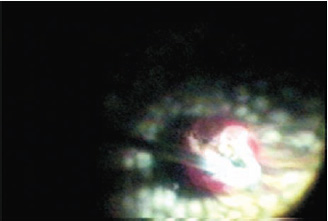
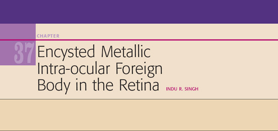
Figure 37.1. Encapsulated metallic foreign body in the retina.
Surgical Technique
- For our case, we adopted the standard pars plana vitrectomy technique.
- After clearing off the dense vitreous haze, we encountered an encysted iron foreign body impacted in the retina, completely surrounded with thick fibrous tissue (Fig. 37.1).
- During vitrectomy the foreign body was located. It was impacted in the retinal surface but cocooned within a layer of fibrosis that seemed to have enveloped it over the years. The fibrosis was so thick that it could not be opened with the tip of a vitrector or the sharp tip of a 24-gauge needle. We even tried using a diamond blade mounted on a 20-gauge tip to cut the shell open but to no avail. Even with the sharpest diamond blade, you need some counterpressure for the knife to work. We decided to use the Fugo blade. Before using the Fugo blade, however, sufficient endo-laser application was done around the point of impact of the foreign body.
- A plasma knife tip long enough to reach the retinal surface was used to expose the foreign body (Fig. 37.2 and 37.3).
- In only three to four repetitive passes of the Fugo blade, the encapsulated foreign body was sufficiently exposed (Fig. 37.4). During this process, no push was generated by the plasma knife, as there would be with a needle and a diamond blade.
- An intraocular foreign body forceps was then used to gently remove the foreign body from the fibrous shell and out of the eye through the sclerotomy opening (Fig. 37.5).
- In only three to four repetitive passes of the Fugo blade, the encapsulated foreign body was sufficiently exposed (Fig. 37.4). During this process, no push was generated by the plasma knife, as there would be with a needle and a diamond blade.
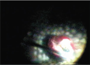
Figure 37.2. Fibrous capsule being cut with the Fugo blade.
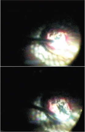
Figure 37.3. Cutting with the Fugo blade continues.
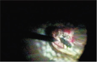
Figure 37.4. The encysted foreign body is sufficiently exposed.
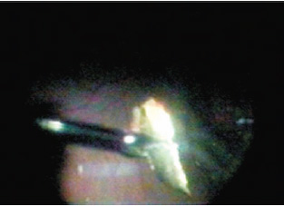
Figure 37.5. The foreign body is extracted from the fibrous capsule.
Summary
Clinical situations such as the case described here may arise only rarely in the course of an ophthalmologist’s career. We can think of no other way to successfully manage an encysted metallic foreign body. This case, and its outcome, strengthens our belief that the Fugo blade is a versatile cutting tool that can prove helpful in the most unlikely situations.
Suggested Reading
Singh IR. Managing proliferative vitreo-retinopathy with the Fugo blade—a case report. Trop Ophthalmol. 2001; 1:24–25.
Singh IR. Vitreo-retinal surgery with plasma blade: a case report. Trop Ophthalmol. 2001;1:13–16.
Singh IR, Singh D, Singh RK, et al. The plasma blade in vitreoretinal surgery. Ann Ophthalmol. 2001;33:280–289.
Stay updated, free articles. Join our Telegram channel

Full access? Get Clinical Tree


