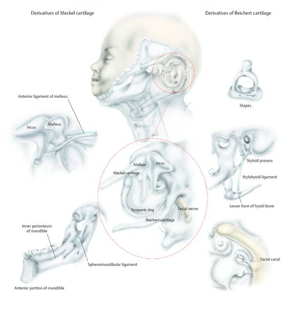
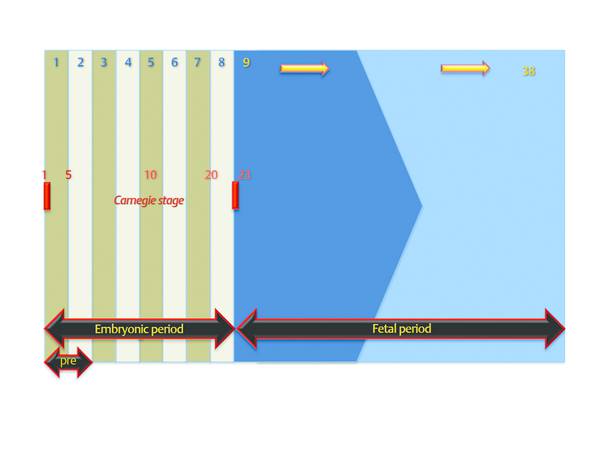
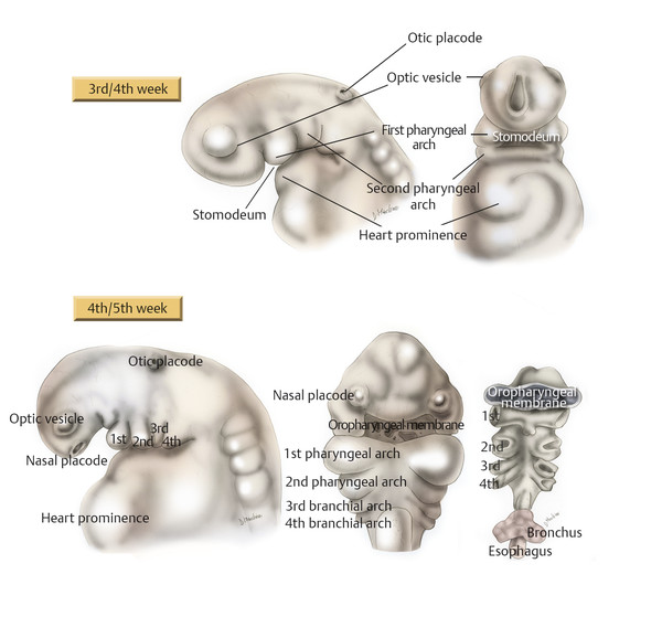
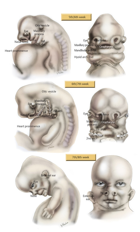
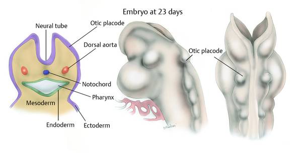
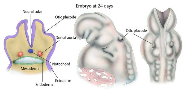
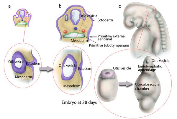
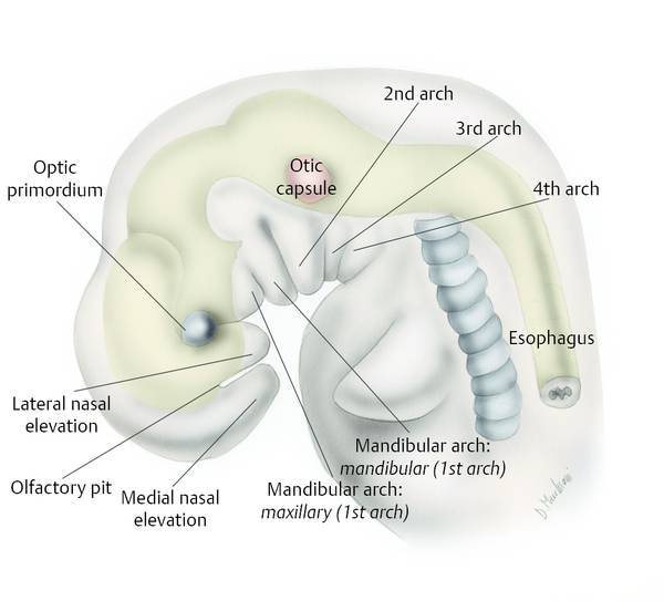
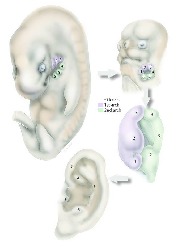
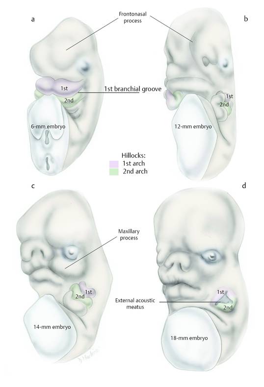
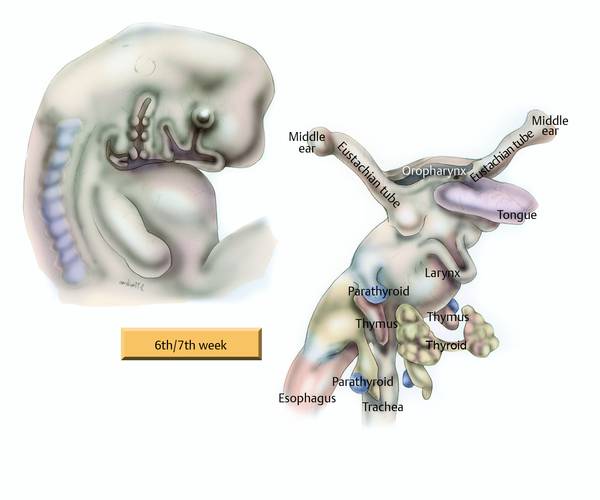
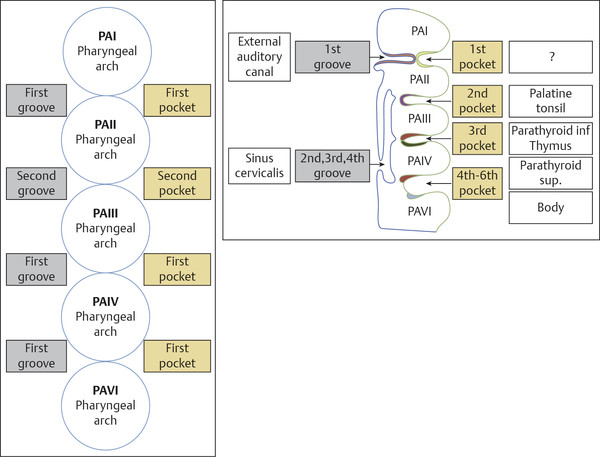
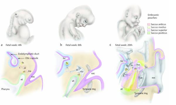
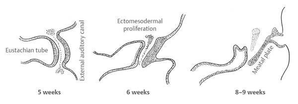
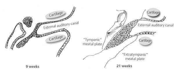
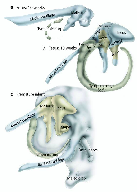
Prenatal development can be divided into a first period (preembryonic period) of cellular zygote division, which includes the 1st and 2nd weeks post conception; a second period, which runs from the 3rd to the 8th week, the definitive embryonic period; and a third period, from the 9th to the 38th week, the fetal period (▶ Fig. 3.1, ▶ Fig. 3.2, ▶ Fig. 3.3, ▶ Table 3.1).1–3 The first stages of ear development are very precocious, during the first half of the embryonic period, and are next only to those involving the formation of the cardiovascular system and the primitive placenta (days 21–22). Already between the 25th and the 28th days neuropores are closed and the neural tube is enclosed inside the cephalic end with the appearance of the otic placode (▶ Fig. 3.4, ▶ Fig. 3.5, ▶ Fig. 3.6, ▶ Fig. 3.7); optic vesicle and nasal placode appear between the 31st and 35th days. The pharyngeal arches appear between the 28th and the 32nd days (▶ Fig. 3.8).4,5 Fig. 3.1 Prenatal development. From the first period (preembryonic period) of cellular zygote division (1st and 2nd weeks post conception), through the embryonic period (from the 3rd to the 8th week), and to the fetal period (from the 9th to the 38th week). Fig. 3.2 The embryonic period. Development of the head from the 3rd to the 5th week. The oral cavity is starting to grow on the cephalic extremity like a superficial invagination, called the “stomodeum.” In the depth of this area the ectodermal layer is in direct contact with the endodermal layer. The connection of these two layers forms the oropharyngeal membrane. At 4 weeks the development of the nasal fossa starts as a nasal placode from two ectodermal thickenings located anteriorly over the embryo’s head. Fig. 3.3 The embryonic period. Development of the head from the 5th to the 8th week. From 5 to 7 weeks a median prominence overhangs the cephalic end of the oral cavity. This prominence is the frontonasal prominence. Nasal pits are located on either side of the frontonasal prominence and are surrounded by horseshoe-shaped eminences. The medial portion of these eminences is called the medial nasal process; the lateral portion is called the lateral nasal process. The lateral nasal process is separated from the maxillary process (the more rostral portion of the first branchial arch) by a furrow that reaches the medial aspect of the developing eye. This furrow is called the nasolacrimal groove. The nasal placodes begin to invaginate by the 5th week, forming the nasal pits. The nasal pits invaginate by forward growth of the medial nasal and lateral nasal processes, and by posteroinferior growth of the pits themselves, the placode tissue comes to line the roof of each pit. The nasal pits grow, approaching the primitive oral cavity. A thin oronasal membrane is located between the pits and the oral cavity. This membrane then ruptures and forms the primitive choanae. At 8 weeks the oral cavity is separated from the nasal cavity and the nasolacrimal groove is reabsorbed; the morphology of the face is developing. Fig. 3.4 Development of the inner ear: embryo at 23 days. Early in the 4th week, a thickening of the surface ectoderm on each side of the hindbrain (rhombencephalon) forms the otic placode. From this thickening will proceed the development of the primordial inner ear. Fig. 3.5 Development of the inner ear: embryo at 24 days. Inductive signals from the paraxial mesoderm and notochord stimulate the surface ectoderm in order to form the placodes. The otic placode soon starts to invaginate, and the ectoderm of the otic placode sinks deep into the mesoderm. Fig. 3.6 Inner ear, developmental stages: 2-week embryo (at 27 days). During this period the auditory placode ectoderm is invaginated into the mesoderm. It appears on the surface as the otic pit continuous with the surface ectoderm from which it was derived. At this time the neuropores are closed and the neural tube is enclosed inside the cephalic end. Fig. 3.7 Development of the ear: embryo at 28 days. Some crucial phenomena occur during this period in order to develop the inner ear. a The aperture of the otic pit narrows into the mesoderm by the meeting of its walls, creating an otic vesicle. This vesicle enlarges, growing chiefly downward. After the original continuity of the vesicle with external ectoderm is lost, for a short time the area of connection remains in the form of apposed thickenings between the superficial layer of the embryo and the lateral aspect of the primordial endolymphatic duct (a, b), after which the vesicle will become independent from the parental external ectoderm (b). At this stage the inner ears are developing. The development of the auditory vesicle continues in the downward direction as the utriculosaccular chamber, and upward as the endolymphatic appendage (c). Fig. 3.8 The branchial arches in a 5-mm human embryo. The development of the outer ear requires consideration regarding the pinna and external auditory canal and will, in fact, be integrated with the discussion of the tympanic ring and the development of the ossicular chain.6–8 The development of the pinna begins early in the second half of the embryonic period, at the 4th week. The first draft of the pinna is represented by a thickening of tissue upstream (mandibular arch) and downstream (hyoid arch), compared to the first branchial groove. Within 2 weeks they are to establish six hillocks, known as the “colliculi of His” (▶ Fig. 3.9, ▶ Fig. 3.10).6,9–11 The contribution of each of the six hillocks to the different components of the ear is still controversial, but knowledge of the position and number of morphological variants of the ear is of great interest in medical genetics. The pinna reaches the configuration of an adult as early as the 5th month, but continues to grow proportionately until the age of 9 years. Fig. 3.9 The development of the external ear commences at 4 weeks as tissue condensations of the mandibular and hyoid arches appear at the distal portion of the first branchial groove. After this time, from the tissue condensations arise six hillocks. At 7 weeks the six hillocks fuse into two folds: the anterior fold arises from the mandibular arch (purple color) and the posterior fold arises from the hyoid arch (green color). The folds unite at the upper end of this groove. From the fusion of these two folds will arise the external ear. Fig. 3.10 Oblique frontal views of the developing face and pharyngeal arches of a human embryo. a At the 6-mm embryo stage the first steps of external ear development are starting in the human embryo. b, c From a 12-mm to a 14-mm embryo: six hillocks form from the first and second branchial arches. d At the 18-mm embryo stage, two folds form from the fusion of the six hillocks: the anterior fold from the first branchial arch, and the posterior fold from the second branchial arch. The pharyngeal apparatus becomes defined early in the first half of the embryonic period. It consists of overlapping rings, called arches, each consisting of all three germ layers (ectoderm, mesoderm, and endoderm).4 In humans, six arches or pharyngeal arches are defined, identified by Roman numerals from I to VI. As well as arches, there are five pockets, and four grooves are recognizable. The pharyngeal apparatus is a fundamental structure in defining the anatomy of the head and neck-cephalic region (▶ Fig. 3.11, ▶ Fig. 3.12). The structures of the pharyngeal apparatus involved in ear development are those that derive from the first and second arches and from the depressions between them—the first groove and the first pocket. From the 4th week the first draft of the external auditory canal appears, in the form of an ectodermal depression of the dorsal portion of the first groove (▶ Fig. 3.13).6–10 Meanwhile, on the endodermal side, the first pocket deepens to form the primitive tubotympanic canal. At the 8th week, the ear becomes defined and from the cavum conchae a considerably deep tunnel shaped ectodermal invagination develops, carving out the primitive external auditory canal (▶ Fig. 3.13). Beginning from the 9th week, from the bottom of the primitive external auditory canal there originates an epithelial proliferation, called a meatal “flat” or “cap,” which is directed medially toward the bottom of the first pocket invagination. The mesoderm (ectomesoderm) that proliferates between the “meatal plate” and the endoderm of the first pocket gives rise to the fibrous tympanic membrane layer (▶ Fig. 3.14). The outer part of the “meatal plate” will form the epidermal tympanic membrane layer. The portion of the external ear canal that surrounds the “meatal plate” will result in the bony external auditory canal (▶ Fig. 3.15). Fig. 3.11 The development of the head and neck-cephalic region from the pharyngeal arches in a 6- to 7-week embryo. Fig. 3.12 The six pharyngeal arches (identified by numerals from I to VI). As well as arches, there are five pockets, and four grooves are recognizable. From the development of these pharyngeal arches it will be possible to define the anatomy of the head and neck-cephalic region. Fig. 3.13 The development of the middle ear. a In the embryo at the 4-week stage the endoderm of the pharyngeal pouch is well formed; the proximal part of the pharyngeal pouch is becoming constricted, forming the primordial eustachian tube, while its distal end is expanding as a flattened sac (which will be the primordial tympanic cavity). At this stage the endoderm of the pharyngeal pouch is almost in contact with the ectodermal branchial groove of the primordial external auditory canal. The primordial ossicles and the facial nerve are appearing. b At the 8-week stage the primordial ossicles are almost fully formed, the facial nerve runs over the stapes. The tympanic ring is forming and the endoderm of the pharyngeal pouch is in an opposing position with respect to the ectodermal branchial groove of the primordial external auditory canal; between these two structures there is the mesoderm. From the fusion of these layers will form the eardrum (the endoderm from the primitive pharynx will form the mucosal layer of the eardrum, from the mesoderm will be formed the fibrous layer, and from the ectoderm of the primordial external auditory canal will be formed the cutaneous layer of the eardrum). c At fetal week 20, the eardrum and the external auditory canal are already formed, also the ossicles are well formed, the facial nerve is in the correct position. Between the 3rd and 7th fetal months, the gelatinous tissue of the middle ear cleft is gradually absorbed. At the 20-week stage the development of the tympanic cavity starts. The tympanic cavity develops by a growth into the cleft of an endothelial-lined fluid pouch or sacci extending from the eustachian tube. There are four of these pouches: saccus anticus, saccus medius, saccus superior, and saccus posticus. During this fetal period the four pouches grow around the ossicular structures; in the course of the growth these pouches will contact each other and from this contact will be created a mucosal fold. Between the mucosal layers of the folds are remnants of the mesoderm, including blood vessels, the “viscera” of the tympanic cavity. At 30 weeks the production of the tympanic cavity will be complete. et, eustachian tube; in, incus; ma, malleus; s, stapes; fn, facial nerve; lsc, lateral semicircular canal; dr, eardrum; eac, external auditory canal; ct, chorda tympani. Fig. 3.14 From the 4th week the first draft of the external auditory canal appears, in the form of an ectodermal depression of the dorsal portion of the first groove. Meanwhile, on the endodermal side, the first pocket deepens to form the primitive tubotympanic canal. Fig. 3.15 The development of eardrum from embryonic week 9 to week 21. Around the 9th week, simultaneously with the formation of the “meatal plate,” a bony structure (the tympanic ring) is created; it provides connection and support for the tympanic membrane in the process of structuring. The tympanic ring is beginning to become clear from the 9th week, with the appearance of four centers of ossification surrounding the primitive tympanic membrane (▶ Fig. 3.16). At the 10th week, the four centers of ossification fuse together, giving rise to a circular structure, incomplete at the top, to form the “Rivinus mark.” At birth, the tympanic ring retains its morphology, reaching its final half-cylinder shape only in adolescence, after having reached its terminal development, called the “foramen of Huschke,” that characterizes this maturation process. Fig. 3.16 Left ear. Developmental anatomy of the tympanic ring from the 10th week to the premature infant. The tympanic ring is formed from four tiny ossification centers (a). Growth proceeds with the fusion of the separate centers of the bony membranes; this is a separate bony structure. During development this skeletal element will begin fusion with the temporal bone with an increase in overall size (b). Fixation of the tympanic ring to the temporal bone will be complete in the newborn, but an opening between the cranial portion of the tympanic ring and the temporal bone will be present forming the tympanic incisure (Rivinus) (c). At 19 weeks (b) ossification of the cartilage starts from the cartilaginous tissues of the malleus and incus; this is made possible at first by two ossification centers on the ossicles. The ossicular chain originates mainly from the mesenchyme of the first and second pharyngeal arches. The mesenchyme of the first arch (mandibular arch) will give rise to the Meckel cartilage and its derivatives, while from the mesenchyme of the second arch (hyoid arch) will arise the Reichert cartilage and its derivatives (▶ Fig. 3.17, ▶ Fig. 3.18).12,13
Week
Inner ear
Middle ear
Outer ear
3
Otic placode
Tubotympanic recess draft
4
Otocyst; vestibulocochlear subdivision
Ectodermal thickening
5
Draft of external auditory canal
6
Utriculosaccular subdivision
7
First cochlear spiral
8
Reunient duct
Cartilaginous incudomalleolar draft
Cartilaginous portion of the external auditory canal
9
Eardrum
11
2.5 cochlear spirals
12
Cochlear sensorial cell membranous labyrinth; otic capsule ossification
15
Cartilage stapes draft
16
Incudomalleolar ossification
18
Stapes ossification
20
Inner ear maturation
21
30
Tympanic cavity cavitation
32
Incudomalleolar ossification
34
Mastoid system
35
Antral cavitation
37
Epitympanic cavitation

3.2 Outer Ear Development
3.2.1 External Auditory Canal, Tympanic Ring, and Eardrum
3.3 Middle Ear Development
3.3.1 Ossicular Chain
Ento Key
Fastest Otolaryngology & Ophthalmology Insight Engine